
Give detailed simplified version of answer on granulomatous conditions of nose including stewarts granuloma, wegners, midline lethal, rhinoscleroma etc and ither such diseases for ms ENT university exam
Reading File
Now let me search the medical library for all the relevant granulomatous conditions of the nose simultaneously.
Finding Sources
Finding Sources
Finding Sources
Good - found relevant sources. Now let me pull the detailed content from the key ENT textbooks.
Finding Sources
Finding Sources
Finding Sources
Let me now read the key sections from Scott-Brown's and K J Lee's ENT textbooks, which are the most relevant for MS ENT exams:
Reading File
Reading File
Excellent! Found the Scott-Brown's Chapter 109 on granulomatous conditions. Let me read its full content now:
Reading File
Finding Sources
Let me continue reading Scott-Brown's chapter and also read Cummings' rhinoscleroma section:
Reading File
Reading File
Excellent! Let me read more of Scott-Brown's chapter (GPA/Wegener's, EGPA, NK/T-cell lymphoma sections) and more of Cummings (rhinoscleroma treatment + other conditions):
Reading File
Reading File
Let me read more of Scott-Brown's NK/T-cell lymphoma and sarcoidosis sections, plus the sarcoidosis diagnosis/treatment section:
Reading File
I now have excellent detailed content from both Scott-Brown's and Cummings. Let me read the sarcoidosis clinical features and treatment section from Scott-Brown's (which I partially skipped), plus the NK/T-cell section more fully:
Reading File
I now have comprehensive data from both Scott-Brown's and Cummings. I have enough material to write a thorough, exam-ready answer. Let me also quickly check the rhinoscleroma stages from Andrews' Diseases of Skin for completeness:
Reading File
I now have all the detailed content needed. Let me search for images to enrich the answer and also quickly look up rhinosporidiosis content:
Searching Images
rhinoscleroma nose Mikulicz cells histology
Finding Sources
I now have all the material needed to write a comprehensive, exam-ready answer. Let me compose the full response:
Granulomatous Conditions of the Nose - MS ENT University Exam Guide
A granuloma is an organized collection of macrophages (epithelioid cells) that fuse to form multinucleated giant cells. In the nose and sinuses, granulomatous conditions are broadly classified as:
| Category | Conditions |
|---|---|
| Infective - Bacterial | TB, Leprosy, Rhinoscleroma, Syphilis, Actinomycosis |
| Infective - Fungal | Rhinosporidiosis, Aspergillosis, Mucormycosis, Histoplasmosis, Blastomycosis |
| Infective - Protozoal | Leishmaniasis |
| Inflammatory / Autoimmune | Sarcoidosis, Wegener's (GPA), Churg-Strauss (EGPA), CIMDL |
| Neoplastic | Extranodal NK/T-cell lymphoma (Stewart's / Midline Lethal Granuloma) |
| Miscellaneous | Eosinophilic granuloma, Giant cell granuloma, Cholesterol granuloma |
1. RHINOSCLEROMA
Aetiology
- Caused by Klebsiella rhinoscleromatis (= Klebsiella pneumoniae subsp. rhinoscleromatis), first isolated by von Frisch
- Gram-negative, short, non-motile, encapsulated rod (2-3 µm), always found in a gelatinous capsule
- Route: direct inhalation of droplets or contaminated material
- Endemic in Central/South America, Mexico, Southeast Asia, Central/Eastern Europe
Pathology - Histology (HIGH YIELD)
- Mikulicz cells: large foamy macrophages containing the bacilli - PATHOGNOMONIC
- Russell bodies: eosinophilic hyaline degenerated plasma cells - also characteristic
- Bacilli are best demonstrated with Warthin-Starry silver stain
- Other cells: plasma cells, spindle cells, fibrosis
Three Stages (HIGH YIELD)
| Stage | Name | Features |
|---|---|---|
| Stage 1 | Catarrhal / Ozaena stage | Persistent mucopurulent rhinitis, crusting, fetor, resembles atrophic rhinitis |
| Stage 2 | Proliferative / Granulomatous stage | Nodular submucous swelling, hard woody/rubbery firm masses; Mikulicz cells present; nasal and lip enlargement |
| Stage 3 | Sclerotic / Cicatricial stage | Fibrosis, stenosis of nostril (alar stenosis), nasopharynx, larynx, trachea; fixed deformity |
Clinical Features
- Bilateral; slow, progressive; no systemic illness
- Most common site: anterior nasal septum and floor of nose
- Advanced stages: complete obstruction of nares, seropurulent exudation, gross facial disfigurement
- Can extend to nasopharynx, oropharynx, larynx
Diagnosis
- Biopsy with histology (Mikulicz cells + Russell bodies)
- Culture (Klebsiella on MacConkey)
- Complement fixation test (heat-killed antigen)
- Warthin-Starry stain for bacilli
Differential Diagnosis
Syphilitic gumma, sarcoidosis, leishmaniasis, yaws (frambesia), keloid, leprosy, hypertrophic TB, rhinosporidiosis
Treatment
- Fluoroquinolones (drug of choice) - prolonged course of at least 3-4 months to prevent relapse (ciprofloxacin / rifampicin)
- Tetracyclines / streptomycin (older regimens)
- Corticosteroids in acute phase
- Surgical/CO2 laser for airway stenosis or deformity correction
- Usually progressive and resistant to short-term therapy
2. GRANULOMATOSIS WITH POLYANGIITIS (GPA) / WEGENER'S GRANULOMATOSIS
Definition
A necrotizing granulomatous vasculitis affecting small-to-medium vessels, classically involving the upper respiratory tract, lower respiratory tract, and kidneys (classic triad).
Epidemiology
- Mean age at diagnosis: 40-50 years
- Male = female; predominantly Caucasian (>90%)
- Rare in children
Pathogenesis
- Autoimmune - strongly associated with c-ANCA (anti-PR3) - cytoplasmic ANCA specific for proteinase-3
- ANCA activates neutrophils → releases oxygen radicals, cytokines, lytic enzymes → kills vascular endothelial cells
- Association with chronic nasal Staphylococcus aureus carriage (more frequent relapses)
Classification
- Localized: respiratory tract only, no systemic vasculitis
- Early systemic / Generalized: multi-organ with features of vasculitis
Clinical Features - ENT (HIGH YIELD)
- Nose and sinuses involved in >80% - most common presenting site (73-93% of all cases)
- Symptoms: nasal obstruction, crusting, purulent/bloody discharge, epistaxis, facial pain
- Destruction of septum, turbinates → single large nasal cavity
- Septal perforation → Saddle nose deformity (collapse of nasal bridge)
- Key distinction: intranasal destruction BUT no gross destructive changes of midfacial skin (unlike NK/T-cell lymphoma)
- Subglottic stenosis in ~20%
- Otitis media with effusion (OME) due to eustachian tube involvement
- Sensorineural or conductive hearing loss
Systemic Features
- Kidneys: focal segmental glomerulonephritis (pauci-immune), may progress to renal failure
- Lungs: nodules, cavities, infiltrates; haemoptysis
- Eyes: proptosis, episcleritis, scleritis, orbital pseudotumour
- Constitutional: malaise, weight loss, fever
Investigations
- c-ANCA (PR3-ANCA): ~90% sensitivity in generalized disease, 60-70% in localized
- Urine: haematuria, proteinuria, red cell casts
- CXR/CT chest: nodules, cavities
- Biopsy: necrotizing granulomatous inflammation - best yield from upper respiratory tract
- CT sinuses: mucosal thickening, bony erosion
Treatment
- Induction: cyclophosphamide + high-dose prednisolone (generalized disease)
- Rituximab (anti-CD20): now preferred for induction in many centres, equivalent to cyclophosphamide
- Methotrexate + prednisolone: for localized/early systemic
- Maintenance: azathioprine or methotrexate
- TMP-SMX (Co-trimoxazole): reduces relapses by suppressing S. aureus carriage
- Surgical: septal reconstruction, subglottic dilatation, OME management
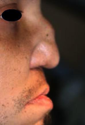
3. EOSINOPHILIC GRANULOMATOSIS WITH POLYANGIITIS (EGPA) / CHURG-STRAUSS SYNDROME
Definition
A systemic small-vessel vasculitis characterized by asthma, eosinophilia, and granulomatous inflammation.
Pathogenesis
- Associated with p-ANCA (MPO-ANCA) in ~40% of cases
- Eosinophilia and IgE deposition in vessels suggests allergic pathogenesis
- Reported after use of zafirlukast (leukotriene receptor antagonist) - actually unmasking of existing vasculitis as steroid dose reduced
Three Clinical Phases (HIGH YIELD)
| Phase | Features |
|---|---|
| Phase 1 - Prodromal | Adult-onset asthma + upper airway inflammation (may persist years) |
| Phase 2 - Eosinophilic | Peripheral eosinophilia + pulmonary infiltrates + eosinophilic gastroenteritis |
| Phase 3 - Vasculitic | Systemic vasculitis: constitutional, neurological, cutaneous involvement |
Clinical Features
- Asthma in 99% - late-onset, precedes vasculitis by average 8 years
- Sinonasal: allergic rhinitis, CRS with nasal polyps in up to 75%; obstruction, rhinorrhoea, anosmia, crusting
- Mononeuritis multiplex in up to 76% - foot drop etc.
- Cutaneous: papules, nodules
- Cardiac involvement = major cause of mortality (granulomatous myocarditis)
- Renal involvement less common than in GPA
Diagnosis
- Peripheral eosinophilia >10% of white cell differential + biopsy
- p-ANCA positive in ~40%
- ACR criteria (≥4 of 6): asthma, eosinophilia >10%, neuropathy, non-fixed pulmonary infiltrates, paranasal sinus abnormality, extravascular eosinophils on biopsy
Treatment
- High-dose corticosteroids (mainstay for less severe disease)
- Cyclophosphamide for severe/refractory
- Mepolizumab (anti-IL-5) - newer biologic approved for relapsing EGPA
4. EXTRANODAL NK/T-CELL LYMPHOMA / MIDLINE LETHAL GRANULOMA / STEWART'S GRANULOMA
Nomenclature History (HIGH YIELD)
This condition has had MANY names over the years:
- Stewart's granuloma (1933) - original description by JW Stewart
- Midline lethal granuloma or Lethal midline granuloma
- Midline malignant reticulosis
- Polymorphic reticulosis
- Current correct term: Extranodal NK/T-cell lymphoma, nasal type (WHO classification)
- Associated with Epstein-Barr virus (EBV)
Key Concept
"Midline lethal granuloma" is NOT a true granuloma - it is a lymphoma (NK/T-cell lineage, EBV-driven). This explains why it was historically mysterious, rapidly fatal, and unresponsive to conventional granuloma treatment.
Epidemiology
- More common in Asia, South America, Mexico (lower frequency in Western countries)
- Male predominance; middle-aged adults
Pathogenesis
- EBV-positive NK/T-cell lymphoma - cells express NK cell markers (CD56) and T-cell markers
- Progressive midface destruction driven by tumour infiltration + necrosis
- Ulcerates through nasal septum, palate, orbit, facial skin
Clinical Features (HIGH YIELD)
- Begins with non-healing nasal ulcer, nasal obstruction, epistaxis, offensive nasal discharge
- Relentlessly progressive midface destruction:
- Destruction of nasal septum, turbinates, lateral nasal wall, palate
- Involvement of orbital floor → proptosis
- Perforation through facial skin → grotesque deformity
- No systemic vasculitis - key distinction from GPA
- Gross midfacial skin destruction - key distinction from GPA (which has only mucosal/bony destruction)
- Constitutional symptoms: fever, weight loss
- Usually fatal within months if untreated
Investigations
- Biopsy (may need multiple attempts): shows lymphomatous infiltrate + necrosis; granuloma-like pattern
- Immunohistochemistry: CD56+, CD3+, TIA-1+, EBV+ (EBER in situ hybridization)
- No ANCA
- CT/MRI: midface destruction, orbital involvement
Treatment
- Radiotherapy (locoregional) - primary modality for localized disease
- Chemotherapy: CHOP or SMILE regimen (Steroid, Methotrexate, Ifosfamide, L-asparaginase, Etoposide) - preferred for disseminated disease
- Combined chemoradiotherapy for advanced stages
- Prognosis: historically very poor; better with modern protocols
5. SARCOIDOSIS (Nasal)
Definition
Systemic non-caseating granulomatous condition of unknown aetiology affecting any organ; nose/sinuses involved in ~20% of systemic cases.
Aetiology
- Unknown; immunological response to unidentified antigen in genetically susceptible individuals
- Possible: mycobacteria, fungi, beryllium, zirconium, pine pollen
- Macrophage + T-cell activation → TNF release → granuloma formation
- Non-caseating granulomas (no central necrosis) - KEY histological feature
Epidemiology
- Young adults, peak 3rd-4th decade
- Slightly more common in women
- 10-20x more common in African-Americans
- Incidence: 6-16/100,000; up to 64/100,000 in Scandinavia
ENT Features
- Lupus pernio: violaceous indurated plaques on nose, cheeks, lips - almost pathognomonic of sarcoidosis; associated with chronic pulmonary sarcoidosis
- Nasal: crusting, obstruction, epistaxis, anosmia
- Intranasal: pale nodular mucosal swellings (especially on septum and inferior turbinate)
- Subglottic stenosis
- Facial palsy (from parotid or temporal bone involvement)
- Bilateral parotid enlargement + uveitis + facial palsy = Heerfordt's syndrome (uveoparotid fever)
- Bilateral parotid enlargement + anterior uveitis + fever = Mikulicz syndrome (used loosely)
Systemic Features
- Lungs: bilateral hilar lymphadenopathy (BHL) - most common finding on CXR
- Eyes: uveitis, keratoconjunctivitis sicca
- Skin: erythema nodosum (acute), lupus pernio (chronic)
- Liver, spleen, lymph nodes
- Cardiac: arrhythmias, sudden death
Investigations
- CXR: bilateral hilar lymphadenopathy (staging: Stage I-IV)
- Serum ACE (angiotensin-converting enzyme): elevated in ~75% - useful for monitoring
- Kveim-Siltzbach test: intradermal injection of sarcoid tissue extract → positive granuloma formation (rarely used now)
- Serum calcium: hypercalcaemia (from 1-alpha-hydroxylase produced by macrophages)
- Biopsy: non-caseating epithelioid granulomas with Langhans giant cells, no acid-fast bacilli
- BAL: lymphocytosis with elevated CD4:CD8 ratio
- Gallium scan / PET: uptake in affected nodes
Treatment
- Many cases resolve spontaneously
- Oral corticosteroids: mainstay for symptomatic/progressive disease
- Methotrexate, azathioprine, hydroxychloroquine for chronic/steroid-sparing
- Anti-TNF (infliximab) for refractory cases
- Nasal: topical steroids, saline douching; endoscopic surgery for significant obstruction
6. NASAL TUBERCULOSIS
Pathology
- Caused by Mycobacterium tuberculosis
- Caseating (central necrosis) granulomas - KEY distinguishing feature from sarcoidosis
- Langhans-type giant cells, epithelioid macrophages, lymphocytes, caseation
- AFB on ZN stain
Clinical Features
- Most common site: cartilaginous part of nasal septum
- Crusting, septal perforation, nodular thickening of mucosa, ulceration
- Lupus vulgaris: painful nodular tuberculoid lesion; characteristic "apple jelly" appearance on diascopy
- Purulent rhinorrhoea, nasal fissures
- Saddle nose deformity with progression
Investigations
- Mantoux/TST or IGRA
- Biopsy: caseating granuloma + AFB
- Culture (gold standard)
- HIV testing
Treatment
- Standard anti-TB therapy (RHEZ for 2 months, RH for 4 months)
7. LEPROSY (Nasal)
- Caused by Mycobacterium leprae
- Nose is the most frequently affected site in leprosy - often the first site involved
- Route of infection: respiratory droplet from nasal secretions
- Features: epistaxis, crusting, nasal obstruction, anosmia
- Early: friable granulomatous intranasal lesions
- Advanced: septal perforation, nasal deformity, atrophy of nasal spine and premaxillary alveolar process, oroantral fistula
- Histology: Virchow cells (foamy macrophages laden with AFB = "globi") in lepromatous; epithelioid granulomas in tuberculoid
- Treatment: WHO multidrug therapy (dapsone + rifampicin ± clofazimine)
8. RHINOSPORIDIOSIS
- Caused by Rhinosporidium seeberi (currently classified as a protist/mesomycetozoea, not true fungus)
- Endemic in India, Sri Lanka, parts of Africa
- Route: contact with stagnant water/sand
- Most common site: nasal cavity (also conjunctiva, nasopharynx)
- Classic presentation: strawberry-like, pedunculated, vascular polyp in nose; bleeds easily on touch; multiple white dots on surface (sporangia)
- Histology: large sporangia (up to 300 µm) containing endospores within a fibrovascular stroma; inflammatory granuloma around ruptured sporangia
- Treatment: surgical excision with base cauterization (dapsone may reduce recurrence rate)
- Recurrence common
9. COCAINE-INDUCED MIDLINE DESTRUCTIVE LESION (CIMDL)
Clinical Importance
- Mimics GPA (Wegener's) and NK/T-cell lymphoma clinically and radiologically
- Caused by chronic intranasal cocaine use (vasoconstriction → ischaemia → necrosis)
Features
- Nasal septal perforation, saddle nose, palatal perforation
- No systemic vasculitis
- May have positive ANCA (due to non-specific inflammation) - can cause confusion with GPA
Key Distinguishing Features from GPA
| Feature | GPA | CIMDL |
|---|---|---|
| ANCA | c-ANCA (PR3) | p-ANCA (MPO) or negative |
| Systemic disease | Yes (kidneys, lungs) | No |
| History | No drug use | Cocaine history |
| Treatment response | Immunosuppression | Cessation of cocaine |
10. SYPHILITIC GUMMA (Tertiary Syphilis)
- Caused by Treponema pallidum
- Nasal gumma: destructive granulomatous lesion of septum/bridge
- Saddle nose deformity (cartilage/bone destruction) - classic
- Histology: gummatous necrosis, obliterative endarteritis, plasma cells, lymphocytes, absence of AFB
- VDRL/TPHA positive
- Treatment: penicillin G
11. EOSINOPHILIC GRANULOMA (Langerhans Cell Histiocytosis)
- Proliferation of Langerhans cells (CD1a+, S100+, Birbeck granules on EM)
- Unifocal bone lesion - can affect nasal bones, sinuses
- Usually young males
- Lytic bone lesion on X-ray
- Histology: Langerhans cells, eosinophils, giant cells
- Treatment: curettage, low-dose radiotherapy, or conservative
12. GIANT CELL GRANULOMA
- Benign giant cell lesion of jaw/sinuses
- Two types: central (within bone) and peripheral (on gingiva)
- Contains multinucleated giant cells in fibrovascular stroma
- May cause nasal obstruction if involving maxilla/sinuses
- Treatment: surgical excision; systemic steroids for aggressive lesions
13. CHOLESTEROL GRANULOMA
- Foreign body reaction to cholesterol crystals precipitating in sinus (from old haemorrhage + poor ventilation)
- Also called "chocolate cyst" or orbital cholesteatoma
- Most common in frontal sinus, also maxillary sinus
- CT: expansile cyst with bony erosion, no enhancement
- MRI: hyperintense on both T1 and T2 (diagnostic)
- Histology: granulation tissue with foreign body-type giant cells surrounding cholesterol clefts
- Treatment: surgical drainage and marsupialization (endoscopic or external)
COMPARATIVE SUMMARY TABLE (HIGH YIELD for Exams)
| Feature | Rhinoscleroma | GPA (Wegener's) | NK/T Lymphoma | Sarcoidosis |
|---|---|---|---|---|
| Cause | Klebsiella rhinoscleromatis | Autoimmune/c-ANCA | EBV + NK/T lymphoma | Unknown |
| Key cell | Mikulicz cell | Neutrophil/macrophage | NK/T lymphocyte | Epithelioid macrophage |
| ANCA | Negative | c-ANCA (PR3) +ve | Negative | Negative |
| Necrosis | Absent | Necrotizing | Extensive necrosis | Non-caseating |
| Skin destruction | No | No | Yes (midfacial) | Lupus pernio |
| Kidneys | No | Yes | No | Rarely |
| Treatment | Fluoroquinolones | Cyclophosphamide/Rituximab | RT ± Chemo (SMILE) | Steroids |
| Prognosis | Good with Rx | Good with Rx | Poor (historically) | Variable |
HIGH-YIELD EXAM POINTS SUMMARY
- Mikulicz cells (foamy macrophages) + Russell bodies (plasma cell derivatives) = Rhinoscleroma
- Warthin-Starry stain demonstrates Klebsiella in rhinoscleroma
- c-ANCA (anti-PR3) = GPA (Wegener's); p-ANCA (anti-MPO) = EGPA / MPA
- Saddle nose = TB, syphilis, leprosy, GPA, CIMDL (cartilage/bone destruction)
- Stewart's granuloma = Midline Lethal Granuloma = Extranodal NK/T-cell lymphoma (EBV-driven)
- Non-caseating granuloma = Sarcoidosis; Caseating = TB
- Asthma + eosinophilia + vasculitis = Churg-Strauss (EGPA)
- Lupus pernio = cutaneous sarcoidosis of the nose/face (nearly pathognomonic)
- Kveim test and elevated serum ACE = Sarcoidosis
- Rhinosporidiosis = strawberry polyp; sporangia on histology; treated by surgical excision
- NK/T-cell lymphoma = destructive midfacial skin lesion + EBV + CD56+ on IHC
- GPA destroys mucosa/bone internally but spares facial skin; NK/T lymphoma destroys facial skin externally
Sources: Scott-Brown's Otorhinolaryngology Head & Neck Surgery, Ch. 109 (Rimmer & Lund); Cummings Otolaryngology Head and Neck Surgery, Ch. 51; K J Lee's Essential Otolaryngology; Andrews' Diseases of the Skin
Internal maxillary artery ligation indication and method, instructions same as above answer
Finding Sources
Finding Sources
Found the key sections. Let me read them all in parallel now:
Reading File
Reading File
Reading File
Excellent! Let me read more of Scott-Brown's IMAL section and also get the Cummings chapter on epistaxis surgical management:
Reading File
Finding Sources
Let me now read the IMAL section that was partially cut off, and also get the Cummings epistaxis management section:
Reading File
Finding Sources
Finding Sources
Now let me get the anatomy of the internal maxillary artery, indications, and the Cummings chapter on epistaxis:
Finding Sources
Finding Sources
Finding Sources
Searching Images
internal maxillary artery anatomy pterygopalatine fossa branches diagram
I have all the key information I need from the textbooks. Let me now compose the comprehensive MS ENT exam answer:
Internal Maxillary Artery (IMAX) Ligation - MS ENT University Exam Guide
ANATOMY OF THE INTERNAL MAXILLARY ARTERY (HIGH YIELD)
The internal maxillary artery (IMAX / IMA) is the larger terminal branch of the external carotid artery, arising in the parotid gland behind the neck of the mandible.
Three Segments (Mnemonic: "DAB" / based on relations)
| Segment | Location | Key Branches |
|---|---|---|
| 1st - Mandibular (Retrocondylar) | Between neck of mandible and sphenomandibular ligament | Deep auricular, anterior tympanic, middle meningeal, accessory meningeal, inferior alveolar |
| 2nd - Pterygoid (Muscular) | Related to lateral pterygoid muscle | Deep temporal (anterior & posterior), pterygoid, masseteric, buccal |
| 3rd - Pterygopalatine | Pterygopalatine fossa | Sphenopalatine (terminal), descending palatine, posterior superior alveolar, infraorbital, artery of pterygoid canal, pharyngeal, greater palatine |
Key Surgical Anatomy Points
- The 3rd (pterygopalatine) segment is the surgical target for IMAX ligation
- Accessed via the posterior wall of the maxillary sinus (antrum)
- The IMAX terminates as the sphenopalatine artery (SPA), which is the main blood supply to the nasal cavity
- Branches identified in pterygopalatine fossa fat: proximal IMAX + descending palatine + sphenopalatine branches
- Infra-orbital nerve runs in the roof of the maxillary sinus and must be preserved
INDICATIONS FOR INTERNAL MAXILLARY ARTERY LIGATION
Primary Indication: Intractable Posterior Epistaxis
IMAX ligation (or its modern equivalent - sphenopalatine artery ligation) is indicated when:
-
Failure of conservative measures for posterior epistaxis:
- First-aid measures (pinching, cold compress) failed
- Anterior nasal packing failed
- Posterior nasal packing (Foley catheter balloon / Brighton balloon / post-nasal pack) failed for 24-48 hours
- Patient unfit to continue with packing
-
Repeated episodes of severe posterior epistaxis despite adequate packing
-
Epistaxis from a specific vascular territory supplied by IMAX / sphenopalatine artery:
- Most posterior and inferior nasal bleeding (Little's area excluded)
- Bleeding from posterior septum or lateral nasal wall
-
Bleeding from nasal tumours supplied by IMAX branches (pre-operative or palliative)
- Juvenile nasopharyngeal angiofibroma (JNA) - as adjunct to surgery
- Other vascular nasal tumours
-
Traumatic epistaxis - post-facial fractures (LeFort, NOE) not controlled by packing
-
Hereditary Haemorrhagic Telangiectasia (HHT / Osler-Weber-Rendu) - recurrent epistaxis refractory to local treatment
-
Failed embolization - when angiographic embolization is unavailable, contraindicated, or has failed
Note on Hierarchy of Surgical Options
The current trend has shifted away from IMAX ligation toward endoscopic sphenopalatine artery ligation (ESPAL) as the first-line surgical procedure, as it:
- Controls bleeding closer to the nasal source
- Higher success rate (approaching 100% in some series)
- Less morbid than open trans-antral approach
- IMAX ligation is now used when ESPAL has failed or when more proximal vascular control is needed
METHODS / SURGICAL APPROACHES TO IMAX LIGATION
Approach 1: TRADITIONAL TRANS-ANTRAL (SUBLABIAL / CALDWELL-LUC) APPROACH
This is the classical described method for MS ENT exams.
Pre-operative Preparation
- General or local anaesthesia with hypotensive technique
- Nasal pack removed just before procedure
- Patient supine, head-up 15° tilt
- Orotracheal intubation preferred
- Topical vasoconstrictor in nose
Step-by-Step Technique
Step 1: Sublabial Incision
- Horizontal incision in the upper gingivobuccal sulcus (sublabial), from canine fossa region extending laterally
- Incision carried down to bone
- Preserve the infra-orbital nerve (exits infra-orbital foramen ~1 cm below infra-orbital rim)
Step 2: Anterior Antrostomy
- Periosteum elevated off anterior wall of maxilla
- Anterior wall of maxillary sinus removed with osteotomes or burr - creating an antrostomy (canine fossa antrostomy)
- Entry into maxillary sinus (antrum) confirmed
Step 3: Exposure of Posterior Wall
- Mucosa of the posterior wall of the maxillary antrum is carefully elevated
- A window is made through the posterior wall of the antrum into the pterygopalatine fossa
- Use fine osteotomes or Kerrison punch to remove this bony wall
Step 4: Identification of IMAX in Pterygopalatine Fossa
- The branches of the internal maxillary artery are identified pulsating within the fat of the pterygopalatine fossa
- The fossa contains: IMAX and branches, maxillary nerve (V2), pterygopalatine ganglion
- Careful blunt dissection in the fat to expose the vessels
- The proximal IMAX, descending palatine artery, and sphenopalatine artery branches are all identified
Step 5: Ligation / Clipping
- Each vessel is carefully dissected out
- Haemostatic clips (silver/titanium) are applied to the proximal IMAX, descending palatine branch, and sphenopalatine branch
- Vessels are ideally clipped and divided
- Multiple clips applied to ensure complete control
- Bipolar diathermy may supplement clipping
Step 6: Closure
- Sinus mucosa repositioned
- Gingivobuccal incision closed with absorbable sutures
- Nasal pack may be replaced
Success Rate
- Trans-antral ligation controls haemorrhage in ~89% of cases
- Comparable to embolization in both cost and efficacy
Approach 2: ENDOSCOPIC TRANS-ANTRAL APPROACH (Modern Variation)
- A middle meatus antrostomy is performed as an instrument port
- A 4 mm endoscope is inserted through a small canine fossa antrostomy
- Posterior antral wall is removed endoscopically
- IMAX branches identified and clipped under endoscopic vision
- Useful when control of a damaged sphenopalatine artery is lost during ESPAL
- Less facial dissection than open sublabial approach
Approach 3: ENDOSCOPIC SPHENOPALATINE ARTERY LIGATION (ESPAL) - Now the Procedure of Choice
Although strictly the SPA rather than IMAX, this is the most commonly performed procedure for intractable posterior epistaxis today:
Technique:
- Under GA or LA
- Incision made approximately 8 mm anterior to and under cover of the posterior end of the middle turbinate
- Incision carried down to bone; mucosal flap elevated posteriorly
- Crista ethmoidalis (small crest of perpendicular plate of palatine bone, anterior to sphenopalatine foramen) - key landmark
- Sphenopalatine foramen lies just inferior to the horizontal attachment of the middle turbinate
- Fibro-neurovascular sleeve arising from the sphenopalatine foramen identified
- Haemostatic clips applied and vessel divided, or bipolar diathermy used
- Important: identify and control all branches (97% specimens have ≥2 branches medial to crista ethmoidalis)
COMPLICATIONS OF IMAX LIGATION (TRANS-ANTRAL)
| Complication | Notes |
|---|---|
| Infra-orbital nerve damage | Numbness / anaesthesia of cheek, upper lip, teeth |
| Dental damage / pain | Root damage during antrostomy |
| Sinusitis | Post-operative maxillary sinusitis |
| Oro-antral fistula | Through sublabial incision into sinus |
| Re-bleeding | From collateral anastomoses (10-15%) |
| Ophthalmoplegia | Rare - from spread of damage or haematoma |
| Blindness | Rare - from ophthalmic artery involvement or haematoma |
| Haematoma of pterygopalatine fossa | If venous plexus injured |
| Failure | If wrong vessels identified or collaterals active |
WHY IMAX LIGATION MAY FAIL
- Collateral circulation from contralateral side via anastomoses
- Ethmoidal artery contribution - anterior and posterior ethmoidal arteries (from internal carotid via ophthalmic artery) supply the superior nasal cavity and are not addressed by IMAX ligation
- Wrong identification of vessels (venous plexus mistaken for artery)
- Incomplete clipping - only one branch clipped; others continue to bleed
- In such cases, anterior/posterior ethmoidal artery ligation or external carotid artery ligation may be needed as adjuncts
COMPARISON OF SURGICAL OPTIONS FOR INTRACTABLE EPISTAXIS
| Procedure | Approach | Target | Success | Notes |
|---|---|---|---|---|
| ESPAL | Endonasal endoscopic | Sphenopalatine artery | ~95-100% | Current procedure of choice |
| IMAX ligation (trans-antral) | Sublabial / Caldwell-Luc | IMAX in pterygopalatine fossa | ~89% | Classical method; now 2nd line |
| Ethmoidal artery ligation | Medial canthal incision | Anterior/posterior ethmoidal arteries | Adjuvant | For superior/ethmoidal bleeding |
| External carotid ligation | Neck incision | ECA at bifurcation | ~93% | For extreme/non-rhinologist cases |
| Embolization | Angiographic | Sphenopalatine / IMAX | ~87-90% | Available 24/7 in specialist centres; prior IMAX ligation hampers this |
HIERARCHICAL MANAGEMENT OF EPISTAXIS (SUMMARY)
STEP 1: First aid - compress Little's area, head-forward, cold compress
STEP 2: Anterior rhinoscopy + cauterization of bleeding point (silver nitrate / electrocautery)
STEP 3: Anterior nasal packing (ribbon gauze / BIPP / Merocel)
STEP 4: Posterior nasal packing (Foley balloon / Brighton balloon / post-nasal pack) 24-48 hrs
STEP 5: SURGICAL OPTIONS (if above fail):
   a. ESPAL (1st choice - endoscopic sphenopalatine artery ligation)
   b. Endoscopic / trans-antral IMAX ligation (if ESPAL fails)
   c. Ethmoidal artery ligation (adjuvant / ethmoidal bleed)
   d. Embolization (if available and packing has failed)
   e. External carotid artery ligation (extreme/non-specialist)
HIGH-YIELD EXAM POINTS
- IMAX = 2nd terminal branch of external carotid artery (larger of the two, the other being superficial temporal)
- Surgical target is the 3rd (pterygopalatine) segment of IMAX
- Classical approach: sublabial (Caldwell-Luc) → canine fossa antrostomy → posterior antral wall removed → pterygopalatine fossa entered → clips on IMAX branches
- Three vessels clipped: proximal IMAX + descending palatine + sphenopalatine branch
- Key landmark in ESPAL: crista ethmoidalis (identifies sphenopalatine foramen)
- IMAX ligation does not control ethmoidal artery bleeding (these come from internal carotid / ophthalmic artery)
- Prior IMAX ligation hampers later embolization (superselective catheterization of SPA becomes impossible)
- Success rate of trans-antral IMAX ligation: ~89%
- Most feared complication: blindness (rare, from ophthalmic artery territory involvement)
- ESPAL has now largely replaced IMAX ligation as the procedure of choice for intractable posterior epistaxis
Sources: Scott-Brown's Otorhinolaryngology Head & Neck Surgery Vol. 1, Ch. 106 (Ligation Techniques); Ch. 87 (Nasal Blood Supply); Cummings Otolaryngology Head and Neck Surgery, Ch. 51; Bailey & Love's Short Practice of Surgery, 28th Ed.
This is a shared conversation. Sign in to Orris to start your own chat.