
Convert the information in the handbook of general anatomy bd chaurasia about the circulatory system in a form of mindmap
A detailed, clean mind map diagram titled "CIRCULATORY SYSTEM - General Anatomy (BD Chaurasia)" with a central node and the following major branches radiating outward: 1. DEFINITION & OVERVIEW - Closed system of tubes (blood vessels + heart) - Heart = central pump (suction & pressure) - Figure-of-eight schematic (pulmonary + systemic) - BP = Cardiac Output × Peripheral Resistance 2. TYPES OF CIRCULATION - Pulmonary Circulation: Right heart → Pulmonary trunk → Lungs → Oxygenation → Pulmonary veins → Left atrium - Systemic Circulation: Left heart → Aorta → Body organs → Capillary exchange → Venae cavae → Right atrium - Portal Circulation: Stomach, intestine, pancreas, spleen → Portal vein → Liver capillaries → Hepatic veins → IVC 3. THE HEART - Right Atrium: receives SVC + IVC + coronary sinus - Right Ventricle: tricuspid valve, pulmonary valve, trabeculae carneae, papillary muscles - Left Atrium: receives 4 pulmonary veins - Left Ventricle: mitral valve, aortic valve, thick muscular wall - Interventricular Septum: muscular part + membranous part 4. BLOOD VESSELS - Arteries: carry blood away from heart; elastic/muscular types - Veins: carry blood toward heart; valves prevent backflow - Capillaries: site of gas + nutrient exchange; arterial side → venous side - Arterioles & Venules: connect capillaries to larger vessels 5. FETAL CIRCULATION - Umbilical vein: oxygenated blood from placenta → fetal liver - Ductus venosus: bypasses liver → IVC - Foramen ovale: shunt between right and left atria - Ductus arteriosus: pulmonary trunk → aorta (bypasses lungs) - Umbilical arteries: deoxygenated blood → placenta - At birth: lungs inflate → foramen ovale closes → ductus arteriosus obliterates 6. LYMPHATIC SYSTEM - Blind-ended lymphatic capillaries - Lymph nodes: biological filters along lymph vessels - Thoracic duct → drains into left subclavian vein (superior vena cava) - Right lymphatic duct → drains right upper body 7. DEVELOPMENT (Embryology) - 3-4 weeks: two-chambered heart + 3 circulatory systems - Aortic arches (I-VI): give rise to major arteries - 3rd arch → common carotid arteries - 4th left arch → definitive aortic arch - 6th arch → pulmonary arteries + ductus arteriosus Use color coding: red for arterial/oxygenated, blue for venous/deoxygenated, green for lymphatics, yellow for fetal circulation, orange for heart chambers. Make it visually appealing, educational, with clear hierarchy and legible text on a white background.
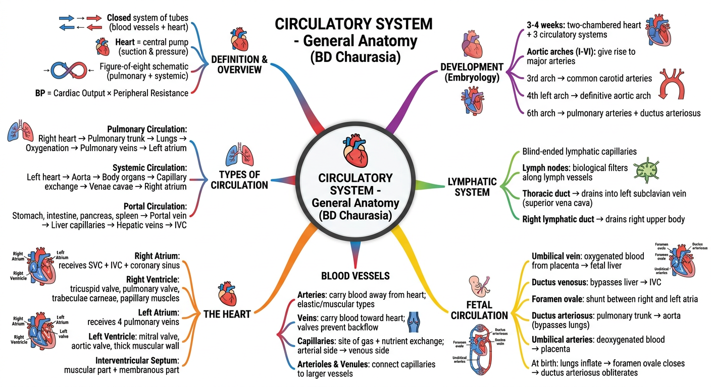
CIRCULATORY SYSTEM - Mind Map Summary
1. DEFINITION & OVERVIEW
- Closed system of tubes (blood vessels) with the heart as a central suction-and-pressure pump
- Schematically represented as a figure-of-eight, with the heart at the intersection
- BP = Cardiac Output × Peripheral Resistance
2. TYPES OF CIRCULATION
| Circuit | Path |
|---|---|
| Pulmonary | Right heart → Pulmonary trunk → Lungs (oxygenation) → Pulmonary veins → Left atrium |
| Systemic | Left heart → Aorta → Organ circuits → Capillary exchange → SVC/IVC → Right atrium |
| Portal | Stomach, intestine, pancreas, spleen → Portal vein → Liver → Hepatic veins → IVC |
| Lymphatic | Lymphatic capillaries → Lymph nodes → Thoracic duct / Right lymphatic duct → SVC |
3. THE HEART
- Right Atrium - receives SVC, IVC, and coronary sinus; contains fossa ovalis
- Right Ventricle - tricuspid valve (3 cusps), pulmonary valve; trabeculae carneae; anterior + posterior + septal papillary muscles; thin wall
- Left Atrium - receives 4 pulmonary veins; smooth walled; valve of foramen ovale
- Left Ventricle - mitral (bicuspid) valve, aortic valve; thick muscular wall (3x RV)
- Interventricular Septum - muscular part (~12 mm thick) + membranous part (~1 mm)
4. BLOOD VESSELS
- Arteries - carry blood away from heart; elastic (large, e.g., aorta) or muscular (medium)
- Veins - carry blood toward heart; valves prevent retrograde flow
- Capillaries - site of O2/CO2 and nutrient/waste exchange
- Arterioles & Venules - resistance vessels connecting capillaries to larger vessels
5. FETAL CIRCULATION (3 Shunts)
| Shunt | Function | Fate at Birth |
|---|---|---|
| Ductus venosus | Umbilical vein bypasses liver → IVC | Obliterates → ligamentum venosum |
| Foramen ovale | Right atrium → Left atrium (bypasses lungs) | Closes → fossa ovalis |
| Ductus arteriosus | Pulmonary trunk → Aorta (bypasses lungs) | Obliterates → ligamentum arteriosum |
- Umbilical vein carries oxygenated blood from placenta
- Umbilical arteries (×2) return deoxygenated blood to placenta
6. LYMPHATIC SYSTEM
- Begins as blind-ended capillaries in the periphery
- Lymph nodes = biological filters interspersed along lymph vessels
- Thoracic duct (left) and Right lymphatic duct drain into the superior vena cava
7. EMBRYOLOGICAL DEVELOPMENT
- 3-4 weeks: functional two-chambered heart; 3 circulatory systems (intraembryonic systemic, vitelline, placental)
- 6 Aortic Arches:
- 1st, 2nd, 5th - regress
- 3rd - common carotid arteries (both sides)
- 4th left - definitive aortic arch; 4th right - proximal right subclavian artery
- 6th - pulmonary arteries + ductus arteriosus