
What are the common complications during twin pregnancy
"twin pregnancy" AND complications
twin pregnancy complications diagram TTTS preterm
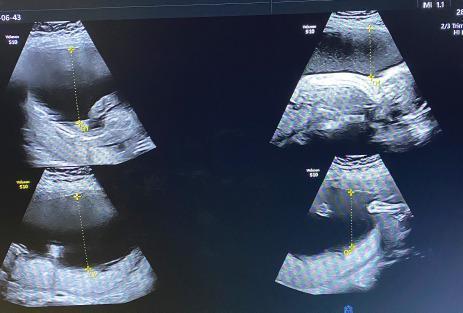
This diagnostic image is an obstetric ultrasound composite displaying four views of the uterus during a twin pregnancy evaluation. The content type is a diagnostic imaging study focusing on amniotic fluid assessment in the context of Twin-to-Twin Transfusion Syndrome (TTTS). The ultrasound shows the recipient twin in a monochorionic diamniotic (MCDA) pregnancy exhibiting severe polyhydramnios. Large, anechoic (black) areas represent significantly increased amniotic fluid volume. Calipers and dotted vertical lines demonstrate the measurement of the Single Deepest Vertical Pocket (SDVP), with the largest pocket exceeding the 8 cm threshold for polyhydramnios. The clinically reported Amniotic Fluid Index (AFI) is approximately 30.9 cm. This visual evidence of excessive liquor volume in one twin, combined with discordant fetal growth, supports a diagnosis of TTTS (Quintero Stage 2). The target audience for this educational material includes radiology and obstetrics students learning to identify ultrasound markers of fetal complications in multiple gestations.
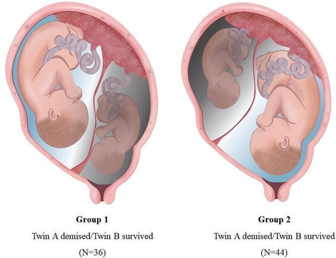
A medical anatomical diagram illustrating two clinical classifications of twin pregnancies complicated by single fetal demise (SFD). Both illustrations depict a monochorionic diamniotic (MCDA) twin pregnancy within a uterus, showing a shared fundal placenta and individual umbilical cords. In 'Group 1 (n=36)', the presenting twin (Twin A, located closest to the cervix) is the demised fetus, visualized as smaller and in a gray tone, while the non-presenting twin (Twin B) is larger and healthy. In 'Group 2 (n=44)', the non-presenting twin (Twin A, located at the fundus) is the demised fetus, while the presenting twin (Twin B) appears healthy. This comparison focuses on the impact of the demised twin's position relative to the birth canal on obstetric outcomes, such as preterm birth and gestational age at delivery. The diagram uses color coding and spatial positioning to differentiate between presenting and non-presenting status in the context of intrauterine fetal death.
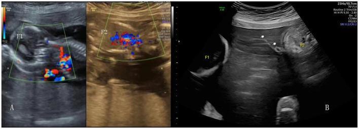
Educational diagnostic ultrasound images demonstrating complications in a monochorionic multifetal pregnancy. Panel A shows a color Doppler ultrasound assessment of surviving fetuses (labeled F1 and F2) following radiofrequency ablation (RFA). The green-bordered boxes highlight vascular flow, with red and blue signals indicating directionality of blood flow in umbilical or placental vessels. Panel B presents a B-mode ultrasound of Twin-Twin Transfusion Syndrome (TTTS), illustrating fetal discordance. Fetus F1 is situated in a polyhydramnios environment (recipient twin), while fetus F2 is shown within a restricted amniotic space. White arrows indicate the 'stuck' donor amnion or dividing membrane floating in the amniotic cavity, a hallmark of TTTS. These images demonstrate key sonographic markers used in fetal medicine to diagnose and monitor complications such as TTTS, selective fetal growth restriction (sFGR), and the post-procedural state after fetal reduction in monochorionic pregnancies.
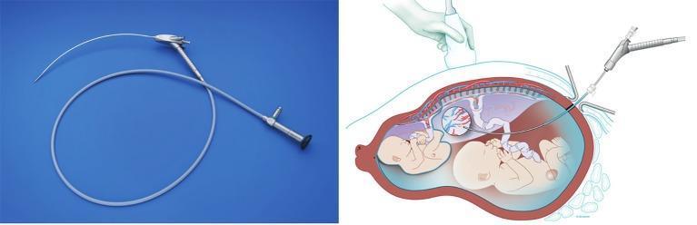
This composite educational resource features a photograph of a curved fetoscope and an anatomical diagram demonstrating its clinical application in fetal surgery. The photograph (left) shows a semi-flexible curved scope with a metallic proximal handle containing control ports and a distal tip designed for angled visualization. The diagram (right) illustrates the use of this curved fetoscope for treating Twin-to-Twin Transfusion Syndrome (TTTS) in a pregnancy with an anterior placenta. It depicts the scope passing through the abdominal and uterine walls into the amniotic cavity under ultrasound guidance (indicated by the transducer on the maternal abdomen). The trajectory of the curved scope is specifically shown to achieve a perpendicular orientation to the anterior placenta, facilitating the identification and laser coagulation of superficial vascular anastomoses on the chorionic plate between the donor and recipient twins. This specialized instrumentation allows for improved surgical access when anatomical positioning of the placenta limits the utility of traditional straight endoscopes.
Complications During Twin Pregnancy
Maternal Complications
1. Preeclampsia and Gestational Hypertension
2. Gestational Diabetes
3. Anaemia
4. Postpartum Haemorrhage
5. Other Maternal Morbidities
- Pruritic urticarial papules and plaques of pregnancy (PUPPP) - more common in multiple gestation
- Acute fatty liver of pregnancy (more common in first pregnancies and twin gestations)
- Higher rates of operative delivery (cesarean section)
Fetal and Perinatal Complications
1. Preterm Birth (Most Common Fetal Complication)
2. Fetal Growth Restriction (FGR)
3. Perinatal Mortality
- Stillbirth rate: 14.1 per 1000 for twins vs. 5.7 per 1000 for singletons
- Infant mortality rate: 23.4 per 1000 for twins vs. 5.1 per 1000 for singletons
- US perinatal mortality rate for twins: 24.4 per 1000 live births
4. Congenital Anomalies
5. Cerebral Palsy
Monochorionic-Specific Complications
6. Twin-to-Twin Transfusion Syndrome (TTTS)
- Donor twin: oligohydramnios, "stuck twin," anuria, growth restriction
- Recipient twin: polyhydramnios, hydrops, cardiac overload
| Stage | Features |
|---|---|
| I | Donor bladder visible, normal Doppler |
| II | Donor bladder not visible, normal Doppler |
| III | Donor bladder not visible, critically abnormal Doppler |
| IV | Hydrops in either twin |
| V | Intrauterine death of one or both twins |
7. Twin Anemia-Polycythemia Sequence (TAPS)
8. Selective Fetal Growth Restriction (sFGR)
9. Single Intrauterine Fetal Death (sIUFD)
10. Cord Entanglement (Monoamniotic Twins)
Summary Table
| Complication | Type | Risk vs. Singleton |
|---|---|---|
| Preterm birth (<34 wk) | Fetal | 10x higher |
| Preeclampsia | Maternal | 4-7x higher |
| Perinatal mortality | Fetal | ~3x higher |
| Cerebral palsy | Fetal | ~3x higher |
| TTTS | Monochorionic only | ~10-15% incidence |
| Congenital anomalies | Fetal | Increased |
| Postpartum haemorrhage | Maternal | Increased |