
Prutitis vulvae 10 marks obgy pg answer
pruritus vulvae lichen sclerosus vulva clinical appearance
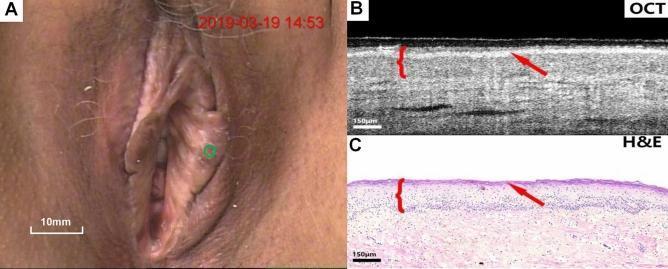
A three-panel figure demonstrating clinical and histological features of lichen sclerosus of the vulva. Panel A is a clinical colposcopy photograph showing the macroscopic appearance of the vulvar region, with a green circle indicating the specific site of interest. Panel B is a cross-sectional optical coherence tomography (OCT) image of the affected tissue. It shows well-defined stratification between the epithelium and stroma with uniform optical scattering. A key diagnostic feature highlighted is the loss of the serrated rete peg architecture at the epithelial-stromal junction. A red arrow indicates a hyper-reflective surface layer corresponding to hyperkeratosis, and a red bracket denotes the epithelial thickness. Panel C is a corresponding hematoxylin and eosin (H&E) stained histological section (150 μm scale) that correlates with the OCT findings. It confirms the presence of surface keratinization (arrow), a thinned or flattened epithelial layer (bracket), and the absence of rete pegs, consistent with lichen sclerosus pathology. This comparison serves as a diagnostic reference for utilizing non-invasive OCT imaging in identifying sub-surface morphological changes in gynecological dermatological conditions.
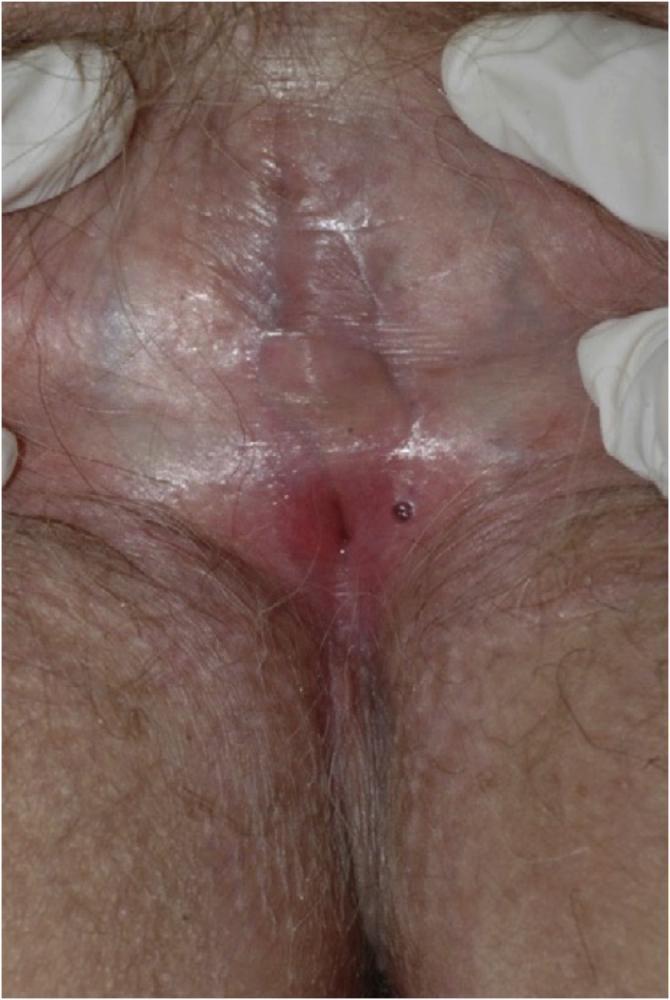
This clinical photograph demonstrates advanced lichen sclerosus affecting the female vulva, characterized by severe architectural distortion. The primary finding is the complete fusion and agglutination of the labia majora and labia minora, resulting in the total obliteration of the interlabial sulci. The skin exhibits hallmark signs of chronic vulvar dermatosis, including diffuse pallor, waxy texture, and areas of shiny, 'parchment-like' scarring (atrophy). The clitoral hood is buried under fused tissue, and the urethral and vaginal openings are significantly narrowed and obscured, leaving only a small central introitus visible. Inflammatory changes are noted by localized erythema and vascular prominence near the residual opening. These features represent the end-stage morphological changes associated with chronic inflammation, leading to extensive scarring and loss of normal anatomical landmarks, which clinically manifest as severe pruritus, pain, and dyspareunia.
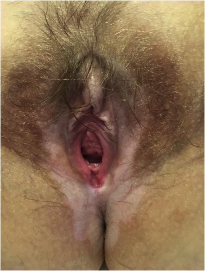
This clinical photograph displays the vulvar and perianal regions of a patient diagnosed with lichen sclerosus. The image illustrates characteristic dermatological changes, primarily marked by porcelain-white patches of hypopigmentation (leukoplakia) and skin thinning. These hypopigmented lesions are distributed in a classic 'figure-of-eight' pattern, encircling both the vulva and the perianal area. There is evidence of architectural distortion, including possible atrophy or resorption of the labia minora and narrowing of the introitus. The surrounding skin shows mottled pigmentation and textural changes consistent with chronic inflammation. The medical specialty involved is gynecology or dermatology, and the image serves as an educational tool for identifying the morphological features of vulvar lichen sclerosus, such as the characteristic whitening, thinning, and anatomical scarring that can lead to symptoms like pruritus and dyspareunia.
Pruritus Vulvae
Definition
Etiology / Causes
A. Local Causes
- Vulvovaginal Candidiasis - the most frequent cause; especially common during pregnancy and with antibiotic use. Candida albicans causes intense itch with curdy white discharge. Microscopy and fungal culture are diagnostic.
- Trichomonas vaginalis - motile protozoan (piriform, slightly larger than a leukocyte) recognized on wet mount of vaginal secretions; causes frothy, offensive discharge with vulvar itch.
- Bacterial Vaginosis (Gardnerella) - thin, fishy-smelling discharge with itch.
- Herpes genitalis - vesicular eruptions causing burning and itch.
- Human Papillomavirus (HPV) / Condyloma accuminata - warty growths; common in adolescents.
- Pinworm (Enterobius vermicularis) - a cause in children; nocturnal perianal/vulvar itch.
- Pubic lice (Phthirus pubis) / Scabies - ectoparasitic infestations.
- Lichen sclerosus - a major cause in middle-aged and elderly women (13% in prospective series). Presents with porcelain-white plaques in a "figure-of-8" pattern around the vulva and perianal region; causes intense itch, atrophy, labial fusion, and narrowing of introitus. Risk of malignant transformation (3-5% to SCC).
- Lichen simplex chronicus (LSC) - end result of chronic scratching; lichenification of vulvar skin.
- Lichen planus - erosive and hypertrophic forms; mucosal erosions, ulcerations, resorption of labia minora.
- Psoriasis - well-defined erythematous plaques (5% of chronic vulvar symptoms).
- Contact dermatitis (allergic or irritant) - from sanitary pads, contraceptives, douche solutions, fragrances, preservatives in moist towelettes, colophony, benzocaine, corticosteroids, partner's condoms. Almost half of women with pruritus vulvae have at least one relevant allergen.
- Atopic dermatitis - chronic relapsing eczematous condition.
- Extramammary Paget's disease - chronic vulvar itch; confluent, raised, erythematous to violet waxy lesion; may be associated with underlying adenocarcinoma or distant cancers (breast, cervix, GI).
- Vulvar Intraepithelial Neoplasia (VIN) - differentiated VIN has high malignant potential; presents with itch and subtle skin changes; requires biopsy.
- Invasive vulvar carcinoma - itch as a presenting symptom.
- Leukoplakia - white plaques; any non-healing lesion should be biopsied.
- Urinary incontinence / chronic moisture
- Vaginal discharge from any cause (fistulae, atrophic vaginitis post-menopause)
- Tight/synthetic clothing, nylon underwear
B. Systemic Causes
| Condition | Mechanism |
|---|---|
| Diabetes mellitus | Glycosuria promotes Candida overgrowth; altered immune response |
| Hepatic disease / Obstructive jaundice | Bile salt deposition in skin |
| Chronic renal failure | Uraemia causing cutaneous nerve stimulation |
| Hypothyroidism / Myxoedema | Dry skin |
| Iron deficiency anaemia | Epithelial changes |
| Polycythaemia vera | Aquagenic pruritus mechanism |
| Drugs | Tamoxifen - causes vulvar itch, vaginal dryness as adverse effect |
C. Psychogenic / Neuropathic
- Purely psychogenic in only 1-10% of cases (often over-diagnosed)
- Neuropathic pruritus from lumbosacral radiculopathy (prolapsed disc, vertebral disease) can cause vulvar itch via compression of lumbosacral nerve roots
- When burning predominates over itch: consider dysesthetic vulvodynia (sensory neuropathy) - accounts for ~9% of chronic vulvar symptoms
Clinical Features
- History: onset, nature (itch vs. burning), timing (nocturnal suggests pinworm or candidiasis), associated discharge, relationship to menstrual cycle, medications, hygiene products
- Symptoms: intense itching often worse at night; secondary excoriation, soreness
- Signs:
- Erythema, excoriation, lichenification (chronic scratching)
- White plaques (lichen sclerosus, leukoplakia)
- Discharge (candidiasis, Trichomonas, BV)
- Vesicles (HSV), warts (HPV)
- Skin atrophy, labial fusion (lichen sclerosus advanced)
- "Figure-of-8" pattern - pathognomonic of lichen sclerosus
Investigation
- Vaginal pH (candidiasis pH <4.5; BV/Trichomonas pH >4.5)
- Wet mount microscopy - Candida pseudohyphae; Trichomonas motility
- KOH preparation - fungal elements
- Swab for culture (bacterial, fungal)
- Scotch tape test / perianal scraping - for Enterobius
- Colposcopy with acetic acid - especially in high-risk women for VIN
- Biopsy - mandatory for any non-healing lesion, white patch, or treatment-resistant case (to exclude malignancy)
- Patch testing - to identify contact allergens (almost half have relevant allergens)
- FBS / Random blood sugar (diabetes)
- CBC (iron deficiency anaemia, polycythaemia)
- LFT, renal function tests
- TFT (thyroid)
- Serum urea/creatinine
Management
General Measures (for all causes)
- Patient education: avoid scratching, tight synthetic clothing
- Silk/cotton underwear to reduce friction and moisture
- Avoid local irritants: scented soaps, douches, moist wipes, latex condoms if allergic
- Keep the area clean and dry; loose-fitting clothing
- Treat any identified systemic cause (e.g., control diabetes)
Specific Treatment
| Cause | Treatment |
|---|---|
| Candidiasis | Topical azole (clotrimazole, miconazole) or oral fluconazole 150 mg single dose; treat partner if recurrent |
| Trichomonas | Metronidazole 400 mg BD × 7 days or 2 g single dose; treat partner |
| BV | Metronidazole or clindamycin |
| Lichen sclerosus | High-potency topical steroids (clobetasol propionate) - pulsed dosing; topical tacrolimus or pimecrolimus as steroid-sparing agents |
| Contact dermatitis | Identify and remove allergen (patch testing); topical corticosteroids |
| Lichen planus | High-potency topical steroids; other options in refractory cases |
| LSC | Topical steroids + breaking the itch-scratch cycle |
| Psoriasis | Topical steroids, calcipotriol, phototherapy |
| Atrophic vaginitis (post-menopausal) | Local estrogen cream/pessary; systemic HRT if appropriate |
| Psychogenic / Neuropathic | Topical lidocaine, pramoxine; oral tricyclic antidepressant (amitriptyline); gabapentin |
| Extramammary Paget's / VIN / Malignancy | Surgical excision, wide local excision; oncology referral |
Additional Options
- Phototherapy (narrowband UVB or comb light device) - effective for some chronic cases
- Acupuncture - reported benefit in some studies
- Biopsy of any resistant case - rule out malignancy
Prognosis
- Treatable when specific cause identified
- Lichen sclerosus: chronic; requires long-term surveillance for SCC (3-5% risk)
- Psychogenic pruritus: responds to TCAs and behavior modification
- Malignant causes: prognosis depends on stage at diagnosis - underscores importance of early biopsy in unresponsive cases
Key Points to Remember (Exam Tips)
- Most common cause: Unspecified dermatitis (54%) > Lichen sclerosus (13%) > Candidiasis (10%) > Dysesthetic vulvodynia (9%) > Psoriasis (5%) - from a prospective series of 141 women
- Candidiasis is the most common infective cause; especially in pregnancy and antibiotic use
- Lichen sclerosus shows "figure-of-8" pattern and carries malignant potential
- Almost half of women with pruritus vulvae have a contact allergen - patch testing is important
- Biopsy any non-healing, white, or treatment-resistant vulvar lesion
- Burning > itching suggests neuropathic/dysesthetic etiology
- Purely psychogenic cause is rare (1-10%) - do not diagnose without excluding organic causes
- Systemic causes (DM, renal failure, hepatic disease) must always be excluded