I now have all I need. Let me compile the complete study guide based on the lecture structure plus textbook enrichment.
🧠 The Telencephalon - Complete Study Guide
MED207 Neuroscience & Behaviour | A-Prof. Royhaan Folarin
Topic: Cerebral Hemispheres, Surface Features, Lobes & White Matter
🏥 OPENING CLINICAL SCENARIO (Lecture Case)
Patient: 58-year-old right-handed man who suddenly develops:
- Weakness of right face and upper limb
- Difficulty producing speech
- Preserved understanding of spoken commands
- No primary visual loss
Answer (from lecture slide 56):
| Finding | Localization |
|---|
| Right facial + upper limb weakness | Left precentral gyrus (motor cortex) |
| Difficulty producing speech | Left inferior frontal region (Broca's area, BA 44/45) |
| Preserved comprehension | Posterior temporal region spared (Wernicke's area intact) |
| No visual loss | Occipital cortex spared |
| Likely lesion | Left frontal lobe |
This is Broca's aphasia (expressive/non-fluent) - speech production impaired, comprehension preserved.
1. WHAT IS THE TELENCEPHALON?
The telencephalon is the most rostral (anterior) part of the brain. It comprises:
- The two cerebral hemispheres (grey cortex + white matter)
- The basal ganglia (deep nuclei)
- The limbic structures (hippocampus, amygdala)
Together with the diencephalon (thalamus + hypothalamus), it forms the prosencephalon (forebrain).
2. HEMISPHERES: BORDERS, SURFACES & POLES
Each cerebral hemisphere has:
3 Surfaces:
- Superolateral (convex, faces skull)
- Medial (faces opposite hemisphere across interhemispheric fissure)
- Inferior (base - rests on skull floor + tentorium)
3 Poles:
- Frontal pole (anterior)
- Occipital pole (posterior)
- Temporal pole (inferior)
Key boundary marker: The preoccipital notch marks the posterior boundary near the occipital lobe on the lateral surface.
The two hemispheres are separated by the interhemispheric (longitudinal) fissure and connected by commissural fibers (mainly the corpus callosum).
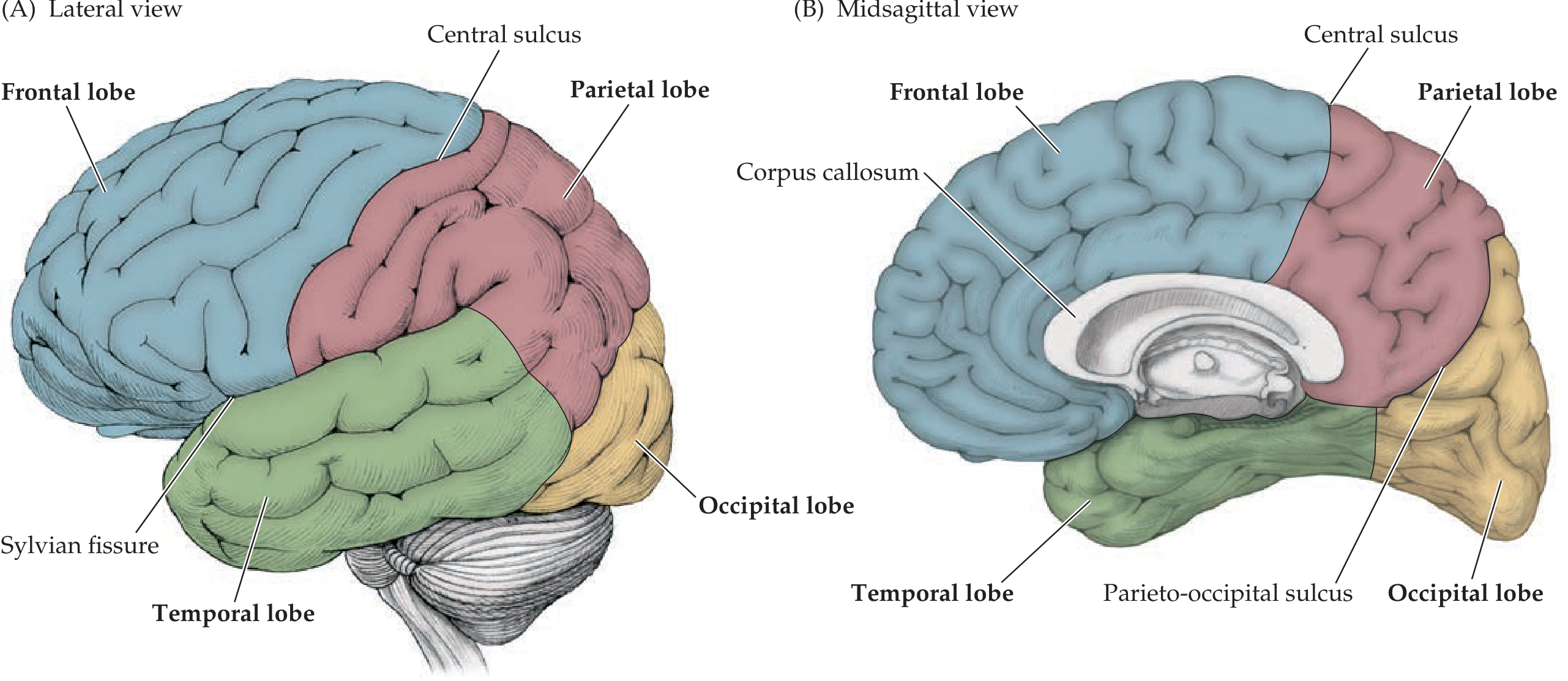
3. THE FOUR LOBES + INSULA
Major Sulci as Lobe Boundaries
| Sulcus / Fissure | What it separates |
|---|
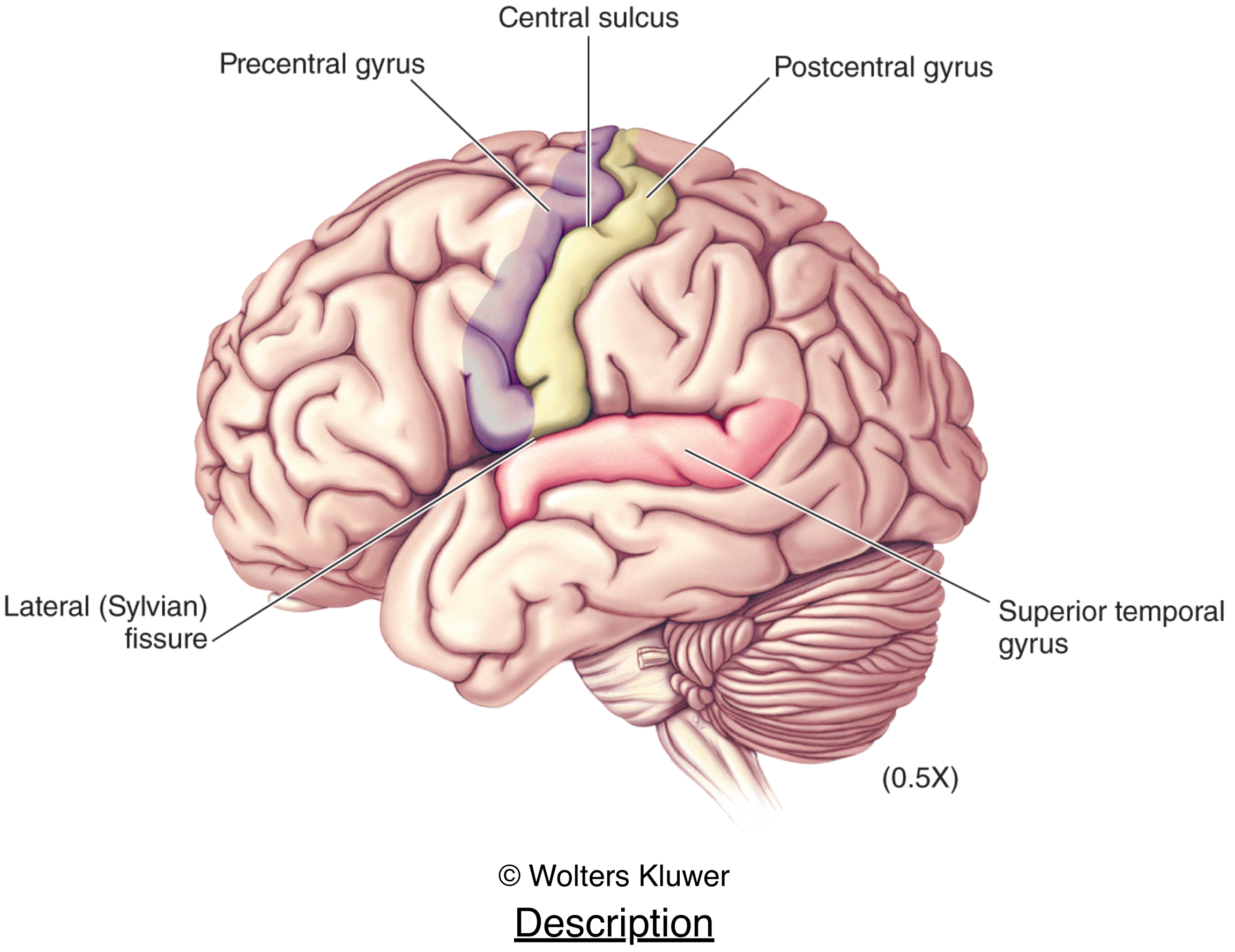
| Central sulcus (of Rolando) | Frontal from Parietal |
| Lateral sulcus (Sylvian fissure) | Frontal/Parietal from Temporal |
| Parieto-occipital sulcus | Parietal from Occipital (best seen medially) |
| Preoccipital notch | Temporal from Occipital (lateral surface) |
The Insula (5th "Hidden" Lobe)
- Buried deep within the lateral (Sylvian) fissure
- Covered by the operculum: frontal operculum (anterior) + parietal operculum (posterior)
- Functions: interoception, pain, taste, autonomic regulation, language
4. KEY GYRI & SULCI (Exam High-Yield)
Frontal Lobe
| Structure | Function |
|---|
| Precentral gyrus (BA 4) | Primary motor cortex - voluntary movement |
| Superior frontal gyrus | Supplementary motor area; working memory |
| Middle frontal gyrus | Prefrontal association; frontal eye fields (BA 8) |
| Inferior frontal gyrus - pars triangularis + opercularis (BA 44/45) | Broca's area - speech production (dominant hemisphere) |
| Prefrontal cortex (BA 9,10,11) | Executive function, personality, judgement |
Parietal Lobe
| Structure | Function |
|---|
| Postcentral gyrus (BA 1,2,3) | Primary somatosensory cortex - touch, proprioception |
| Superior parietal lobule (BA 5,7) | Stereognosis, visuospatial integration |
| Angular gyrus (BA 39) | Reading, writing, mathematics |
| Supramarginal gyrus (BA 40) | Language processing; part of Wernicke's area |
Temporal Lobe
| Structure | Function |
|---|
| Superior temporal gyrus (BA 22) | Wernicke's area (posterior, dominant hemisphere) - speech comprehension |
| Heschl's gyrus (BA 41, 42) | Primary auditory cortex (buried in lateral sulcus) |
| Middle + inferior temporal gyri (BA 20, 21) | Semantic memory, object recognition |
| Parahippocampal gyrus (BA 28) | Memory encoding, primary olfactory cortex |
| Hippocampus (medial temporal) | Long-term memory formation |
Occipital Lobe
| Structure | Function |
|---|
| Calcarine fissure / striate cortex (BA 17) | Primary visual cortex |
| Lingual + cuneate gyri (BA 18, 19) | Secondary + tertiary visual cortex |
Medial Surface Landmarks
- Cingulate gyrus - emotion, pain, attention (part of limbic system)
- Paracentral lobule - motor/sensory for the lower limb (the leg representation of the homunculus wraps around the medial surface)
- Precuneus - visuospatial imagery, self-consciousness
- Cuneus + lingual gyrus - visual cortex (above and below calcarine sulcus)
5. BRODMANN AREAS - Key Numbers to Know
| BA | Area | Location | Function |
|---|
| 1, 2, 3 | Primary somatosensory | Postcentral gyrus | Touch |
| 4 | Primary motor | Precentral gyrus | Voluntary movement |
| 6 | Premotor / Supplementary motor | Pre-precentral | Movement planning |
| 8 | Frontal eye fields | Middle frontal gyrus | Saccadic eye movements |
| 17 | Primary visual | Calcarine fissure | Vision |
| 18, 19 | Secondary/tertiary visual | Occipital | Visual processing |
| 22 | Wernicke's area (posterior) | Superior temporal gyrus | Speech comprehension |
| 41, 42 | Primary auditory | Heschl's gyrus | Hearing |
| 44, 45 | Broca's area | Inferior frontal gyrus | Speech production |
| 9-12 | Prefrontal | Superior/middle frontal | Cognition, planning |
Mnemonic for Broca vs Wernicke: Broca = Boken speech (production), Wernicke = Word soup (comprehension)
6. WHITE MATTER FIBERS - The 3 Types
The lecture specifically tests these. They are grouped by which areas they connect:
A. Association Fibers - connect areas WITHIN the same hemisphere
| Fiber Tract | Connects | Clinical note |
|---|
| Arcuate fasciculus (superior longitudinal fasciculus) | Broca's ↔ Wernicke's areas | Damage = conduction aphasia (can't repeat, but can speak & understand) |
| Uncinate fasciculus | Frontal lobe ↔ anterior temporal lobe | Personality, behavior |
| Cingulum | Cingulate gyrus + other limbic areas | Emotion/memory |
| Inferior longitudinal fasciculus | Occipital ↔ temporal lobe | Visual-object recognition |
| Inferior fronto-occipital fasciculus | Frontal ↔ occipital | |
B. Commissural Fibers - connect BETWEEN the two hemispheres
| Commissure | Connects |
|---|
| Corpus callosum (largest) | Homologous cortical areas of both hemispheres |
| Anterior commissure | Temporal lobes (olfactory, anterior temporal) |
| Posterior commissure | Midbrain structures |
| Hippocampal commissure (fornix commissure) | Hippocampi |
Corpus Callosum Parts (rostral to caudal):
Rostrum → Genu → Body → Splenium
- Genu - connects prefrontal cortices
- Body - connects motor + somatosensory cortices
- Splenium - connects occipital/posterior parietal cortices
- Mnemonic: "Really Great Big Spoons" (Rostrum, Genu, Body, Splenium)
C. Projection Fibers - connect CORTEX to deeper/distant structures
- Internal capsule - the main highway carrying motor (corticospinal) and sensory (thalamocortical) fibers
- Anterior limb - frontopontine + anterior thalamic radiations
- Genu - corticobulbar fibers (to cranial nerve nuclei)
- Posterior limb - corticospinal tract (most important!) + sensory radiations
- Corona radiata - fan-shaped spread above internal capsule to cortex
- Optic radiations (from lateral geniculate → primary visual cortex)
7. CEREBRAL DOMINANCE & LATERALIZATION
- Left hemisphere dominant in ~95% of right-handed people (and ~70% of left-handers)
- Language (Broca's + Wernicke's) - left hemisphere dominant
- Spatial processing, prosody (emotional tone of speech) - right hemisphere
- Handedness does NOT equal dominance in all functions
8. CLINICAL CORRELATES (Lecture Slide 55 Content)
| Lesion | Syndrome | Key features |
|---|
| Corpus callosum (surgical/tumor/MS) | Disconnection syndrome / Alien hand syndrome | Left hand acts autonomously; can't transfer info between hemispheres |
| Internal capsule stroke | Dense hemiplegia + hemisensory loss | Contralateral face, arm, leg all affected |
| Multiple sclerosis | White matter demyelination | Episodic, multifocal neurological deficits |
| Temporal lobectomy (dominant) | Memory + language deficits | Used for epilepsy surgery |
| Frontal lobe damage (prefrontal) | Executive dysfunction, disinhibition, flat affect | Phineas Gage personality |
| Parietal lobe damage (non-dominant) | Hemineglect, dressing apraxia, constructional apraxia | |
| Parietal lobe damage (dominant) | Gerstmann syndrome: agraphia, acalculia, finger agnosia, L-R confusion | Angular gyrus (BA 39) |
| Occipital lobe damage | Cortical blindness, homonymous hemianopia | |
| Temporal lobe damage (dominant) | Wernicke's aphasia, anterograde amnesia | |
9. LANGUAGE AREAS - APHASIA SUMMARY (High Yield!)
| Type | Fluency | Comprehension | Repetition | Lesion |
|---|
| Broca's (expressive) | Non-fluent | Intact | Impaired | Inferior frontal gyrus (BA 44/45) |
| Wernicke's (receptive) | Fluent (but paraphasic) | Impaired | Impaired | Posterior superior temporal gyrus (BA 22) |
| Conduction | Fluent | Intact | Severely impaired | Arcuate fasciculus |
| Global | Non-fluent | Impaired | Impaired | Large left MCA territory |
10. THE HOMUNCULUS (Motor & Sensory)
- The precentral gyrus (motor) and postcentral gyrus (sensory) are somatotopically organized
- Body representation: toe/leg on medial surface → trunk → arm → hand (large representation - most dexterous) → face/tongue at the bottom of lateral surface
- Large cortical area = greater fine motor control (hands, lips >> trunk)
11. QUICK Q&A DRILL
Q1. What are the 3 types of white matter fibers?
Association (within hemisphere), Commissural (between hemispheres), Projection (cortex ↔ deep structures)
Q2. Which fiber connects Broca's to Wernicke's area? What happens if it's cut?
Arcuate fasciculus → Conduction aphasia (fluent speech, intact comprehension, poor repetition)
Q3. What are the 4 parts of the corpus callosum from front to back?
Rostrum, Genu, Body, Splenium
Q4. A patient has non-fluent speech, intact comprehension, after a stroke. What area was damaged?
Broca's area (inferior frontal gyrus, BA 44/45, left hemisphere) - Broca's aphasia
Q5. The internal capsule's posterior limb carries which major tract?
Corticospinal tract (and somatosensory radiations from thalamus)
Q6. Where is primary visual cortex? Primary auditory cortex?
Visual: calcarine fissure, occipital lobe (BA 17). Auditory: Heschl's gyrus, buried in lateral sulcus (BA 41/42)
Q7. What sulcus separates the frontal and parietal lobes?
Central sulcus (of Rolando)
Q8. What is the insula, and where is it?
Buried cortex ("island") within the depths of the lateral (Sylvian) fissure; covered by the operculum
Q9. Patient can't name fingers, can't do arithmetic, confuses left vs right, can't write. What syndrome?
Gerstmann syndrome - lesion at left angular gyrus (BA 39) of dominant parietal lobe
Q10. Name the 5 surfaces/aspects used to describe the cerebral hemisphere.
Superolateral, medial, inferior; Poles: frontal, occipital, temporal
📋 LECTURE LEARNING OBJECTIVES CHECKLIST
Sources: Neuroanatomy through Clinical Cases 3rd ed. (9781605359625); Neuroscience: Exploring the Brain 5th ed. (9781284286878)