
Maxillary premolars first year bds students dont skip any main points for exams point of view make a summary imp points
maxillary premolars BDS dental anatomy first year exam important points morphology roots cusps
https://pocketdentistry.com/9-the-permanent-maxillary-premol…
Maxillary Premolars - BDS First Year Exam Summary
OVERVIEW (General Points)
- Premolars are succedaneous teeth - they replace the deciduous molars
- There are 4 maxillary premolars (2 on each side): first premolar and second premolar
- FDI notation: 14, 15 (right) and 24, 25 (left)
- Universal notation: 5, 4 (upper right) and 12, 13 (upper left)
- Position: between canine (anteriorly) and first molar (posteriorly)
- Function: tearing and grinding (transitional between canines and molars)
MAXILLARY FIRST PREMOLAR
Chronology (High-Yield!)
| Event | Timing |
|---|---|
| Calcification begins | 1.5 years |
| Enamel completed | 5-6 years |
| Eruption | 10-11 years |
| Root completed | 12-13 years |
Crown - Buccal Aspect
- Resembles a maxillary canine from buccal view - but crown is shorter (1.5-2 mm shorter than canine) and narrower mesiodistally
- Two cusps: buccal and lingual (both sharply defined)
- Buccal cusp is ~1 mm longer than lingual cusp
- Mesial slope of buccal cusp is longer than distal slope (opposite of the canine)
- Mesial and distal contact areas are at the same level (unlike canine where they differ)
- Buccal line angles are prominent - crown appears angular
- Buccal cervical ridge present
Crown - Mesial Aspect (VERY IMPORTANT)
- Mesial concavity (mesial developmental depression) - a characteristic feature that distinguishes maxillary first premolar from the second premolar
- This depression extends from the cervical line onto the mesial root surface
- Mesial marginal developmental groove - another hallmark; groove cuts across the mesial marginal ridge from the occlusal surface onto the mesial surface
- Buccolingual width is greater than mesiodistal width
Crown - Occlusal Aspect
- Outline is roughly hexagonal (6-sided)
- Widest in the buccal third
- Central developmental groove runs mesiodistally
- Mesial triangular fossa and distal triangular fossa present
- Buccal cusp is slightly offset toward the distal
- Two major cusps: buccal and lingual, separated by the central groove
- Marginal ridges are in a horizontal plane (part of the occlusal surface - unlike incisors/canines where they are part of the lingual surface)
Crown - Lingual Aspect
- Lingual cusp is shorter and narrower than buccal cusp
- Crown tapers toward the lingual
Root (HIGH YIELD!)
- Most maxillary first premolars have TWO roots (bifurcated) - buccal and lingual (palatal)
- This is the only premolar that normally has two roots
- Buccal root is larger and longer than the palatal root
- Even when only one root is present, two pulp canals are usually found - key exam point
- Single-rooted form has a prominent mesial root groove (giving appearance of root trying to divide)
- Root trunk = area between cervical line and bifurcation
- Bifurcation = junction of buccal and lingual roots
Pulp Cavity
- Two pulp horns (one for each cusp)
- Two root canals (one for each root)
- If single root: usually still two canals, often combining into one apical foramen
- Rarely: one canal in a single-rooted tooth
MAXILLARY SECOND PREMOLAR
Chronology
| Event | Timing |
|---|---|
| Calcification begins | 2-2.5 years |
| Enamel completed | 6-7 years |
| Eruption | 10-12 years |
| Root completed | 12-14 years |
Crown
- Slightly smaller than maxillary first premolar overall
- Buccal cusp is shorter and less pointed than in first premolar
- Cusps are more equal in size (buccal and lingual cusps are more symmetrically sized) - key differentiating feature from first premolar
- Markings (grooves, ridges) are less defined than in first premolar
- No mesial concavity - smooth mesial surface (vs. first premolar which has mesial depression)
- No mesial marginal groove cutting across the marginal ridge
- More efficient as a grinding tooth - functions like a molar
- Occlusal surface: cusps articulate with opposing teeth more efficiently
Root (HIGH YIELD!)
- Usually ONE root (single rooted) - main difference from first premolar
- Root is longer than that of the first premolar
- Occasionally two roots can occur - not uncommon
- Root grooves present (less prominent than first premolar)
Pulp Cavity
- Usually one root canal (corresponding to single root)
- Two root canals if two roots present
KEY DIFFERENCES: First vs Second Premolar (Exam Favourite!)
| Feature | Maxillary 1st Premolar | Maxillary 2nd Premolar |
|---|---|---|
| Cusp size | Buccal >> lingual (~1 mm longer) | Buccal ≈ lingual (nearly equal) |
| Roots | Usually 2 (bifurcated) | Usually 1 (single) |
| Mesial concavity | Present | Absent |
| Mesial marginal groove | Present (cuts across marginal ridge) | Absent |
| Crown sharpness | Cusps sharply defined | Cusps less sharp |
| Markings | Well defined | Less defined |
| Root length | Shorter | Longer |
| Pulp canals | Usually 2 | Usually 1 (2 if 2 roots) |
RELATIONS & CLINICAL IMPORTANCE
- Maxillary first premolar is close to the maxillary sinus (floor) - extractions can lead to oroantral fistula
- The posterior superior alveolar nerve (branch of maxillary nerve, V2) supplies molar and premolar teeth
- The middle superior alveolar nerve supplies premolar teeth primarily
- Important landmark for infraorbital nerve block and posterior superior alveolar nerve block
- The junction of first molar and second premolar is a landmark for the maxillary nerve approach
INNERVATION
- Maxillary premolars are supplied by the posterior superior alveolar nerve and middle superior alveolar nerve (branches of the maxillary division of trigeminal nerve, CN V2)
BLOOD SUPPLY
- Posterior superior alveolar artery (branch of maxillary artery) - descends on the infratemporal surface of the maxilla to supply molar and premolar teeth, adjacent bone, and maxillary sinus
ADDITIONAL EXAM POINTS
- Premolars are the only permanent teeth with no deciduous predecessors - FALSE; they replace deciduous molars. Premolars are succedaneous to deciduous molars.
- Maxillary first premolar is the only premolar with 2 roots as a common occurrence
- Even with a single root, 2 canals are almost always present in maxillary first premolar
- Buccal root of maxillary first premolar is larger and longer than palatal root
- The mesial concavity of the first premolar makes it the most difficult to clean and predisposed to periodontal disease on the mesial surface
- Crown is wider buccolingually than mesiodistally in both maxillary premolars
- Maxillary premolars are shorter in crown height than maxillary canines
- Root length of maxillary premolars equals root length of molars
Anatomy of maxillary premolars
maxillary premolar detailed anatomy crown root pulp cavity surfaces cusps dental anatomy Wheeler's
https://pocketdentistry.com/9-the-permanent-maxillary-premol…
https://lib.bpums.ac.ir/UploadedFiles/xfiles/File/library/2.…
Anatomy of Maxillary Premolars
General Introduction
- FDI notation: 14 & 15 (upper right), 24 & 25 (upper left)
- Location: Between the maxillary canine anteriorly and the maxillary first molar posteriorly
- Function: Tearing and grinding - transitional between canines (tearing) and molars (grinding)
- Crowns are shorter than maxillary canines but longer than maxillary molars
- Crown is wider buccolingually than mesiodistally (a posterior tooth characteristic)
- Marginal ridges lie in a horizontal plane, forming part of the occlusal surface (unlike anterior teeth where marginal ridges are part of the lingual surface)
MAXILLARY FIRST PREMOLAR
Chronology
| Event | Time |
|---|---|
| Calcification begins | 1.5 years |
| Enamel completed | 5-6 years |
| Eruption | 10-11 years |
| Root completed | 12-13 years |
Measurements (Wheeler's)
| Dimension | Value |
|---|---|
| Crown length (cervico-occlusal) | 8.5 mm |
| Crown diameter - mesiodistal | 7.0 mm |
| Crown diameter - cervical (mesiodistal) | 5.0 mm |
| Crown diameter - buccolingual | 9.0 mm |
| Root length | 14.0 mm |
Note: Buccolingual (9 mm) > Mesiodistal (7 mm) - wider BL than MD
Crown Anatomy - Surface by Surface
1. Buccal (Facial) Aspect
- Resembles the maxillary canine from the buccal view - but differences:
- Crown is shorter by 1.5-2 mm than the canine
- Crown is narrower mesiodistally than the canine
- Contact areas mesially and distally are at the same level (in canine, mesial contact is more cervical)
- Mesial slope of buccal cusp is longer than the distal slope (opposite of canine)
- Crown is angular with prominent buccal line angles
- Buccal cusp is tipped slightly toward the distal
- Shoulders: Prominent
- Buccal cervical ridge present at the cervical third
- Developmental depressions on either side of the buccal ridge demarcate the three buccal lobes (mesiobuccal, central buccal, distobuccal)
- Roots are 3-4 mm shorter than those of the maxillary canine but the buccal root outline resembles the canine root
2. Lingual Aspect
- Gross outline is the reverse of the buccal aspect
- Lingual cusp is shorter and narrower than the buccal cusp
- Crown tapers toward the lingual - lingual surface is narrower than buccal
- Lingual cusp tip is offset toward the mesial
- From lingual aspect, the buccal profile is visible (unlike the second premolar where the buccal profile is not visible from lingual - a siding aid)
- Lingual surface is smooth and convex
3. Mesial Aspect (Most Distinctive Surface)
- Mesial developmental depression (concavity) - a hallmark feature of the first premolar; a hollow or depression on the mesial surface of the crown and root
- Mesial marginal developmental groove - a groove that crosses the mesial marginal ridge from the occlusal surface onto the mesial surface (cuts across the marginal ridge)
- Mesial root depression present on root surface
- Cervical line shows more curvature on the mesial than on the distal
- Both buccal and lingual cusp tips are centered over the root
- Crown outline is roughly rhomboidal from this view
4. Distal Aspect
- Similar to mesial view but:
- No groove crosses the distal marginal ridge (no distal marginal groove - or much less prominent)
- No developmental depression on the distal surface
- Crown surface appears more rounded and smooth
- Cervical line is less curved than on the mesial
- Some specimens show a faint distal marginal groove but it is shallower than the mesial marginal groove
5. Occlusal Aspect
- Outline form: Hexagonal (6-sided)
- Crown is widest in the buccal third and tapers toward the lingual
- Central developmental groove (CDG) runs mesiodistally across the occlusal surface
- Mesial triangular fossa (MTF) - depression on the mesial side where the mesiobuccal developmental groove lies
- Distal triangular fossa (DTF) - depression on the distal side where the distobuccal developmental groove lies
- Mesiobuccal developmental groove and distobuccal developmental groove radiate from the central groove
- Supplemental grooves: Usually NOT present (vs. second premolar which commonly has many)
- Lobes: 4 developmental lobes (3 buccal, 1 lingual)
- 2 pulp horns corresponding to 2 cusps
- Buccal triangular ridge and lingual triangular ridge meet to form the transverse ridge
- Marginal ridges: mesial and distal marginal ridges are sharp and prominent
Root
- Most commonly 2 roots (bifurcated) - one buccal, one palatal (lingual)
- The maxillary first premolar is the only premolar that normally has two roots
- Buccal root is larger and longer than the palatal root
- Root trunk = area between the cervical line and the point of bifurcation
- Single-rooted form also occurs; the single root shows prominent longitudinal grooves (especially on the mesial surface) - "root trying to divide itself"
- Root grooves: Present on both buccal and lingual aspects, most prominent on the mesial
- Root length equals that of the molars
Pulp Cavity
- Pulp chamber: Well-defined, with a distinct pulp chamber before the canals divide
- 2 pulp horns - buccal pulp horn (larger, under buccal cusp tip) and lingual pulp horn (smaller, under lingual cusp tip)
- 2 root canals in most teeth: buccal root canal and lingual (palatal) root canal
- Even when only 1 root is present, 2 pulp canals are usually still found - sometimes fusing to a single apical foramen
- Rarely: only 1 canal in a single-rooted specimen
MAXILLARY SECOND PREMOLAR
Chronology
| Event | Time |
|---|---|
| Calcification begins | 2-2.5 years |
| Enamel completed | 6-7 years |
| Eruption | 10-12 years |
| Root completed | 12-14 years |
Measurements (Wheeler's)
| Dimension | Value |
|---|---|
| Crown length (cervico-occlusal) | 8.5 mm |
| Crown diameter - mesiodistal | 7.0 mm |
| Crown diameter - cervical (mesiodistal) | 5.0 mm |
| Crown diameter - buccolingual | 9.0 mm |
| Root length | 14.0 mm |
Overall measurements are similar to the first premolar, but the second premolar has variations in crown features
Crown Anatomy - Surface by Surface
1. Buccal (Facial) Aspect
- Similar general appearance to the first premolar
- Buccal cusp is shorter and less pointed than in the first premolar
- Cusp ridges are similar in length (mesial and distal cusp ridges are more equal)
- Buccal cusp is NOT tipped toward the distal (unlike the first premolar)
- Shoulders are narrow (first premolar shoulders are prominent)
- Markings (grooves, ridges, developmental depressions) are less defined than in the first premolar
- Contact areas: both mesial and distal contacts in the middle third
2. Lingual Aspect
- Buccal profile is NOT visible from the lingual view (unlike first premolar) - a useful siding feature
- Lingual cusp is more nearly the same size as the buccal cusp
- Cusps are more equal in height - less difference between buccal and lingual cusp lengths
3. Mesial Aspect
- No mesial developmental depression (concavity) - crown surface is convex on the mesial (key differentiating feature from first premolar)
- No mesial marginal developmental groove crossing the marginal ridge
- Mesial root depression is present (shallow groove on root surface)
- Buccal and lingual cusp tips are shorter, with greater distance between them - giving a wider buccolingual occlusal surface
4. Distal Aspect
- No outstanding variation from mesial aspect
- No distinct groove crossing the distal marginal ridge
5. Occlusal Aspect
- Outline form: Ovoid (vs. hexagonal in first premolar)
- Central groove is short (vs. long in first premolar)
- Many supplemental grooves often present - giving the occlusal surface a wrinkled appearance
- Lobes: 4 developmental lobes
- 2 pulp horns
- Cusps are more nearly equal in size - the tooth functions more efficiently as a grinding tooth
- Contact areas: centered at the middle third both mesially and distally
Root
- Usually 1 root (single-rooted) - the major difference from the first premolar
- Root is longer than that of the first premolar
- Occasionally may have 2 roots - not uncommon
- No distinct root grooves (vs. first premolar with prominent mesial groove)
- Root tapers to a pointed apex
Pulp Cavity
- Usually 1 root canal (when single root)
- 2 pulp horns (one buccal, one lingual)
- When 2 roots present: 2 root canals
- Pulp cavity is less complex than the first premolar
KEY COMPARISON TABLE: First vs. Second Maxillary Premolar
| Feature | 1st Premolar | 2nd Premolar |
|---|---|---|
| Eruption | 10-11 yrs | 10-12 yrs |
| Cusp sharpness | Sharply defined | Less sharp |
| Cusp size ratio | Buccal > Lingual (~1 mm longer) | Buccal ≈ Lingual (nearly equal) |
| Buccal cusp tip | Tipped toward distal | Not tipped |
| Occlusal outline | Hexagonal | Ovoid |
| Central groove | Long | Short |
| Supplemental grooves | Usually absent | Many, often present |
| Mesial concavity | Present | Absent (convex) |
| Mesial marginal groove | Crosses marginal ridge | Does not cross ridge |
| Buccal profile from lingual | Visible | Not visible |
| Shoulders | Prominent | Narrow |
| Roots | Usually 2 (bifurcated) | Usually 1 |
| Root grooves | Prominent mesial groove | No distinct grooves |
| Root length | 14 mm | 14 mm (slightly longer) |
| Pulp canals | 2 (even with 1 root) | Usually 1 |
| Markings | Well defined | Less defined |
NERVES & BLOOD SUPPLY
Innervation
- Middle superior alveolar nerve (MSAN) - primary supply to upper premolars; a branch of the maxillary nerve (V2) that descends along the lateral wall of the maxillary sinus, ending in branches that unite with the superior dental plexus and supply rami to the upper premolar teeth
- Posterior superior alveolar nerve (PSAN) - also contributes
- Anterior superior alveolar nerve - minor contribution
- All branches of CN V2 (maxillary division of trigeminal nerve)
Blood Supply
- Posterior superior alveolar artery (branch of the third part of the maxillary artery in the pterygopalatine fossa) - descends on the infratemporal surface of the maxilla to supply molar and premolar teeth, adjacent bone, and the maxillary sinus
RELATIONS
- Roots of maxillary premolars are in close proximity to the floor of the maxillary sinus - clinically important in extractions (risk of oroantral communication)
- The mental foramen of the mandible is located approximately opposite the mandibular first premolar (not maxillary, but a common association question)
- The second premolar-first molar junction is a landmark for the maxillary nerve block approach
Anatomy of mandibular pre molars anatomy bds first year students
mandibular first second premolar detailed anatomy BDS dental anatomy Wheeler's crown root pulp cusps surfaces
https://lib.bpums.ac.ir/UploadedFiles/xfiles/File/library/2.…
https://www.intechopen.com/chapters/62386
mandibular premolar tooth anatomy diagram surfaces
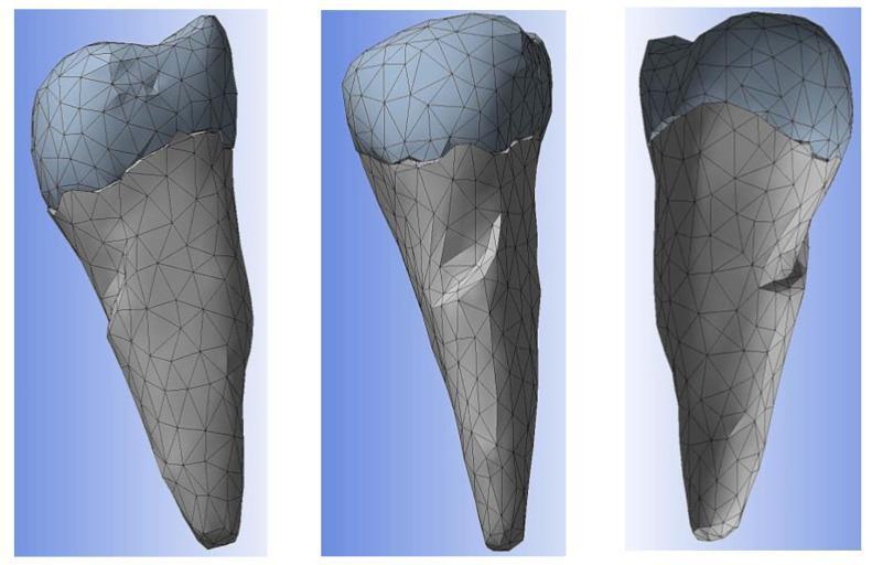
This diagnostic 3D computer-aided design (CAD) model depicts a mandibular premolar, specifically reconstructed for biomechanical analysis. The model is presented in three separate views showing various surfaces of the tooth. The reconstruction utilizes a triangular mesh, characteristic of Finite Element Analysis (FEA) preparation. The dental anatomy is differentiated by color and surface density: the coronal portion representing the enamel is shaded in light blue, while the root portion representing the cementum is shaded in gray. A significant clinical feature visible in the buccal cervical zone is an abfraction lesion, appearing as a wedge-shaped indentation at the enamel-cementum junction. The model illustrates the anatomical relationship between the crown and the tapered root, with surface contours designed to simulate volumetric consistency for stress and strain distribution studies. This visualization serves as an educational tool for understanding non-carious cervical lesions and the mechanical behavior of dental tissues under occlusal loading.
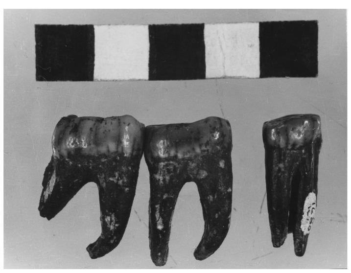
A clinical photograph of three isolated mandibular teeth from a paleoanthropological specimen (Kromdraai Paranthropus TM 1600), demonstrating significant occlusal wear and dental anatomy. The set includes a premolar, a first molar, and a second molar. The molars exhibit a characteristic multi-rooted structure with clear root bifurcation, while the premolar shows a more compact morphology. All teeth display severe abrasive wear and attrition on the occlusal surfaces, resulting in the flattening of cusps and exposure of underlying dentin, which is indicative of a coarse, high-fiber diet. The specimen shows significant extrinsic staining and discoloration consistent with fossilization. This image serves as an educational example of comparative odontological morphology and the effects of chronic mechanical stress on enamel and tooth structure across human evolutionary history. A scale bar is included for metric reference of crown and root dimensions.
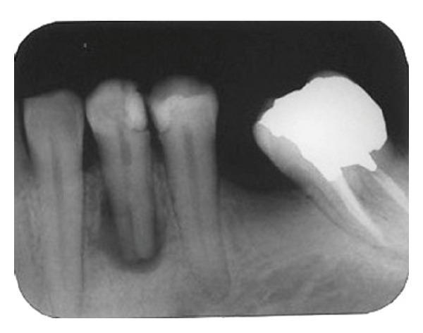
A diagnostic periapical radiograph of the mandibular left first premolar and adjacent dentition. The primary tooth of interest shows a complex endodontic anatomy characterized by a single coronal canal that bifurcates or trifurcates at the mid-root level into multiple canal systems. Intracanal calcifications are visible, appearing as radiopaque structures within the pulp chamber and canal space. Apical and periradicular pathology is evidenced by a large, well-defined radiolucency surrounding the root apex, consistent with a periapical granuloma or cyst secondary to pulp necrosis. Coronally, the tooth presents with a large, radiopaque restoration involving multiple surfaces. The surrounding alveolar bone shows a normal trabecular pattern outside the area of the apical lesion, and the lamina dura of the affected tooth appears interrupted apically. This radiographic finding is significant for endodontists as it demonstrates anatomical variation in mandibular premolars and the importance of thorough radiographic assessment for successful root canal therapy.
Anatomy of Mandibular Premolars
BDS First Year - Complete Notes
GENERAL INTRODUCTION
- Mandibular premolars are permanent succedaneous teeth replacing the mandibular deciduous molars
- There are 2 mandibular premolars per quadrant (4 total): mandibular first premolar and mandibular second premolar
- FDI notation: 44 & 45 (lower right), 34 & 35 (lower left)
- Universal notation: 28, 29 (lower right), 20, 21 (lower left)
- Location: Between the mandibular canine (anteriorly) and mandibular first molar (posteriorly)
Key General Features
- Both develop from lobes but differ: first premolar = 4 lobes, second premolar = 4 or 5 lobes
- The term "bicuspid" is misleading for mandibular premolars - the first premolar has a nearly non-functional lingual cusp, and the second can have 2-3 cusps
- Crown tilts lingually relative to the root axis (a hallmark of ALL mandibular premolars)
- Crown is wider buccolingually than mesiodistally is less pronounced here - the crown is nearly as wide BL as MD (making it look more round compared to maxillary premolars which are very oblong)
- Second premolar is larger than the first premolar (opposite to maxillary jaw where 1st > 2nd)
MANDIBULAR FIRST PREMOLAR
Chronology
| Event | Time |
|---|---|
| Calcification begins | 1.7-2 years |
| Enamel completed | 5-6 years |
| Eruption | 10-12 years |
| Root completed | 12-13 years |
Measurements (Wheeler's)
| Dimension | Value |
|---|---|
| Crown length (cervico-occlusal) | 8.5 mm |
| Crown diameter - mesiodistal | 7.0 mm |
| Crown diameter - cervical (MD) | 5.0 mm |
| Crown diameter - buccolingual | 7.5 mm |
| Root length | 14.0 mm |
Note: BL (7.5 mm) is nearly equal to MD (7.0 mm) - this gives the crown a round/square appearance on occlusal view, unlike maxillary premolars
Crown Anatomy - Surface by Surface
1. Buccal (Facial) Aspect
- Resembles the mandibular canine from the buccal view - but:
- Crown is shorter cervico-occlusally
- Crown is narrower mesiodistally than the canine
- Crown is nearly bilaterally symmetrical from buccal view
- Buccal cusp is sharp, long, and well-formed - the dominant feature
- Buccal cusp is offset slightly toward the mesial (both buccal and lingual cusps are offset to the mesial)
- Mesial and distal contact areas are at about the middle third
- Buccal cervical ridge present (height of contour of buccal surface is in the cervical third)
- Mesiobuccal and distobuccal line angles are prominent
2. Lingual Aspect
- Lingual cusp is very small and rudimentary - often described as "no larger than the cingulum of a canine"
- Lingual cusp is nonfunctional (as this is a mandibular tooth - lingual cusps of mandibular teeth do not occlude with maxillary teeth in normal occlusion)
- Crown converges markedly toward the lingual - much more than the second premolar
- Because of this lingual convergence, much of the mesial and distal surfaces are visible from the lingual view
- Lingual surface is narrow compared to the buccal surface
- Height of contour of lingual surface: middle third
3. Mesial Aspect (Important)
- Marked lingual tilt of crown relative to root axis - seen clearly from mesial view
- This is the most prominent feature from this aspect
- Buccal cusp is much longer than the lingual cusp - lingual cusp appears short and stump-like
- Mesial marginal groove may be present (crosses the mesial marginal ridge)
- Canine fossa (mesial developmental depression) - a concavity on the mesial crown surface near the cervical line, resembling the canine fossa
- Longitudinal grooves on the mesial and distal root surfaces (deepest on mesial)
- Mesial contact area: middle third
- No transverse ridge visible from this aspect (unlike the second premolar)
4. Distal Aspect
- Similar to mesial view
- Less prominent groove than the mesial
- Distal contact area: middle third
- Crown surface is generally smoother than the mesial
5. Occlusal Aspect (Most Distinctive)
- Outline form: Diamond-shaped / Rhomboidal
- Crown converges sharply toward the lingual - the narrowest posterior tooth from the lingual side
- 2 cusps: large buccal cusp + small lingual cusp
- Transverse ridge - formed by the union of the buccal triangular ridge and the lingual triangular ridge crossing the central groove area - a key feature of the mandibular first premolar
- Mesiolingual groove - a groove that separates the mesial marginal ridge from the lingual cusp; runs from the central groove toward the mesiolingual area
- Central groove runs mesiodistally
- Mesial triangular fossa and distal fossa present
- Lobes: 4 (3 buccal + 1 lingual)
- Mesiobuccal, distobuccal, mesiolingual, and distolingual cusp ridges present
- Lingual cusp tip is offset slightly toward the mesial
Root
- Single, straight root with a sharp apex
- Root is 3-4 mm shorter than the mandibular canine root
- Tapers from cervical to apex; often curves distally at the apex
- Buccolingual width > Mesiodistal width (root is broader BL than MD)
- Mesial root surface: Prominent longitudinal groove (deepest of all root surfaces) - occasionally a second root or bifurcation can occur here (rare)
- Very rarely: two roots (buccal + lingual) or three roots - these are uncommon variants
Pulp Cavity
- Pulp chamber: Well-defined
- 2 pulp horns - buccal pulp horn (large, prominent) and lingual pulp horn (small, less prominent)
- 1 root canal typically (single root)
- Occasionally 2 canals can be present (clinically important for root canal treatment)
- The buccal pulp horn is the largest, corresponding to the dominant buccal cusp
MANDIBULAR SECOND PREMOLAR
Chronology
| Event | Time |
|---|---|
| Calcification begins | 2.3-2.5 years |
| Enamel completed | 6-7 years |
| Eruption | 11-12 years |
| Root completed | 13-14 years |
Measurements (Wheeler's)
| Dimension | Value |
|---|---|
| Crown length (cervico-occlusal) | 8.0 mm |
| Crown diameter - mesiodistal | 7.0 mm |
| Crown diameter - cervical (MD) | 5.0 mm |
| Crown diameter - buccolingual | 8.0 mm |
| Root length | 14.5 mm |
Note: Second premolar is larger than first premolar overall; root is longer (14.5 vs 14 mm)
Crown Anatomy - Surface by Surface
1. Buccal (Facial) Aspect
- Larger crown and longer root than the first premolar
- Buccal cusp is shorter and less sharp than in the first premolar
- Buccal cusp tip is approximately at the center of the tooth mesiodistally (distobuccal and mesiobuccal slopes are equal) - unlike first premolar where cusp is offset mesially
- Crown form is bilaterally symmetrical from the buccal view
- Contact areas: both mesial and distal at the middle third
- Less convergence toward the lingual compared to the first premolar
2. Lingual Aspect
- Buccal profile is NOT visible from the lingual view (unlike the first premolar where the lingual convergence is so severe that buccal surface peeks through)
- Lingual cusp height is nearly equal to buccal cusp - a major distinguishing feature from the first premolar
- Lingual surface is smooth and spheroidal (bulbous) above the constricted cervical area
- Much less convergence of the crown toward the lingual compared to the first premolar
- If 3 cusps: mesiolingual and distolingual cusps visible, with mesiolingual being slightly larger
3. Mesial Aspect
- No marked lingual tilt of crown - occlusal plane is essentially horizontal (vs. marked lingual tilt in first premolar)
- Buccal and lingual cusp heights are nearly equal (vs. very unequal in first premolar)
- No transverse ridge present when viewed from mesial (vs. first premolar which has a prominent transverse ridge visible from mesial)
- No developmental depression or canine fossa on the mesial surface - crown surface is convex
- No mesial marginal groove crossing the marginal ridge
- Shallow developmental groove may be present on the root mesial surface
- Contact area and marginal ridge are at a slightly more cervical level than on the first premolar
4. Distal Aspect
- No outstanding variation compared to mesial
- Crown is smooth and rounded distally
5. Occlusal Aspect (Most Important for Distinguishing)
Form 1: Three-Cusp Type (Most Common ~60%) - "Y" groove pattern
- 3 cusps: 1 large buccal cusp + 2 lingual cusps (mesiolingual and distolingual)
- 5 developmental lobes (3 buccal + 2 lingual)
- Mesiolingual cusp is larger than the distolingual cusp
- Lingual groove separates mesiolingual and distolingual cusps, offset to the distal
- Groove pattern forms a "Y" shape
- 3 fossae: mesial, central, distal
- 2-3 pulp horns
Form 2: Two-Cusp Type - "H" or "U" groove pattern
- 2 cusps: 1 buccal + 1 lingual
- 4 developmental lobes
- Groove pattern forms either a "U" shape or "H" shape
- More similar in appearance to the mandibular first premolar but with more equal cusp sizes
Common Occlusal Features (Both Forms)
- Outline form: Square
- Lobes: 4 or 5 depending on form
- Much less lingual convergence of crown - appears more square than the first premolar
- Central groove present
- Supplemental grooves often present
Root
- Single root, longer and wider than the first premolar's root
- Root is wider than first premolar root
- Apex is inclined (curved) toward the distal
- Buccal surface: convex
- Longitudinal grooves present on the proximal surfaces (mesial and distal), mainly in the middle third on the distal; longitudinal depression rarely seen on mesial
- Lingual surface of root is slightly convex and narrower than buccal surface
Pulp Cavity
- 2 pulp horns (buccal and lingual; if 3-cusp type: 3 pulp horns)
- 1 root canal (single root in most cases)
- Pulp horns are less sharply defined than in the first premolar
- Pulp chamber corresponds to cusp arrangement
KEY COMPARISON: Mandibular First vs. Second Premolar
| Feature | Mandibular 1st Premolar | Mandibular 2nd Premolar |
|---|---|---|
| Eruption | 10-12 yrs | 11-12 yrs |
| Crown size | Smaller | Larger |
| Root length | 14.0 mm | 14.5 mm |
| Lobes | 4 | 4 or 5 |
| Buccal cusp | Very long, sharp, mesially offset | Shorter, less sharp, centered |
| Lingual cusp | Very small, rudimentary, nonfunctional | Well-formed, nearly equal to buccal |
| Number of cusps | 2 (one nonfunctional) | 2 or 3 |
| Crown shape (occlusal) | Diamond/rhomboidal | Square |
| Lingual tilt of crown | Marked | Slight / essentially horizontal |
| Transverse ridge | Present | Absent |
| Mesiolingual groove | Present | Absent |
| Lingual convergence | Severe | Mild |
| Buccal profile from lingual | Visible (due to convergence) | Not visible |
| Mesial depression/canine fossa | Present | Absent (convex) |
| Mesial marginal groove | May be present | Absent |
| Groove pattern | Simple (central groove) | Y / H / U pattern |
| Pulp canals | 1 (rarely 2) | 1 |
| Resembles | Canine | Molar |
NERVES & BLOOD SUPPLY
Innervation of Mandibular Premolars
- Inferior alveolar nerve (IAN) - a branch of the mandibular division (V3) of the trigeminal nerve (CN V)
- The IAN runs in the mandibular canal and supplies branches to the molar and premolar teeth
- At the level of the first premolar, the IAN divides into two terminal branches:
- Mental nerve - exits through the mental foramen (located inferior to the second premolar), supplies labial gingiva of incisors, canine, and premolars
- Incisive nerve - continues anteriorly in the mandibular canal, supplies the first premolar, canine, and incisors
- Mental foramen is typically located inferior to the mandibular second premolar - clinically important landmark
Blood Supply
- Inferior alveolar artery (branch of maxillary artery) - runs with the inferior alveolar nerve in the mandibular canal, supplies mandibular teeth and associated bone
Gingival Innervation
- Buccal gingiva of mandibular incisors, canines, and premolars: innervated by the mental branch of the inferior alveolar nerve
- Lingual gingiva of mandibular premolars: innervated by the lingual nerve
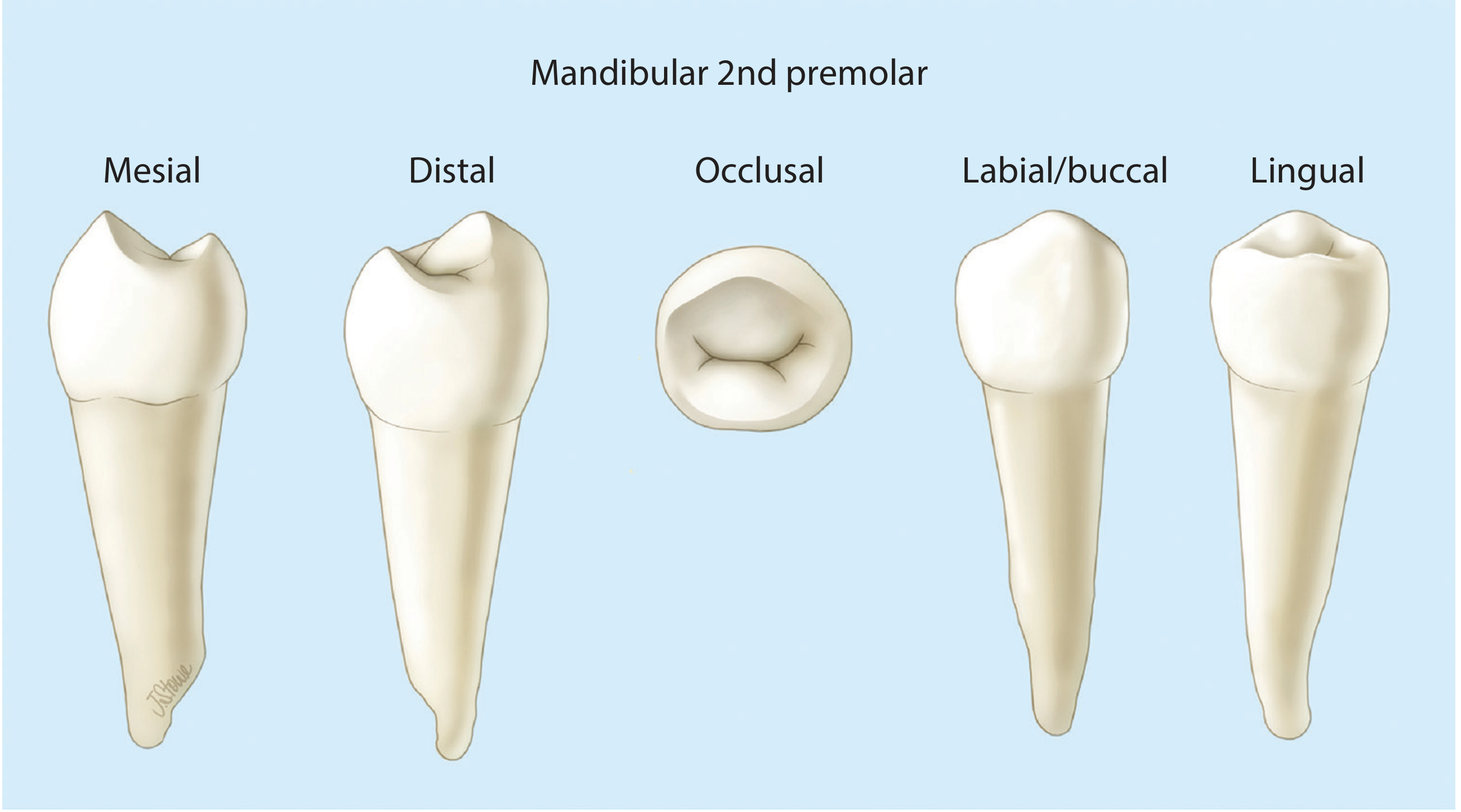
DIAGRAM OF MANDIBULAR SECOND PREMOLAR
CLINICAL IMPORTANCE (Exam Points)
- Mental foramen is located inferior to the 2nd mandibular premolar - important for inferior alveolar nerve block (mental nerve block)
- The mandibular first premolar's transverse ridge is a unique identifying feature
- The lingual cusp of mandibular first premolar is nonfunctional - this is an exam classic
- Mandibular second premolar is the only tooth with 3 cusps and 1 root (the three-cusp form)
- The second premolar is larger than the first (opposite to the maxillary arch)
- Both mandibular premolars have single roots - unlike the maxillary first premolar (which has 2 roots)
- The crown of mandibular premolars shows a lingual tilt relative to root axis - this is a posterior mandibular tooth characteristic
- Root canal treatment of mandibular first premolar can be challenging due to occasional canal bifurcation despite a single external root
Nose anatomy easy summary one shot revision dont leave anything important for exam point of view bds students 1 st year
🫀 NOSE ANATOMY - BDS First Year One-Shot Revision
Complete Exam-Oriented Summary
PART 1: OVERVIEW
- External Nose - projects from the face; skeleton of bone + cartilage
- Nasal Cavities - two internal air spaces; the actual functional spaces
- Nasal cavities = uppermost parts of the respiratory tract
- Contain olfactory receptors
- Shape: elongated wedge-shaped spaces - large inferior base, narrow superior apex
- Separated from each other by the nasal septum (midline)
- Separated from oral cavity below by the hard palate
- Separated from cranial cavity above by the cribriform plate
- Lateral to nasal cavities = orbits
PART 2: EXTERNAL NOSE
Framework (Skeleton)
- Nasal bones (form the bridge)
- Parts of the maxillae (form the side walls below nasal bones)
- Frontal bone (nasal spine, forms the root of nose)
| Cartilage | Position | Notes |
|---|---|---|
| Septal cartilage | Midline | Forms anterior part of nasal septum |
| Lateral processes of septal cartilage | Lateral, on each side | Support upper lateral walls |
| Major alar cartilages | On each side | Support the nostrils (nares) |
| Minor alar cartilages (3-4) | On each side | Additional support |
Memory tip: From top to bottom: Nasal bone → Lateral process of septal cartilage → Major alar cartilage
Surface Features
- Root = upper attachment to forehead
- Dorsum = bridge (upper surface)
- Apex/Tip = lower free end
- Alae = wing-like lateral parts of nostril
- Nares (nostrils) = oval openings at base; held open by alar cartilages
Muscles of the External Nose
- Nasalis - widens/compresses the nostrils
- Depressor septi nasi - depresses the septum
- Levator labii superioris alaeque nasi - elevates the upper lip and alae
PART 3: NASAL CAVITIES - WALLS
FLOOR (Bottom Wall)
- Smooth and concave
- Much wider than the roof
- Formed by:
- Palatine process of maxilla (anterior 3/4)
- Horizontal plate of palatine bone (posterior 1/4)
- Together = hard palate
- The naris opens anteriorly into the floor
- Incisive canal opens through the floor (lateral to the nasal septum, near the central incisor) - transmits:
- Nasopalatine nerve (downward, into oral cavity)
- Terminal end of greater palatine artery (upward, into nasal cavity)
ROOF (Top Wall)
- Narrow - highest in the center
- Divided into 3 parts (anterior → posterior):
| Part | Bones |
|---|---|
| Anterior slope (toward nares) | Nasal spine of frontal bone + nasal bones + lateral processes of septal cartilage + major alar cartilages |
| Middle (horizontal - highest part) | Cribriform plate of ethmoid ← MOST IMPORTANT; perforated for olfactory nerve fibers |
| Posterior slope (toward choana) | Anterior surface of sphenoid + ala of vomer + sphenoidal process of palatine bone + vaginal process of medial pterygoid plate |
- Cribriform plate perforations → CN I (olfactory nerve fibers) pass through
- Foramen for anterior ethmoidal nerve and vessels (just anterolateral to crista galli)
- Foramen cecum (between crista galli and frontal bone) → emissary vein connecting nasal veins to superior sagittal sinus
- Sphenoidal sinus opening → opens onto the posterior slope of the roof
MEDIAL WALL - NASAL SEPTUM (High Yield!)
| Component | Position |
|---|---|
| Septal nasal cartilage | Anteroinferior |
| Perpendicular plate of ethmoid | Posterosuperior |
| Vomer | Posteroinferior |
| Nasal spine of frontal bone | Superoanterior |
| Nasal crest of maxilla | Inferior anterior |
| Nasal crest of palatine bone | Inferior posterior |
Memory: "Septal cartilage + Perpendicular plate (ethmoid) + Vomer" = the 3 main parts
LATERAL WALL (Most Complex and Exam-Important!)
- Ethmoidal labyrinth (superior & middle conchae + uncinate process)
- Lacrimal bone
- Medial surface of maxilla
- Inferior concha (separate bone)
- Perpendicular plate of palatine bone
- Medial pterygoid plate of sphenoid
THE CONCHAE (Turbinates)
| Concha | Bone | Notes |
|---|---|---|
| Inferior concha | Separate bone (one of facial bones) | Largest; most developed |
| Middle concha | Part of ethmoid bone | Has ethmoidal bulla below it |
| Superior concha | Part of ethmoid bone | Smallest; most posterior |
| Air Channel | Location |
|---|---|
| Inferior meatus | Below inferior concha |
| Middle meatus | Between inferior and middle conchae |
| Superior meatus | Between middle and superior conchae |
| Sphenoethmoidal recess | Above/behind superior concha |
KEY LATERAL WALL STRUCTURES (Middle Meatus - Very High Yield!)
- Ethmoidal bulla - dome-shaped bulge formed by middle ethmoidal cells; just inferior to attachment of middle concha
- Semilunar hiatus - curved gutter (groove) between ethmoidal bulla above and uncinate process below; where most sinuses drain
- Ethmoidal infundibulum - channel at anterior end of semilunar hiatus, continues as the frontonasal duct into the frontal sinus
- Uncinate process - curved process of ethmoid bone; forms lower border of semilunar hiatus
PART 4: OPENINGS ON THE LATERAL WALL
| Structure | Opens into | Notes |
|---|---|---|
| Nasolacrimal duct | Inferior meatus (under anterior lip of inferior concha) | Drains tears from eye |
| Frontal sinus | Middle meatus (via frontonasal duct + ethmoidal infundibulum into anterior semilunar hiatus) | |
| Anterior ethmoidal cells | Middle meatus (into frontonasal duct or ethmoidal infundibulum) | |
| Middle ethmoidal cells | Middle meatus (onto or just above ethmoidal bulla) | |
| Maxillary sinus | Middle meatus (semilunar hiatus, just inferior to center of ethmoidal bulla) | Opening near roof of sinus - poor drainage |
| Posterior ethmoidal cells | Superior meatus | |
| Sphenoidal sinus | Sphenoethmoidal recess (posterior slope of roof) | Only sinus NOT draining on lateral wall |
Memory trick for what drains into middle meatus: "FMA drains into the Middle meatus" = Frontal, Maxillary, Anterior + Middle ethmoidal
PART 5: REGIONS OF THE NASAL CAVITY
| Region | Location | Lining | Function |
|---|---|---|---|
| Vestibule | Just inside the naris (anterior) | Skin + hair follicles (vibrissae) | Filters large particles |
| Respiratory region | Largest part of cavity | Ciliated respiratory epithelium (ciliated + mucous cells) | Warms, humidifies, filters air |
| Olfactory region | Apex (roof area) | Olfactory epithelium | Smell receptors (CN I) |
PART 6: ETHMOID BONE (High Yield Bone!)
- Cribriform plate - horizontal, perforated; separates nasal cavity from cranial cavity; CN I fibers pass through
- Crista galli - bony projection above cribriform plate; anchors the falx cerebri (fold of dura mater)
- Perpendicular plate - vertical; forms upper part of nasal septum
- Two ethmoidal labyrinths (one on each side) - contain:
- Orbital plate (flat, forms medial wall of orbit)
- Superior concha (process)
- Middle concha (process)
- Ethmoidal bulla (bulge from middle ethmoidal cells)
- Ethmoidal infundibulum (groove/channel for frontonasal duct)
- Uncinate process (hook-shaped projection)
PART 7: GATEWAYS INTO NASAL CAVITIES
| Gateway | Structures Passing Through |
|---|---|
| Cribriform plate | CN I (olfactory nerve fibers); anterior ethmoidal nerve + vessels; emissary vein (foramen cecum) |
| Sphenopalatine foramen | Sphenopalatine artery (terminal branch of maxillary artery); nasopalatine nerve (V2); superior nasal branches (V2) |
| Incisive canal | Nasopalatine nerve (from nasal cavity → oral cavity); terminal end of greater palatine artery (from oral cavity → nasal cavity) |
| Small foramina in lateral wall | Posterior inferior nasal nerves (from greater palatine nerve) |
| Around nares | Internal nasal branches of infra-orbital nerve (V2); alar branches of lateral nasal artery |
PART 8: BLOOD SUPPLY (High Yield!)
Arteries - Two sources: Internal + External Carotid
| Artery | Source | Area supplied |
|---|---|---|
| Sphenopalatine artery | Terminal branch of maxillary artery; enters via sphenopalatine foramen | Largest vessel supplying nasal cavity; posterior lateral wall + nasal septum |
| Greater palatine artery | Branch of maxillary artery; enters via incisive canal | Anterior floor + anterior septum |
| Superior labial artery | Branch of facial artery | Anterior septum (septal branch) + anterior nasal cavity |
| Lateral nasal artery | Branch of facial artery | External nose + nasal vestibule |
| Artery | Area supplied |
|---|---|
| Anterior ethmoidal artery | Anterior medial + lateral walls; frontal sinus; continues as external nasal branch on skin of nose |
| Posterior ethmoidal artery | Upper medial + lateral walls; ethmoidal cells; sphenoidal sinus |
Little's Area (Kiesselbach's Plexus) - VERY HIGH YIELD!
On the anteroinferior part of the nasal septum, 4 arteries anastomose:
- Sphenopalatine artery (septal branch)
- Greater palatine artery
- Superior labial artery (septal branch)
- Anterior ethmoidal artery
This area is closest to the surface → most common site of epistaxis (nosebleed)
Veins
| Vein | Drains to |
|---|---|
| Anterior nasal veins | Facial vein |
| Posterior nasal veins (with maxillary artery branches) | Pterygoid plexus (infratemporal fossa) |
| Ethmoidal veins | Superior ophthalmic vein → cavernous sinus |
| Via foramen cecum | Superior sagittal sinus (emissary vein - route for intracranial spread of infection) |
PART 9: NERVE SUPPLY (High Yield!)
Three cranial nerves supply the nasal cavities:
1. CN I - Olfactory Nerve
- Olfaction (smell) only
- Cell bodies in olfactory epithelium (roof of nasal cavity)
- Axons pass through cribriform plate perforations → synapse in olfactory bulb
2. CN V1 - Ophthalmic Division of Trigeminal
- General sensation to ANTERIOR nasal cavity
- Via nasociliary nerve → gives:
- Anterior ethmoidal nerve - supplies anterior nasal cavity (medial + lateral walls), frontal sinus, anterior ethmoidal cells; terminates as external nasal nerve on skin of nose
- Posterior ethmoidal nerve - supplies ethmoidal cells, sphenoidal sinus (does NOT reach nasal cavity itself)
3. CN V2 - Maxillary Division of Trigeminal
- General sensation to POSTERIOR nasal cavity
- Branches enter via sphenopalatine foramen (from pterygopalatine fossa) or via small foramina:
- Posterior superior lateral nasal nerves - lateral wall
- Posterior superior medial nasal nerves - roof + septum
- Nasopalatine nerve (largest V2 nasal branch) - medial wall (septum) → passes through incisive canal → oral mucosa behind incisors
- Posterior inferior nasal nerves (from greater palatine nerve) - inferior lateral wall and floor
- Internal nasal branches of infra-orbital nerve - around nares
4. CN VII - Facial Nerve (NOT sensory, but glandular)
- Parasympathetic secretomotor to nasal glands
- Route: Greater petrosal nerve (branch of CN VII) → pterygopalatine ganglion → postganglionic fibers join V2 branches → nasal glands
- Also: Sympathetic fibers (from T1 via superior cervical ganglion) reach nasal glands via blood vessels or V2 branches in pterygopalatine fossa - cause vasoconstriction
PART 10: PARANASAL SINUSES - Summary Table
| Sinus | Bone | Shape | Drains into | Nerve | Artery |
|---|---|---|---|---|---|
| Frontal | Frontal bone | Triangular | Middle meatus (via frontonasal duct + ethmoidal infundibulum) | Supra-orbital nerve (V1) | Anterior ethmoidal artery |
| Maxillary | Maxilla | Pyramidal | Middle meatus (semilunar hiatus) | Infra-orbital + alveolar nerves (V2) | Infra-orbital + superior alveolar branches of maxillary artery |
| Anterior ethmoidal cells | Ethmoid labyrinth | Variable | Middle meatus (ethmoidal infundibulum) | V1 (anterior ethmoidal) + V2 | Anterior ethmoidal artery |
| Middle ethmoidal cells | Ethmoid labyrinth | Variable | Middle meatus (on/above ethmoidal bulla) | V1 + V2 | Anterior ethmoidal artery |
| Posterior ethmoidal cells | Ethmoid labyrinth | Variable | Superior meatus | V1 (posterior ethmoidal) + V2 | Posterior ethmoidal artery |
| Sphenoidal | Sphenoid body | Variable | Sphenoethmoidal recess (only sinus on roof) | V1 (posterior ethmoidal) + V2 | Pharyngeal branches of maxillary artery |
- Develop as outgrowths from nasal cavities
- Lined by ciliated respiratory mucosa
- All open into nasal cavities
- All innervated by branches of CN V (trigeminal)
- Roof = floor of orbit
- Floor = roots of upper molar + premolar teeth (dental infections can cause sinusitis; extractions risk oroantral fistula)
- Posterior wall = infratemporal fossa
- Opening is near the ROOF of the sinus - poor drainage (leading to sinusitis)
- Above = cranial cavity, pituitary gland, optic chiasm
- Laterally = cavernous sinus
- Below and front = nasal cavities
- Surgical access to pituitary gland is via transsphenoidal route through the nose
PART 11: ANTERIOR APERTURE (NARES) vs POSTERIOR APERTURE (CHOANAE)
| Feature | Nares (Nostrils) | Choanae (Posterior nasal apertures) |
|---|---|---|
| Location | Inferior surface of external nose | Between nasal cavities and nasopharynx |
| Shape | Oval | Oval |
| Borders | Flexible - cartilage + soft tissue | Rigid - bone only |
| Boundary (inferior) | Maxilla + nasal spine | Horizontal plate of palatine bone |
| Boundary (lateral) | Alar cartilages | Medial pterygoid plate |
| Boundary (medial) | Septal cartilage | Posterior border of vomer |
| Boundary (roof) | - | Ala of vomer + sphenoid body |
| Clinical condition | - | Choanal atresia (congenital blockage) |
PART 12: FUNCTIONS OF NASAL CAVITY
- Olfaction - smell via olfactory receptors
- Warming of air - rich blood supply (erectile/cavernous submucosa of conchae)
- Humidification of air - mucus secretion
- Filtration - hair (vibrissae) in vestibule trap large particles; cilia move mucus posteriorly to be swallowed
- Resonance - for voice
PART 13: CLINICAL POINTS (Exam Favorites!)
| Condition | Anatomy involved |
|---|---|
| Epistaxis (nosebleed) | Little's area (Kiesselbach's plexus) on anteroinferior septum - anastomosis of 4 arteries |
| Deviated nasal septum | Shift of vomer/perpendicular plate/septal cartilage; causes obstruction |
| Sinusitis | Maxillary most common (poor drainage); middle meatus blocked |
| CSF rhinorrhea | Fracture of cribriform plate → CSF leaks into nasal cavity |
| Anosmia | Damage to CN I or olfactory region (trauma, cribriform plate fracture) |
| Choanal atresia | Congenital bony/membranous blockage of choana; neonate cannot breathe through nose |
| Pituitary surgery | Transsphenoidal approach through nasal cavity → sphenoidal sinus → sella turcica |
| Intracranial infection | Via emissary vein (foramen cecum) → superior sagittal sinus; or via ophthalmic veins → cavernous sinus |
| Dental-sinus relation | Upper premolar/molar roots close to maxillary sinus floor |
QUICK REVISION CHART
NASAL CAVITY
├── ROOF → Cribriform plate (center) ← CN I passes through
├── FLOOR → Hard palate (maxilla + palatine bone)
├── MEDIAL WALL → Nasal septum (cartilage + ethmoid perpendicular plate + vomer)
└── LATERAL WALL
    ├── Inferior concha → Inferior meatus → NASOLACRIMAL DUCT
    ├── Middle concha → Middle meatus → FRONTAL + MAXILLARY + ANT/MID ETHMOIDAL
    │       (contains: ethmoidal bulla, semilunar hiatus, ethmoidal infundibulum)
    ├── Superior concha → Superior meatus → POSTERIOR ETHMOIDAL CELLS
    └── Above superior concha → Sphenoethmoidal recess → SPHENOIDAL SINUS