
Explain this PDF to me as easily as possible.
I'll read the PDF skill first, then extract and explain the content for you.
Reading File
Now let me extract the PDF content:
Running Command
python3 -c " import pdfplumber pdf_path = '/home/daytona/workspace/attachments/483a5276-7d31-4774-98ce-255ab700733e/BIO-201 Carbohydrates.pdf' with pdfplumber.open(pdf_path) as pdf: print(f'Total pages: {len(pdf.pages)}') for i, page in enumerate(pdf.pages): print(f'\n=== PAGE {i+1} ===') text = page.extract_text() if text: print(text) else: print('[No extractable text]') "
Here's a simple breakdown of everything in your BIO-201 Carbohydrates lecture (from Lehninger Principles of Biochemistry, Chapter 7):
🍞 What Are Carbohydrates?
Carbohydrates are the most abundant biomolecules on Earth. Chemically, they are molecules made of carbon, hydrogen, and oxygen - specifically polyhydroxy aldehydes or ketones. You find them in bread, potatoes, milk, pasta, fruits, and vegetables.
Their 4 main jobs in the body:
- Provide energy (immediate fuel)
- Store energy (for later use)
- Build other large molecules (like DNA, cell walls)
- Spare proteins and fats from being burned for energy
📦 The 3 Big Classes of Carbohydrates
| Class | What it is | Examples |
|---|---|---|
| Monosaccharides | Single sugar unit | Glucose, fructose |
| Oligosaccharides (especially disaccharides) | 2 sugar units joined together | Sucrose (table sugar), lactose (milk sugar), maltose |
| Polysaccharides | Hundreds/thousands of sugar units chained together | Starch, glycogen, cellulose |
🔬 Monosaccharides in Detail
These are the building blocks of all carbohydrates. Key facts:
- Water-soluble, sweet-tasting
- Two families:
- Aldoses - the carbonyl (C=O) group is at the end of the chain (an aldehyde)
- Ketoses - the carbonyl group is in the middle of the chain (a ketone)
- Named by how many carbons they have: triose (3C), tetrose (4C), pentose (5C), hexose (6C)
Special terms to know:
- Epimers - two sugars that are identical except at ONE carbon. Example: glucose vs. galactose (differ at C-4), glucose vs. mannose (differ at C-2).
- Anomers - cyclic sugars that differ only at the anomeric carbon (C-1 for aldoses). Example: α-D-glucopyranose vs. β-D-glucopyranose.
- Mutarotation - when a sugar in solution spontaneously switches between its α and β forms until it reaches an equilibrium mixture.
- Pyranoses - 6-membered ring sugars (like glucose in ring form)
- Furanoses - 5-membered ring sugars (like fructose in ring form)
- Hemiacetal/Hemiketal - the chemical reaction that forms these rings: an aldehyde + alcohol → hemiacetal; a ketone + alcohol → hemiketal.
- Chair conformation - the actual 3D shape of a pyranose ring (it's not flat - it looks like a chair). The 3D shape matters for biological function.
🔗 Disaccharides
Two monosaccharides joined by a glycosidic bond (specifically an O-glycosidic bond - formed when a hydroxyl group reacts with the anomeric carbon of another sugar).
| Disaccharide | Made of | Found in |
|---|---|---|
| Sucrose | Glucose + Fructose | Table sugar |
| Lactose | Glucose + Galactose | Milk |
| Maltose | Glucose + Glucose | Malt/germinating grains |
🌾 Polysaccharides
Long chains of sugars. Two types:
- Homopolysaccharides - made of only ONE type of sugar
- Heteropolysaccharides - made of TWO or more different sugars
Key Homopolysaccharides (all made of glucose!):
| Polysaccharide | Where found | Branching | Function |
|---|---|---|---|
| Starch (amylose + amylopectin) | Plants | Moderately branched | Energy storage |
| Glycogen | Animals (liver, muscle) | Highly branched | Energy storage |
| Cellulose | Plant cell walls | None (unbranched) | Structural support |
- Humans can digest starch and glycogen but NOT cellulose (we lack the enzyme). Ruminants (cows, etc.) can digest it.
- Chitin - a linear polysaccharide made of N-acetylglucosamine. It's the second most abundant polysaccharide in nature (after cellulose) and makes up insect exoskeletons and fungal cell walls.
Key Heteropolysaccharide:
- Peptidoglycan - found in bacterial cell walls. Made of alternating GlcNAc and MurNAc units. It gives bacteria their rigid shape and protects them. (This is actually the target of penicillin-type antibiotics!)
🔗 Glycoconjugates - Carbs Linked to Other Molecules
Carbohydrates don't always act alone - they can be covalently attached to proteins or lipids:
| Type | What it is | Example |
|---|---|---|
| Proteoglycans | Protein + many glycosaminoglycan (GAG) chains | Aggrecan (in cartilage) |
| Glycoproteins | Protein + one or a few oligosaccharide chains | Mucin (in mucus), Collagen |
| Glycolipids | Lipid + carbohydrate attached by glycosidic bond | Gangliosides (in cell membrane) |
Their roles include cell recognition, membrane stability, and structural support.
🔑 Quick Exam Tips (the slides tell you directly what's on the exam!)
The lecturer explicitly flagged these as important:
- Definition and examples of mono-, di-, and polysaccharides
- Drawing structures of glucose, fructose, and disaccharides (Haworth formulas)
- Epimers vs. anomers (know the difference!)
- What mutarotation is
- Chair conformation of α-D-glucopyranose
- Homo vs. heteropolysaccharides with examples
- Differences between starch, glycogen, and cellulose (bonding and branching)
- Glycoconjugates (proteoglycans, glycoproteins, glycolipids) with examples
Some slides specifically said "No question in the exam from the images of this slide" for polysaccharide images, the pyranose/furanose formation from glucose, and the chitin polymer structure - so those diagrams are lower priority.
The next lecture covers Lipids. Hope this breakdown helps!
explain the difference between aldotriose and ketotriose
Both are 3-carbon sugars (trioses), but they differ in where the carbonyl group (C=O) sits on the carbon chain.
Searching sources... Let me pull the textbook content to give you the most accurate explanation.
Reading File
Finding Sources
There's a textbook diagram of exactly this. Let me fetch it and read the surrounding text:
Loading Image
Reading File
Here's a clear explanation, with the actual textbook diagram:
Aldotriose vs. Ketotriose
Both are 3-carbon sugars (trioses). The only difference is where the carbonyl group (C=O) is located on the chain.
Aldotriose - Glyceraldehyde
The carbonyl group is an aldehyde - it sits at carbon 1, at the very end of the chain. An aldehyde always has a hydrogen attached to it (C=O-H).
  C=O  ← aldehyde group at the END (C-1)
   |
H-C-OH
   |
 CH₂OH
Name: Glyceraldehyde
Ketotriose - Dihydroxyacetone
The carbonyl group is a ketone - it sits at carbon 2, in the middle of the chain. A ketone has carbons on both sides of it (no hydrogen).
 CH₂OH
   |
 C=O   ← keto group in the MIDDLE (C-2)
   |
 CH₂OH
Name: Dihydroxyacetone
Here's the textbook diagram showing both side by side:
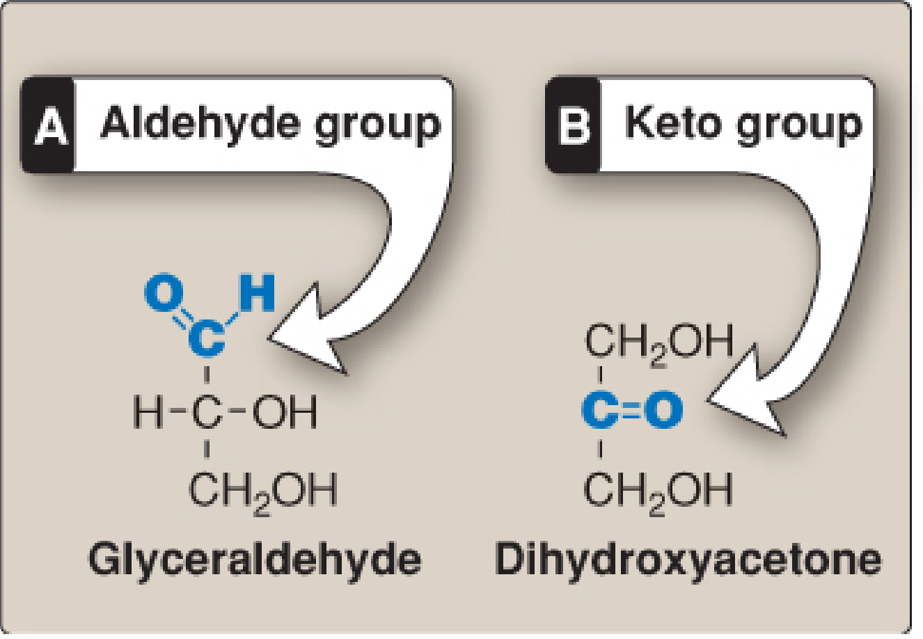
(Lippincott's Illustrated Reviews: Biochemistry, 8th Ed., Fig. 7.2)
Summary Table
| Feature | Aldotriose | Ketotriose |
|---|---|---|
| Carbonyl type | Aldehyde (C=O with H) | Ketone (C=O between 2 carbons) |
| Position of C=O | Carbon 1 (end of chain) | Carbon 2 (middle of chain) |
| Name | Glyceraldehyde | Dihydroxyacetone |
| Has a chiral center? | Yes (C-2 is chiral) | No (no asymmetric carbon) |
The last point is important: glyceraldehyde has a chiral carbon (C-2), so it can exist as D- or L-glyceraldehyde. Dihydroxyacetone is symmetric, so it has no D/L forms - it's the simplest ketose and the only monosaccharide with no chiral center.
explain the difference between aldohexose and ketohexose
Both are 6-carbon sugars (hexoses), differing in where the carbonyl group sits - just like the triose pair, but with more carbons and more complexity.
Searching sources...
Finding Sources
Reading File
Loading Image
Finding Sources
Finding Sources
Finding Sources
Reading File
Loading Image
Great - I have everything needed for a thorough explanation. Here it is:
Aldohexose vs. Ketohexose
Both are 6-carbon sugars (hexoses). The difference is exactly the same principle as the trioses - it's all about where the carbonyl group (C=O) is located.
Aldohexose - Glucose
The carbonyl group is an aldehyde sitting at carbon 1 (the very top/end) of the chain.
   C=O-H    ← aldehyde at C-1 (top of chain)
    |
H - C - OH   ← C-2
    |
HO- C - H    ← C-3
    |
H - C - OH   ← C-4
    |
H - C - OH   ← C-5
    |
   CH₂OH     ← C-6
Name: D-Glucose (also called dextrose - your blood sugar)
Because the aldehyde is at the end, C-2 through C-5 are all chiral centers (4 chiral carbons). This gives 2⁴ = 16 possible stereoisomers for an aldohexose (e.g., glucose, galactose, mannose, etc. - they're all aldohexoses, just arranged differently).
Ketohexose - Fructose
The carbonyl group is a ketone sitting at carbon 2 of the chain (one carbon in from the end).
Here is the textbook structure:
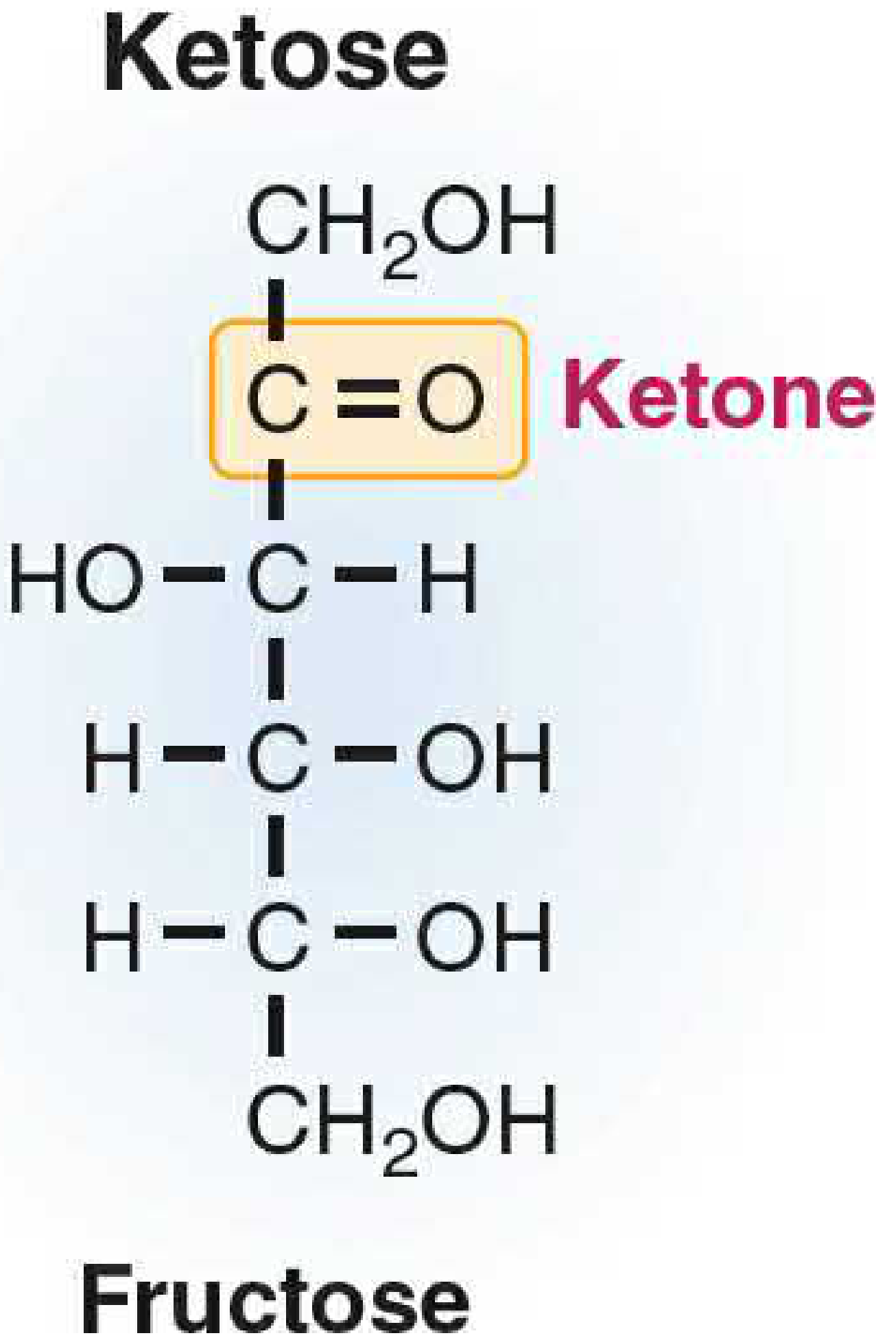
(Basic Medical Biochemistry, 6th Ed., Fig. 5.9)
  CH₂OH     ← C-1 (just an alcohol, no carbonyl here)
    |
   C=O      ← ketone at C-2
    |
HO- C - H   ← C-3
    |
H - C - OH  ← C-4
    |
H - C - OH  ← C-5
    |
  CH₂OH     ← C-6
Name: D-Fructose (fruit sugar, also found in table sugar)
Because C-1 is no longer a chiral carbon (it's just CH₂OH), only C-3 through C-5 are chiral (3 chiral carbons). This gives 2³ = 8 possible stereoisomers for a ketohexose.
Side-by-Side Comparison
| Feature | Aldohexose | Ketohexose |
|---|---|---|
| Carbonyl type | Aldehyde (C=O with H) | Ketone (C=O between 2 carbons) |
| Position of C=O | C-1 (end of chain) | C-2 (one from the end) |
| Number of chiral carbons | 4 | 3 |
| Possible stereoisomers | 16 | 8 |
| Common example | Glucose | Fructose |
| Other examples | Galactose, Mannose | Sorbose, Tagatose |
| Ring form it prefers | Pyranose (6-membered ring) | Furanose (5-membered ring) |
| Suffix naming rule | Ends in "-ose" (e.g., glucose) | Often has "-ul-" (e.g., xylulose) - though fructose is an exception |
The Key Takeaway
Think of it this way:
- Aldo- = aldehyde at the end → like glucose (your blood sugar)
- Keto- = ketone in the middle → like fructose (fruit sugar)
They have the same molecular formula (C₆H₁₂O₆) but different structures - making them isomers of each other. This small structural difference has big biological consequences: glucose is the primary fuel for cells and gets metabolized directly, while fructose must first be converted (mainly in the liver) before it can be used for energy.
This is a shared conversation. Sign in to Orris to start your own chat.