
Deltoid muscle
Reading File
Now let me search the medical library for deltoid muscle content in parallel.
Finding Sources
Finding Sources
Good results. Let me now read the key sections in parallel - Gray's Anatomy for Students (deltoid section), Rockwood & Green's (deltoid musculature), and the Imaging Anatomy atlas.
Reading File
Reading File
Reading File
Excellent content. Now let me fetch the key anatomical images and also read Campbell's section on deltoid dysfunction, all in parallel.
Loading Image
Loading Image
Loading Image
Reading File
I now have excellent content from multiple authoritative textbooks plus high-quality anatomy images. Let me compile the comprehensive answer.
Deltoid Muscle
The deltoid is the large, triangular muscle forming the rounded contour of the shoulder. Its name comes from its resemblance to the Greek letter delta (Δ) - triangular in shape.
Anatomy Overview
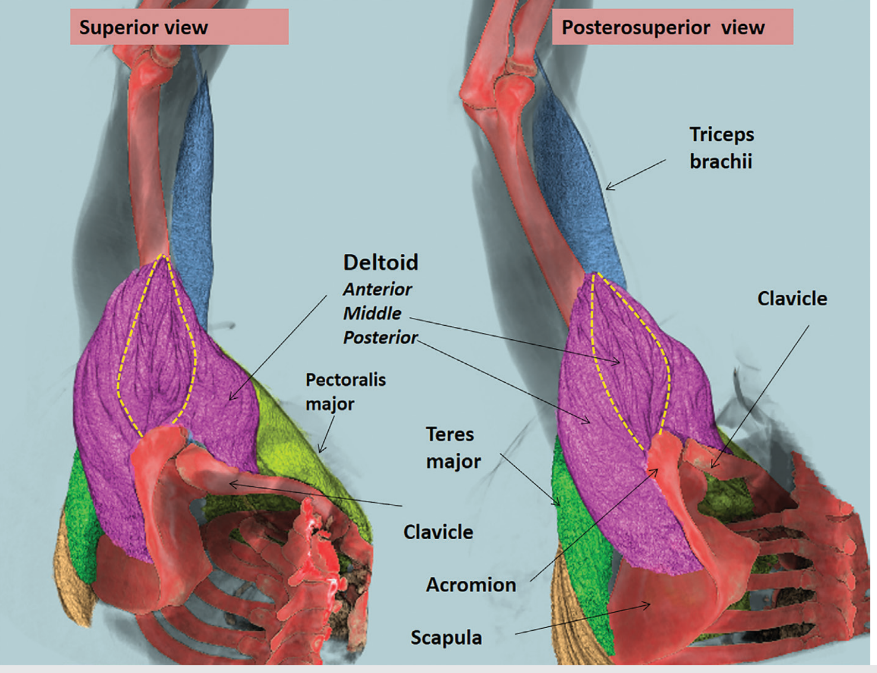
Deltoid muscle from superior and posterosuperior views showing all three portions (Imaging Anatomy, Vol. 3)
Three Parts: Origin, Course, and Insertion
The deltoid is divided into three anatomical portions - anterior, middle, and posterior - each with a distinct origin but a common insertion:
| Part | Origin | Action |
|---|---|---|
| Anterior | Anterior border and superior surface of the lateral third of the clavicle; anterior aspect of the lateral acromion | Flexion, medial rotation, horizontal adduction |
| Middle | Mid-third of the lateral margin of the acromion (relatively narrow) | Abduction (prime mover) |
| Posterior | Posterior third of the lateral acromion and the spine of the scapula | Extension, lateral rotation, horizontal abduction |
Insertion (all parts): Deltoid tuberosity - a roughened V-shaped impression on the lateral surface of the midshaft of the humerus.
The origin of the deltoid mirrors the adjacent insertion sites of the trapezius along a continuous U-shaped line on the clavicle and scapula. The deltoid contributes approximately 20% of all shoulder muscle mass (Rockwood & Green's Fractures in Adults, 10th ed).
Innervation
The deltoid is innervated by the axillary nerve (C5, C6), a branch of the posterior cord of the brachial plexus. The axillary nerve and the posterior circumflex humeral artery and vein pass posteriorly around the surgical neck of the humerus through the quadrilateral (quadrangular) space to enter the deep surface of the deltoid.
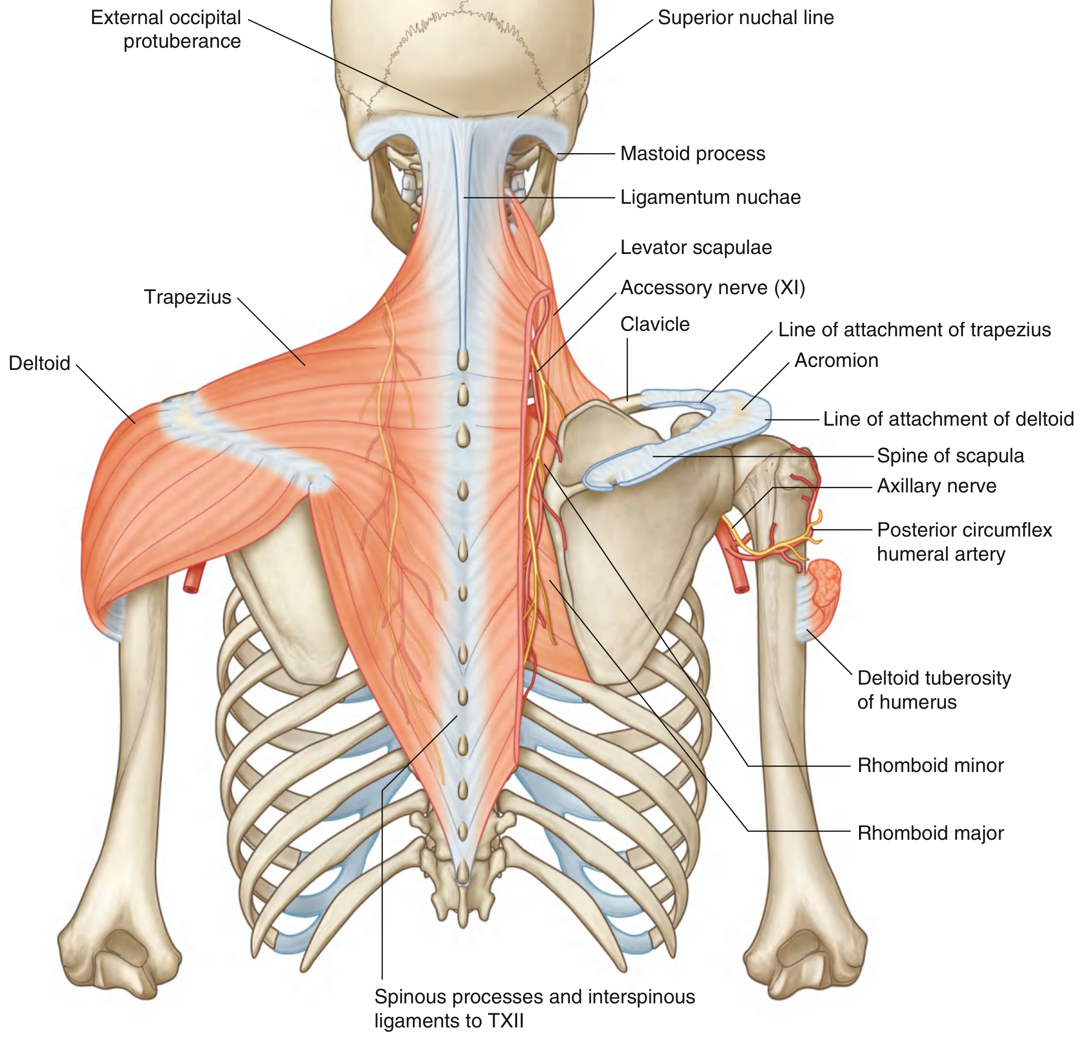
Attachments and neurovascular supply of the trapezius and deltoid muscles (Gray's Anatomy for Students)
Blood Supply
The deltoid is supplied by the posterior circumflex humeral artery (from the axillary artery) along with anastomotic branches from adjacent arteries. The artery accompanies the axillary nerve around the surgical neck.
Functions
- Abduction of the arm - the primary and most important function; the middle deltoid is the prime mover for abduction from approximately 15° to 90°
- Flexion (anterior part) - raising the arm forward
- Extension (posterior part) - pulling the arm backwards
- Medial rotation (anterior part)
- Lateral rotation (posterior part)
- Dynamic glenohumeral stabilization - the deltoid contributes to shoulder stability through:
- Passive tension from its muscle bulk
- Compression of the humeral head on the articular surface
- Joint motion that tightens passive ligaments
- Barrier effect of the contracted muscle
The middle and posterior deltoid provide more stability than the anterior deltoid by generating greater compressive forces and lower shear forces. Deltoid compressive force increases when the arm is elevated compared to neutral position (Rockwood & Green's).
Clinical Relevance
Intramuscular Injections
The deltoid is a common site for intramuscular injections (e.g., vaccines). The injection is given into the thickest part of the muscle, avoiding the lower portion where the axillary nerve travels close to the surface.
Axillary Nerve Injury
The axillary nerve is vulnerable to injury in:
- Fractures of the surgical neck of the humerus
- Anterior shoulder dislocations
- Poorly placed IM injections
Axillary nerve damage causes deltoid paralysis, resulting in loss of arm abduction and a flattened shoulder contour. This is a catastrophic functional loss.
Deltoid Muscle Dysfunction (Post-Surgical)
Deltoid dysfunction can result from axillary nerve injury or detachment of the deltoid muscle (as seen after open rotator cuff repair or open acromioplasty). After reverse total shoulder arthroplasty, deltoid degeneration has been attributed to increased moment arm in the anterior and middle heads, which while reducing the muscle effort required for most activities, can result in attritional stretching of the deltoid with eventual loss of function. Postoperative fatty infiltration of the deltoid is also a recognized complication leading to inferior clinical outcomes (Campbell's Operative Orthopaedics, 15th ed, 2026).
Deltoid Tears
Tears of the deltoid muscle, or avulsion of the tendon from its acromial origin, are rare but can occur in association with rotator cuff tears. Rupture or dehiscence at the acromial origin is a known complication of open shoulder surgery (Imaging Anatomy, Vol. 3).
Key Anatomical Relationships
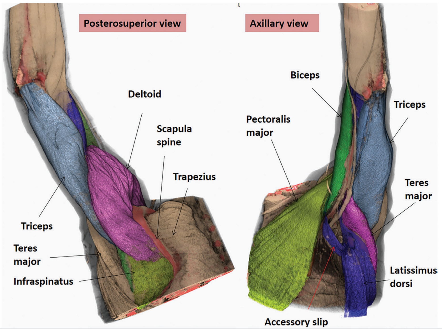
Shoulder region: the deltoid forms the superior aspect of the shoulder. The posterior axillary fold is formed by latissimus dorsi and teres major; the anterior axillary fold by pectoralis major (Imaging Anatomy, Vol. 3)
- Superficially: Skin and subcutaneous tissue
- Deep to deltoid: Rotator cuff muscles (supraspinatus, infraspinatus, teres minor, subscapularis), glenohumeral joint, subacromial bursa
- Medial relations: Pectoralis major (anteriorly), trapezius (posteriorly) - note the mirror-image origin/insertion relationship between trapezius and deltoid
- The quadrilateral space (bounded by teres minor superiorly, teres major inferiorly, long head of triceps medially, and surgical neck of humerus laterally) transmits the axillary nerve and posterior circumflex humeral vessels to the deltoid
Sources:
- Gray's Anatomy for Students, p. 834
- Rockwood and Green's Fractures in Adults, 10th ed 2025, p. 1273
- Imaging Anatomy Text and Atlas, Vol. 3 (Bones, Joints, Vessels and Nerves), p. 100
- Campbell's Operative Orthopaedics, 15th ed 2026
This is a shared conversation. Sign in to Orris to start your own chat.