
JE vector
Japanese encephalitis Culex mosquito vector lifecycle
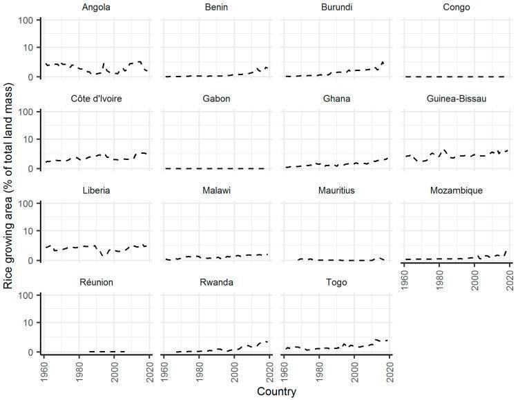
This figure is a multi-panel line graph illustrating the percentage of total land mass used for rice cultivation across 15 African countries from 1960 to 2020. This data serves as a secondary indicator for Japanese Encephalitis Virus (JEV) transmission risk, as rice paddies provide essential breeding habitats for Culex mosquito vectors. The countries displayed include Angola, Benin, Burundi, Congo, Côte d'Ivoire, Gabon, Ghana, Guinea-Bissau, Liberia, Malawi, Mauritius, Mozambique, Réunion, Rwanda, and Togo. The y-axis uses a logarithmic scale to show the percentage of land mass, while the x-axis represents time in years. Key trends reveal that most countries maintain low rice-growing areas (below 10% of total land), with notable fluctuations in Liberia and Guinea-Bissau. Several nations, specifically Burundi, Rwanda, and Togo, exhibit a distinct upward trend in rice production starting around the year 2000, signifying potentially increasing ecological suitability for JEV vectors. In contrast, countries like Gabon, Congo, and Réunion show negligible rice-growing area (near 0%) throughout the study period.
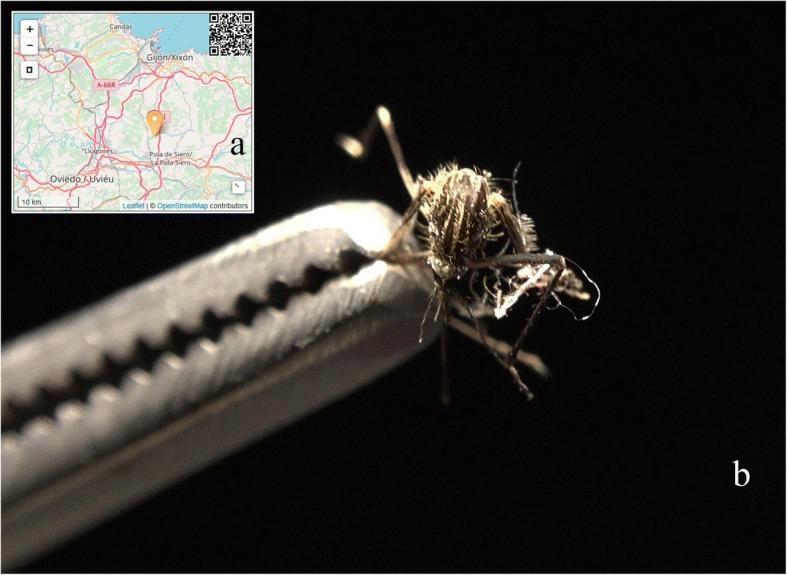
This composite educational graphic details a field specimen of a medically significant disease vector. Image (a) provides a geographic locator map focused on the Asturias region in Spain, identifying the reporting site for an invasive mosquito population. Image (b) is a close-up clinical photograph of an adult female mosquito specimen, identified as Aedes japonicus (Asian bush mosquito), held by metallic serrated forceps. The specimen exhibits diagnostic morphological features including a dark body with distinctive pale longitudinal stripes on the dorsal thorax and spindly, hirsute legs. The image illustrates the physical appearance used for taxonomic identification in medical entomology. This material is designed for public health surveillance and epidemiology training, highlighting the role of citizen science and expert verification in monitoring potential vectors of zoonotic diseases such as West Nile virus and Japanese encephalitis.
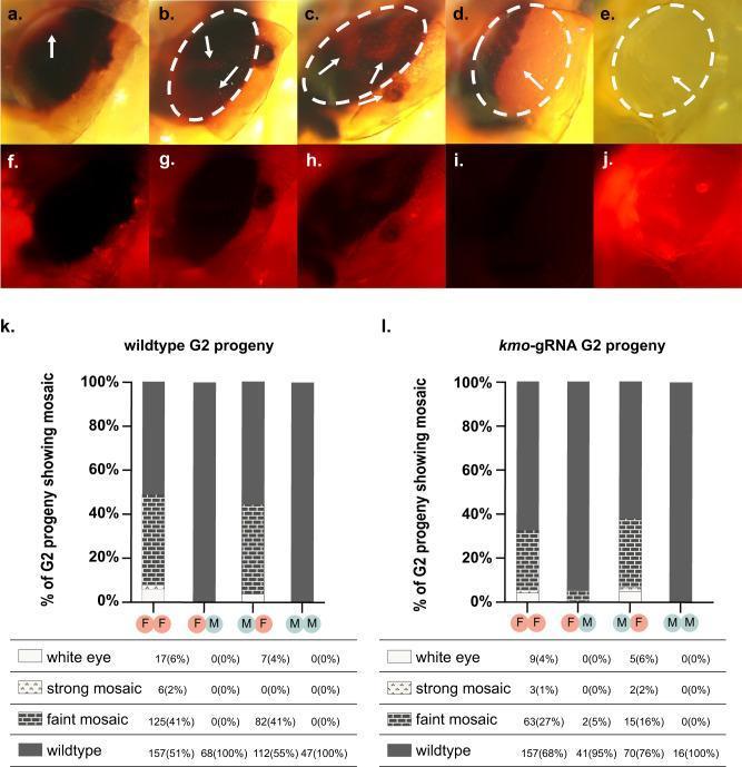
This composite educational resource illustrates the phenotypic outcomes of CRISPR-based gene drive targeting the kmo gene in Culex mosquitoes, a common vector in medical entomology research. Panels a–j present clinical-style close-up photographs of mosquito eyes across a spectrum of pigmentation loss. Under white light (a–e), 'faint mosaics' (a–c) show small localized depigmented patches (white arrows). 'Strong mosaics' (d) exhibit larger, coalescing areas of reduced pigmentation, while 'white eyes' (e) demonstrate total loss of endogenous eye pigment. Panels f–j provide corresponding mCherry fluorescence filter views, used to track transgenes via fluorescent markers. Panels k and l contain comparison stacked bar charts detailing the penetrance of these phenotypes in G2 progeny (wildtype vs. kmo-gRNA groups). The charts compare four experimental crosses (FF, FM, MF, MM), quantifying the distribution of wildtype, faint mosaic, strong mosaic, and white eye phenotypes. These data visualize how parental sex and transgene inheritance influence somatic mosaicism through transgenerational deposition of Cas9/gRNA complexes. This material is used to teach concepts of gene-drive mechanisms, inheritance bias, and genetic vector control strategies in medical research.
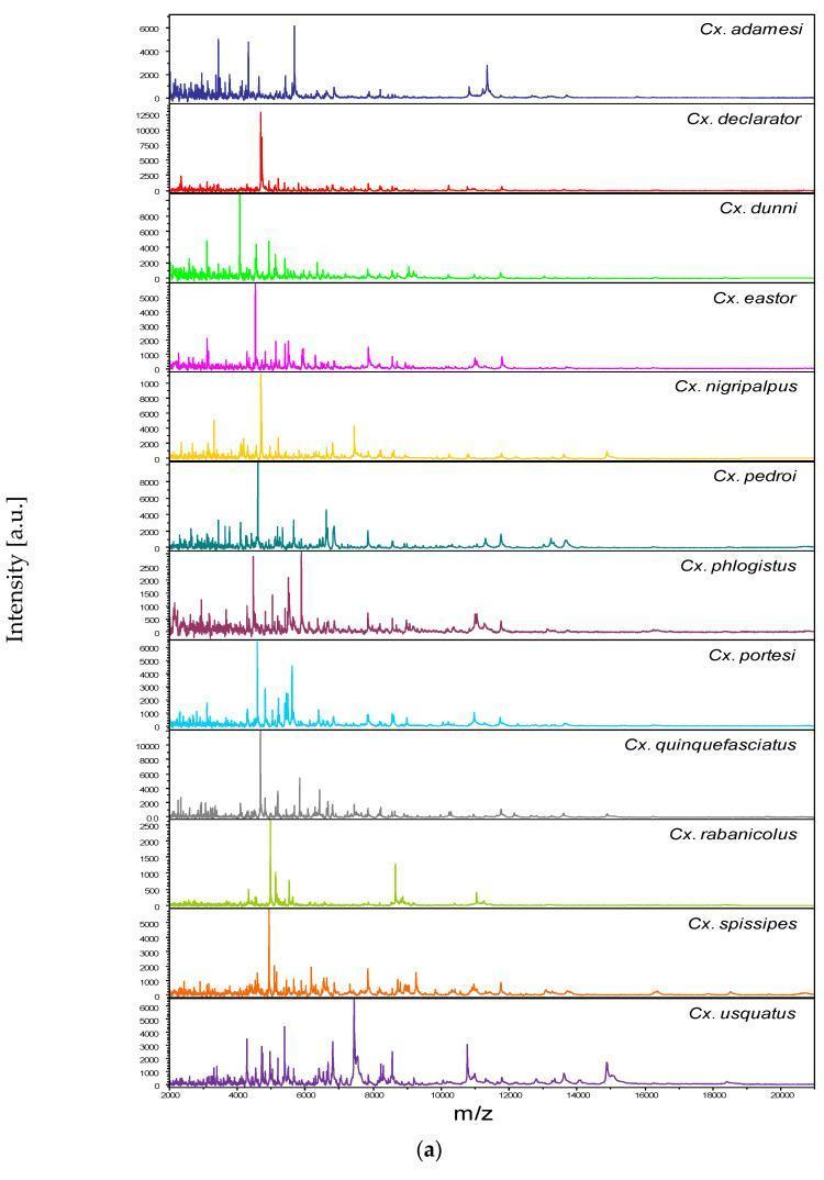
This diagnostic graphic presents a series of Matrix-Assisted Laser Desorption/Ionization Time-of-Flight Mass Spectrometry (MALDI-TOF MS) spectra used for the rapid identification and classification of Culex mosquito species, which are significant vectors for human diseases such as West Nile virus and lymphatic filariasis. The image displays twelve distinct protein mass spectra, each corresponding to a specific Culex species (e.g., Cx. adamesi, Cx. declarator, Cx. quinquefasciatus). The horizontal axis represents the mass-to-charge ratio (m/z) ranging from 2,000 to 20,000, while the vertical axis indicates signal intensity in arbitrary units (a.u.). Each spectrum serves as a unique 'protein fingerprint' characterized by specific peak distributions. For instance, Cx. declarator shows a sharp, high-intensity peak near 3,000 m/z, whereas Cx. usquatus exhibits a more complex distribution between 4,000 and 8,000 m/z. This proteomic profiling method is an essential tool in medical entomology for epidemiological surveillance and the control of vector-borne illnesses, allowing for higher taxonomic resolution than traditional morphological identification.
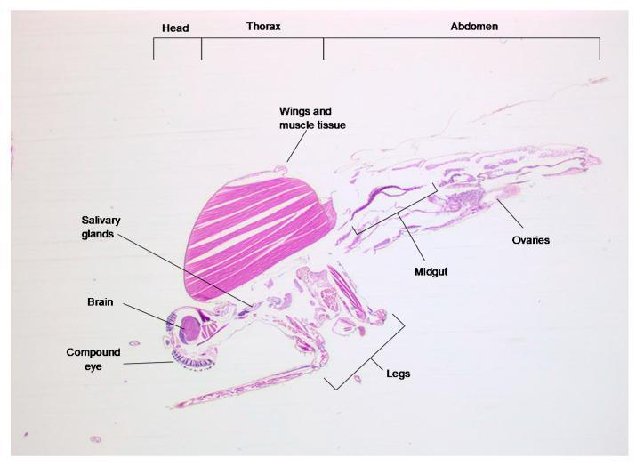
This diagnostic image shows a 3 µm longitudinal-transverse histological section of a Culex pipiens mosquito, a primary vector for Rift Valley fever virus (RVFV). The specimen is stained with haematoxylin and eosin (H&E), illustrating the anatomical organization relevant to arbovirus transmission and pathogenesis. The image is divided into three labeled segments: Head, Thorax, and Abdomen. Key structures identified include: 1) Head: The compound eye and brain (cerebral ganglion), showing dark nuclear staining; 2) Thorax: Dominated by large, eosinophilic bundles of dorsal longitudinal wing muscles and salivary glands, which are critical sites for virus replication and secretion; 3) Abdomen: Visualizes the midgut epithelium—the initial site of viral entry—and the ovaries. The lower portion displays the leg structures. This section serves as an educational reference for identifying mosquito anatomy during vector competence studies to determine tissue-specific viral distribution and dissemination barriers.
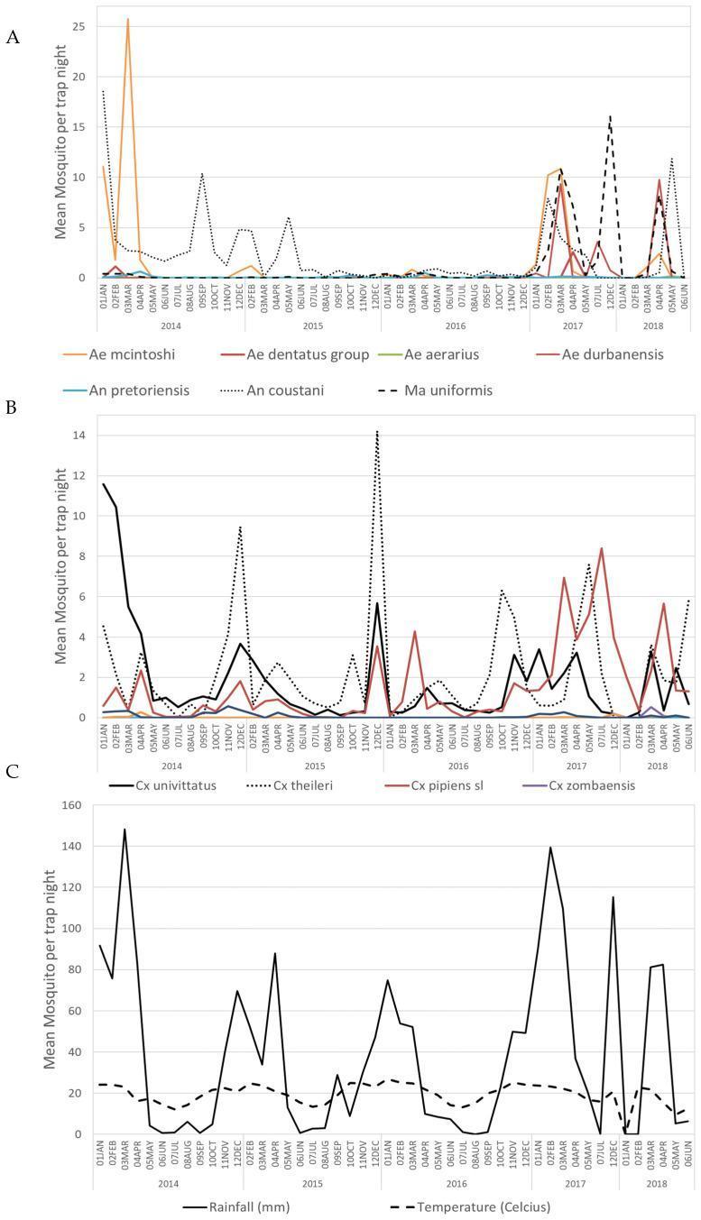
A multi-panel line graph illustrating the temporal abundance of mosquito vectors associated with alphavirus transmission, alongside environmental data. Panel A tracks the mean abundance per trap night for Aedes (Ae. mcintoshi, Ae. dentatus, Ae. aerarius, Ae. durbanensis), Anopheles (An. pretoriensis, An. coustani), and Mansonia (Ma. uniformis) species from January 2014 to May 2018. Panel B displays abundance data for Culex species, including Cx. univittatus, Cx. theileri, Cx. pipiens s.l., and Cx. zombaensis. Panel C provides contextual environmental data, showing monthly rainfall in millimeters and mean temperature in degrees Celsius over the same period. The data demonstrates seasonal fluctuations in vector density, highlighting specific peaks in species like Ae. mcintoshi and Cx. univittatus that correlate with high rainfall events and specific temperature ranges. This infographic serves as an epidemiological tool for understanding the ecological drivers of mosquito-borne diseases like Sindbis, Middelburg, and Ndumu virus outbreaks in South Africa.
Japanese Encephalitis (JE) - Vector
Primary Vector
Key Features of the Vector
| Feature | Detail |
|---|---|
| Genus/Species | Culex tritaeniorhynchus (and related ground-dwelling Culex spp.) |
| Breeding habitat | Flooded/irrigated rice fields; areas with standing water attracting aquatic/wading birds |
| Biting time | Twilight (dusk/dawn); outdoors |
| Geographic range | Asia (India, China, Japan, Southeast Asia, Russian Far East, Pakistan), western Pacific Islands, and recently documented in parts of Australia (2021-22 outbreak) |
Transmission Cycle
Amplifying hosts (Pigs + Aquatic/wading birds)
        ↕ (mosquito bites → blood meal)
Culex tritaeniorhynchus (VECTOR)
        ↓ (infective bite)
Humans (dead-end host - no onward transmission)
- Amplifying hosts: Pigs (viremia without illness; also associated with pregnancy loss) and aquatic birds are the main reservoir amplifiers
- Horses also develop encephalitis and can act as amplifying hosts
- Humans are incidental/dead-end hosts - they develop insufficient viremia to infect a feeding mosquito
- Vaccinating domestic pigs and horses can reduce viral transmission to humans
Why Rice Fields Matter
Epidemiology
- ~68,000 cases and ~13,600-20,400 deaths reported annually - Harrison's 22E
- Endemic throughout Asia; causes epidemic or sporadic outbreaks across the western Pacific
- Rarely affects travelers to endemic regions because the vector breeds primarily in rural rice fields - Tintinalli's Emergency Medicine
- In India, JE falls under the National Vector Borne Disease Control Programme (NVBDCP) alongside malaria, filariasis, dengue, Kala-azar, and chikungunya - Park's Textbook of Preventive and Social Medicine
Quick Summary Table: JE vs Other Culex-Borne Diseases
| Disease | Vector | Reservoir/Amplifier |
|---|---|---|
| Japanese Encephalitis | Culex tritaeniorhynchus | Pigs, aquatic birds |
| West Nile Encephalitis | Culex spp. | Birds |
| St. Louis Encephalitis | Culex tarsalis / pipiens / quinquefasciatus | Birds |
| Bancroftian Filariasis | Culex spp. | Humans |
Prevention (Vector-Related)
- Insecticide-treated bed nets
- DEET-containing repellents
- Permethrin-embedded clothing
- Avoidance of mosquito breeding grounds (rice paddies at dusk)
- Indoor residual spraying
- Vaccination (killed or live-attenuated JE vaccines available; two IM doses 28-45 days apart; second dose ≥1 week before travel) - Goldman-Cecil Medicine; Harrison's 22E
Malaria chemo prophylaxis for travelling
"malaria chemoprophylaxis" AND "travelers"
"malaria prophylaxis" AND travelers
Malaria Chemoprophylaxis for Travelers
Step 1 - Determine the Risk Area (Drug Resistance Pattern)
| Zone | Examples |
|---|---|
| Chloroquine-sensitive only | Caribbean, parts of Central America (west of Panama Canal), some Middle East areas |
| Chloroquine-resistant (most of the world) | Sub-Saharan Africa, South/Southeast Asia, South America, Oceania |
Risk in travelers not taking prophylaxis ranges from 3.4% per month in West Africa to ~0.034% per month in South America. - Goldman-Cecil Medicine
Drugs Available for Prophylaxis
Drug-by-Drug Summary
1. Chloroquine (+ Hydroxychloroquine)
- Use: Chloroquine-sensitive areas only
- Dose: 500 mg salt (300 mg base) orally once weekly; pediatric: 5 mg/kg base weekly (max adult dose)
- Timing: Start 1-2 weeks before travel, continue 4 weeks after leaving
- Adverse effects: GI disturbance, headache, dizziness, blurred vision, pruritus, insomnia; can exacerbate psoriasis
- Notes: Finite dose limit due to ocular (retinal) toxicity with prolonged use; take with meals
2. Mefloquine
- Use: Chloroquine-resistant areas; best for long-stay travelers
- Dose: 250 mg salt once weekly; pediatric: 4.6 mg/kg salt weekly (>5 kg body weight)
- Timing: Start 2-3 weeks before travel (to allow time to identify side effects before departure), continue 4 weeks after leaving
- Adverse effects: GI disturbance, headache, insomnia, vivid dreams, visual disturbance, anxiety, dizziness
- Black box warning: Neurologic (dizziness, tinnitus) and psychiatric (anxiety, paranoia, depression, hallucinations) side effects that may persist months to years after stopping
- Contraindications: History of seizures, psychiatric disorder, cardiac conduction abnormalities, known hypersensitivity
- Notes: Best-documented for long-term travelers; weekly dosing improves adherence; ~5% discontinue due to side effects - Goldman-Cecil Medicine
3. Doxycycline
- Use: All chloroquine-resistant areas; good alternative when mefloquine is contraindicated
- Dose: 100 mg/day orally; pediatric: 2.2 mg/kg/day (max 100 mg/day)
- Timing: Start 1-2 days before travel, continue 4 weeks after leaving
- Adverse effects: Photosensitivity (use sunscreen), GI disturbance (take with meals), esophageal irritation
- Contraindications: Children <8 years (when prophylaxis >21 days), pregnancy, lactation
- Notes: Also active against rickettsiae and leptospirae (useful for hikers, campers, freshwater swimmers); complete oral typhoid vaccine >72 h before starting; ~5% discontinue - Red Book 2021; Goldman-Cecil
4. Atovaquone-Proguanil (Malarone)
- Use: All chloroquine-resistant areas; best for short-stay travelers (<6 months)
- Dose: 250/100 mg once daily (adult); pediatric dosing by weight
- Timing: Start 1-2 days before travel, continue only 7 days after leaving (shortest post-exposure period of all drugs - very convenient)
- Adverse effects: GI upset, headache; hepatitis is rare and not common despite concerns
- Contraindications: Severe renal impairment (CrCl <30 mL/min); not for prophylaxis in pregnancy (limited data)
- Notes: Most convenient for short trips (1-3 week itineraries); daily dosing and higher cost limit use long-term; if malaria develops on atovaquone-proguanil, do NOT use it for treatment - Goldman-Cecil; Red Book 2021
5. Primaquine
- Use: All areas; especially useful where P. vivax predominates; also causal prophylaxis (acts on liver stages)
- Dose: 30 mg base (52.6 mg salt) daily; pediatric: 0.5 mg/kg base daily
- Timing: Start 1-2 days before travel, continue 7 days after leaving
- Contraindications: G6PD deficiency (causes hemolysis - MUST test before prescribing); pregnancy; lactation (unless infant tested G6PD-normal)
- Notes: Also used as terminal/antirelapse prophylaxis for P. vivax and P. ovale hypnozoites; licensed for 4 months but may be given up to 2 years
6. Tafenoquine (FDA approved 2018)
- Use: Short-duration travel (<6 months) to all areas; also antirelapse
- Dose: Loading: 200 mg daily x 3 days before departure; Maintenance: 200 mg weekly during travel; Post-trip: single 200 mg dose 7 days after last weekly dose
- Contraindications: G6PD deficiency (quantitative test mandatory before use); pregnancy; lactation; not approved <18 years for prophylaxis
- Notes: Addresses P. vivax/P. ovale liver stages like primaquine; single post-trip dose is an advantage over primaquine's 7-day course - Red Book 2021
Quick Comparison Table
| Drug | Area | Timing (Start) | Post-exposure | Frequency | Best For |
|---|---|---|---|---|---|
| Chloroquine | Sensitive only | 1-2 wk before | 4 weeks | Weekly | Long stay, sensitive areas |
| Mefloquine | Resistant | 2-3 wk before | 4 weeks | Weekly | Long-stay travelers |
| Doxycycline | All resistant | 1-2 days before | 4 weeks | Daily | Mefloquine contraindicated |
| Atovaquone-Proguanil | All resistant | 1-2 days before | 7 days | Daily | Short trips (1-3 wk) |
| Primaquine | All (esp. vivax) | 1-2 days before | 7 days | Daily | P. vivax areas, G6PD-normal |
| Tafenoquine | All | 3 days before | 1 week (single dose) | Weekly | Short stay, G6PD-normal |
Special Populations
- Chloroquine: safe in all trimesters for sensitive areas
- Mefloquine: recommended by CDC in all trimesters when chloroquine-resistant P. falciparum exposure is unavoidable
- Doxycycline: contraindicated in pregnancy
- Primaquine & tafenoquine: contraindicated (fetal G6PD status unknown) - Red Book 2021
- Dosing calculated by current weight (never exceed adult dose)
- Doxycycline: avoid in children <8 years when prophylaxis >21 days
- Tafenoquine: not approved <18 years for prophylaxis
- Mefloquine or doxycycline preferred
- Atovaquone-proguanil cost and daily dosing become impractical
Terminal Prophylaxis (Antirelapse)
- Primaquine 30 mg base daily x 14 days OR
- Tafenoquine 300 mg single dose (antirelapse formulation, ≥16 years)
- Always test for G6PD deficiency before prescribing either drug - Harrison's 22E; Red Book 2021
Key Reminders for Travelers
- No drug is 100% effective - always combine with personal protection (DEET 20-50%, permethrin clothing, bed nets, avoid outdoor activity dusk to dawn)
- Continue drugs for the full post-exposure period - most failures are due to early discontinuation
- Any febrile illness within 3 months of return from a malarious area requires immediate evaluation with blood smear or rapid diagnostic test
- Do not obtain prophylactic drugs overseas - quality cannot be assured - Red Book 2021