
Direct and indirect hernia from the book of surgery, bellies and love.
Reading File
Now let me search the library for the book "Surgery, Bellies and Love" and find content on direct and indirect hernias.
Finding Sources
Finding Sources
The book is Bailey and Love's Short Practice of Surgery. Now let me search specifically for direct and indirect hernia content from that book.
Finding Sources
Finding Sources
Now let me read the main hernia sections from Bailey and Love's block 9 and also search for direct/indirect specific content:
Reading File
Finding Sources
Excellent! Now let me read the key sections in detail:
Reading File
Reading File
Reading File
Reading File
I now have enough rich content from Bailey and Love's. Let me also fetch the key anatomical images referenced in the text:
Loading Image
Loading Image
Loading Image
I now have all the content needed for a thorough answer. Here is the complete, well-organized response from Bailey and Love's:
Direct and Indirect Inguinal Hernia
Source: Bailey and Love's Short Practice of Surgery, 28th Edition
Overview
Inguinal hernia, often called a "rupture" by patients, is the most common hernia in men - around 10 times more common in men than in women. There are two basic types that are fundamentally different in anatomy, causation, and complications: indirect (lateral/oblique) and direct (medial). Despite these differences, surgical repair techniques are very similar, so they are often discussed together.
Anatomy of the Inguinal Canal
To understand both types, the inguinal canal anatomy is key:
- Deep inguinal ring - a defect in the transversalis fascia, located midway between the anterior superior iliac spine (ASIS) and the pubic tubercle, ~2-3 cm above and just lateral to the femoral artery pulse
- Inferior epigastric vessels - lie just medial to the deep inguinal ring (this is the key landmark separating the two hernia types)
- Conjoint tendon - formed by fusion of transversus abdominis and internal oblique muscles, arches over the deep ring and attaches to the pubic tubercle; it forms the roof of the canal
- Walls of the canal: anterior = external oblique aponeurosis; posterior = transversalis fascia; roof = conjoint tendon; floor = inguinal (Poupart's) ligament
- Contents in males: testicular artery, veins, lymphatics, vas deferens (covered in cremasteric muscle), and three nerves - ilioinguinal, iliohypogastric, and genital branch of genitofemoral nerve
- Superficial inguinal ring - an inverted V-shaped defect in the external oblique aponeurosis, through which the testis descends into the scrotum
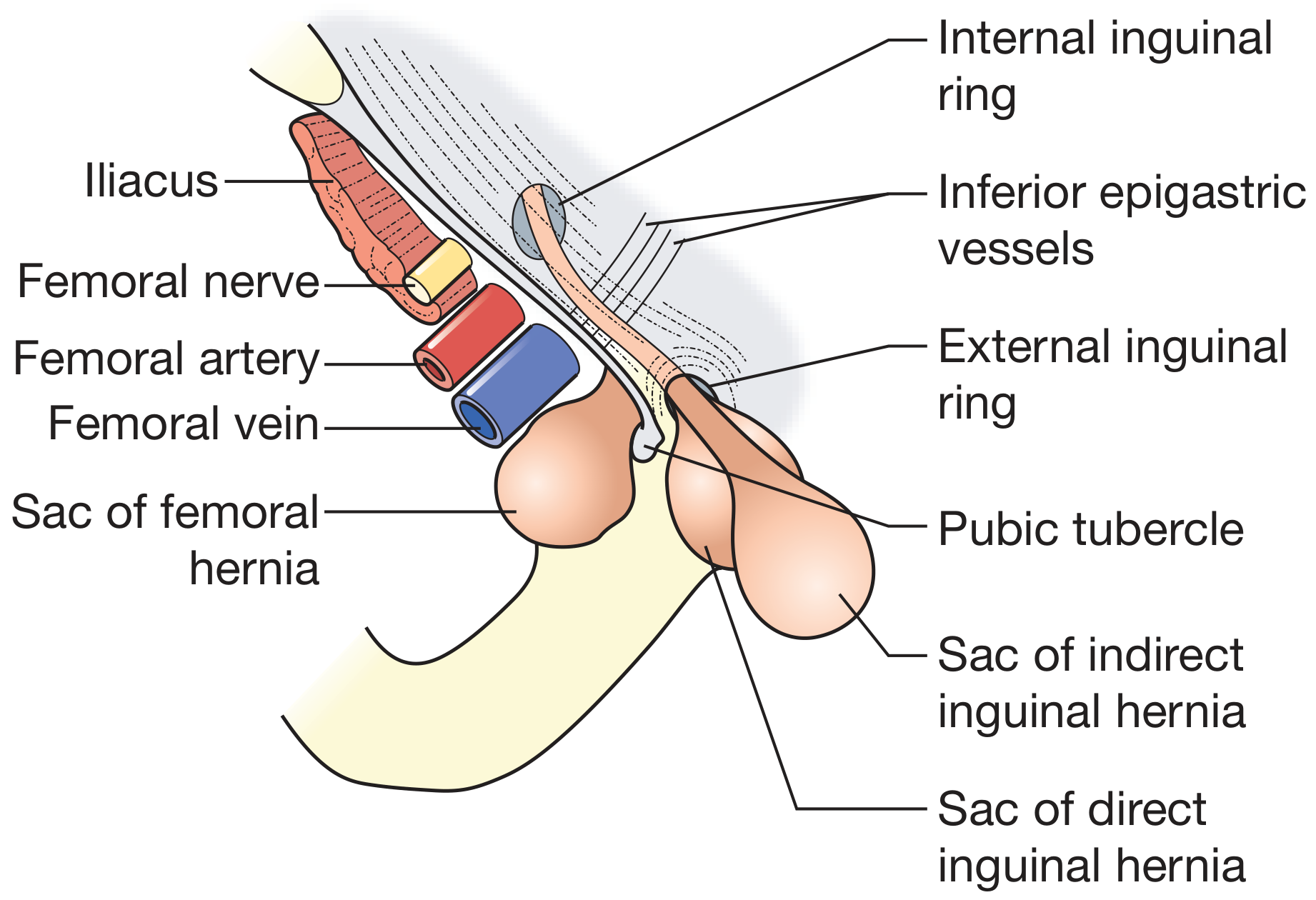
Indirect (Lateral / Oblique) Inguinal Hernia
Anatomy and Pathogenesis
As the testis descends from the abdominal cavity, it pulls a tube of peritoneum with it, which ultimately forms the tunica vaginalis. This peritoneal tube, the processus vaginalis, should obliterate after birth - but commonly fails to do so completely.
- Congenital type: When the processus vaginalis remains patent, bowel within the peritoneal cavity can pass inside the tube towards the scrotum. All inguinal hernias in neonates and children are of this congenital indirect type.
- Acquired type: In older patients, muscles around the deep inguinal ring can stretch under constant positive abdominal pressure, and a hernia becomes apparent over time.
Key Features
| Feature | Detail |
|---|---|
| Position relative to epigastric vessels | Lateral to the inferior epigastric vessels |
| Direction | Oblique - passes obliquely from lateral to medial through abdominal muscle layers |
| Entry point | Enters via the deep inguinal ring |
| Extent | Can pass all the way down into the scrotum (following the processus vaginalis line) |
| Origin | Congenital or acquired |
| Strangulation risk | Higher - narrow neck at deep ring makes it prone to strangulation |
Sliding Hernia (a subtype of indirect)
Acquired indirect hernias where retroperitoneal fatty tissue is pushed down the inguinal canal; as more tissue enters, peritoneum is pulled with it creating a sac secondarily. On the left, sigmoid colon may descend; on the right, caecum or bladder may be involved.
Direct (Medial) Inguinal Hernia
Anatomy and Pathogenesis
The direct hernia results from stretching and weakening of the abdominal wall just medial to the inferior epigastric vessels, in the region known as Hesselbach's triangle.
Hesselbach's Triangle - the three boundaries are:
- Laterally - the inferior epigastric vessels
- Medially - the lateral edge of rectus abdominis muscle
- Below - the inguinal ligament (iliopubic tract)
This area is inherently weak because the abdominal wall at this point consists of only transversalis fascia covered by the external oblique aponeurosis (no muscle layer).
Key Features
| Feature | Detail |
|---|---|
| Position relative to epigastric vessels | Medial to the inferior epigastric vessels |
| Direction | Pushes directly forward through Hesselbach's triangle |
| Entry point | Does NOT enter through the deep inguinal ring |
| Extent | Cannot descend into the scrotum |
| Origin | Always acquired (never congenital) |
| Patient profile | More likely in elderly patients |
| Base | Broadly based |
| Strangulation risk | Unlikely to strangulate (broad-based neck) |
| Special note | The urinary bladder can be pulled into a direct hernia |
Laparoscopic View of Hernia Defects
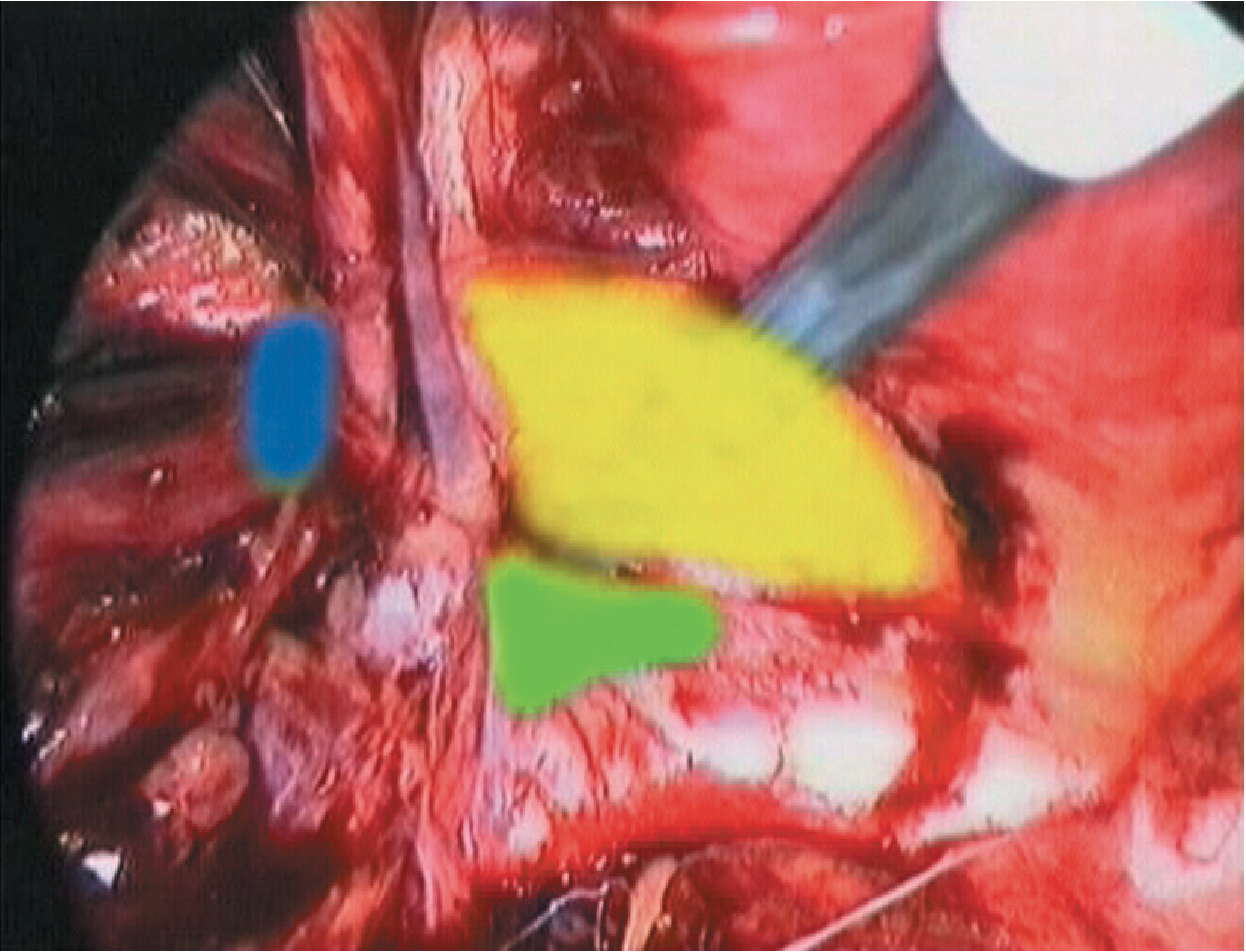
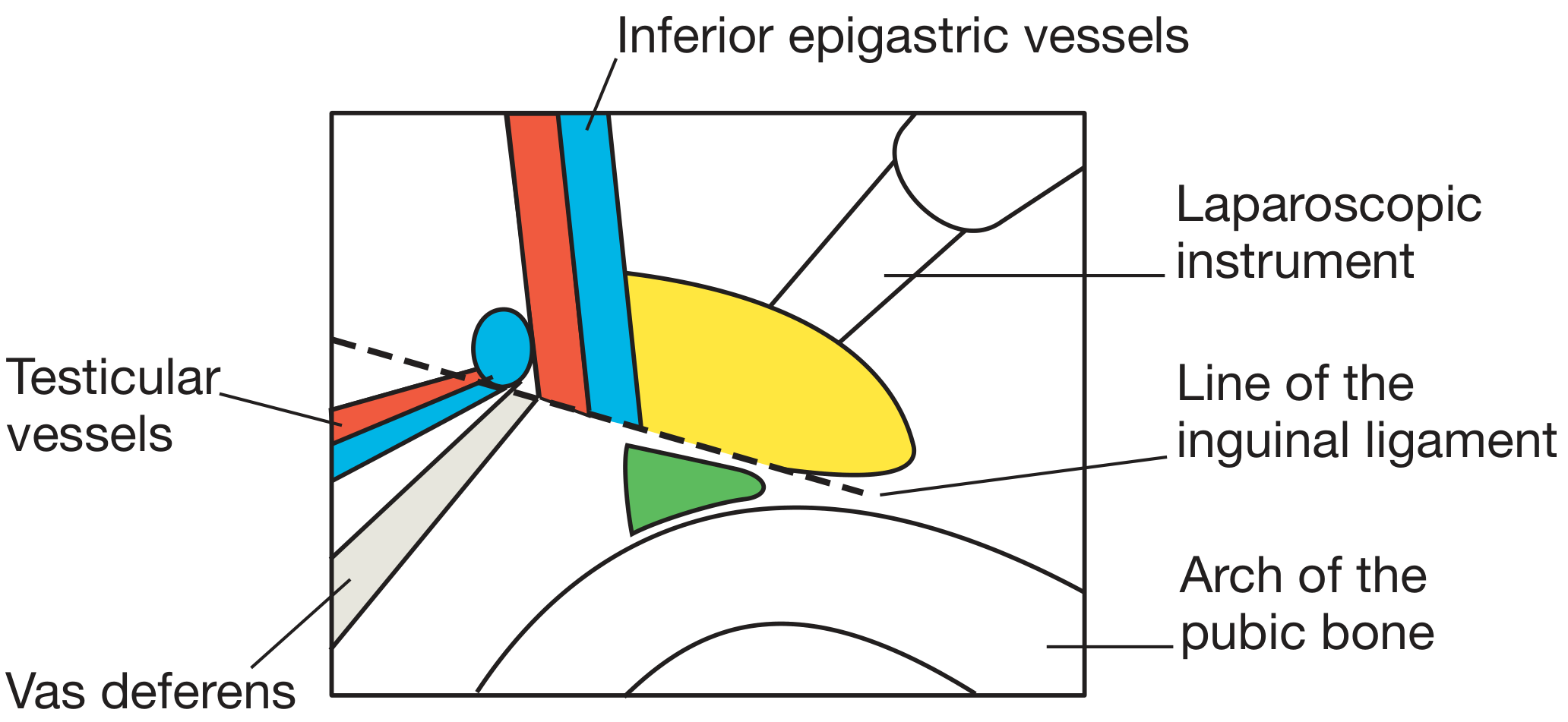
Side-by-Side Comparison
| Feature | Indirect (Lateral) | Direct (Medial) |
|---|---|---|
| Relation to inferior epigastric vessels | Lateral | Medial |
| Entry point | Deep inguinal ring | Hesselbach's triangle (no ring) |
| Origin | Congenital or acquired | Always acquired |
| Direction | Oblique | Direct/forward |
| Can reach scrotum? | Yes | No |
| Common age | Young (congenital) or any age | Elderly |
| Broad base? | No - narrow neck | Yes - broadly based |
| Strangulation risk | Higher | Lower |
| Sac | True peritoneal sac | Peritoneum pushed forward |
| Bladder involvement | Less common | Can occur |
Classification (EHS System)
Bailey and Love describes the European Hernia Society (EHS) classification:
- P = primary, R = recurrent
- Location: L = lateral (indirect), M = medial (direct), F = femoral
- Defect size: 1 = ≤1 fingerbreadth, 2 = 1-3 fingerbreadths, 3 = ≥3 fingerbreadths
Example: A primary indirect hernia with a 3 cm defect = PL2
Clinical Diagnosis
- With the patient lying down, hernia is reduced manually
- The deep inguinal ring is located at the midpoint of the inguinal ligament
- Gentle pressure is applied at the deep ring; patient is asked to cough
- If the hernia is controlled by pressure on the deep ring → likely indirect/lateral
- If the hernia appears medial to this point despite pressure → likely direct/medial
- Note: even experienced surgeons find it difficult to distinguish the two with certainty clinically
Management
Asymptomatic Direct Hernia
It is safe to observe (no active treatment) in early asymptomatic direct hernias, especially in elderly patients who do not wish for surgery. These patients must be warned to seek advice promptly if the hernia enlarges or becomes symptomatic. Surgical trusses are not recommended.
Herniotomy (Children)
In children with indirect hernias (patent processus vaginalis), herniotomy alone (excision and closure of the sac) is sufficient. In adults, herniotomy alone has a high recurrence rate.
Open Suture Repair (Bassini)
Bassini (1890) described the foundational repair: the external oblique is opened, the cord is freed, and the hernia type is confirmed. For a lateral hernia, the sac is separated, opened, reduced, and closed at the neck. For a medial hernia, the sac is inverted and transversalis fascia is plicated. Sutures are then placed between the conjoint tendon (above) and inguinal ligament (below) to strengthen the posterior wall.
The Shouldice modification opens the transversalis fascia centrally and closes it in two layers ("double breasting"), reporting lifetime failure rates below 2% at expert centres.
Most modern Bassini-type repairs use continuous non-absorbable nylon or polypropylene (Maloney darn technique).
All content from: Bailey and Love's Short Practice of Surgery, 28th Edition - Chapter 64, pp. 1087-1090
This is a shared conversation. Sign in to Orris to start your own chat.