
Differences of mri angio ct angio and dosa md radio long answer
MR angiography CT angiography DSA digital subtraction angiography comparison vascular imaging
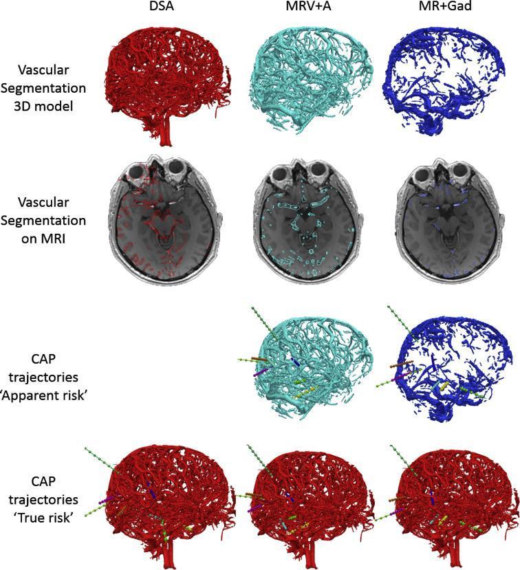
A comparison grid illustrating cerebrovascular segmentation across different imaging modalities for neurosurgical computer-assisted planning (CAP). The modalities compared include Digital Subtraction Angiography (DSA) in red, Magnetic Resonance Angiography and Venography (MRV+A) in light blue, and Gadolinium-enhanced MRI (MR+Gad) in dark blue. The top row displays 3D reconstructed models of the intracranial vasculature, demonstrating that DSA provides the highest vessel density and detail. The second row shows axial MRI slices at the level of the middle cerebral artery origin, with segmented vessels overlaid to highlight modality-specific resolution. The bottom rows illustrate CAP trajectories, distinguishing between 'apparent risk' (calculated using MR-based models) and 'true risk' (re-evaluated against the DSA gold standard). This visualization highlights the clinical significance of modality selection in avoiding vascular injury during electrode placement, as MR-based segmentations may underrepresent small vessels, leading to a falsely low apparent risk score compared to the true risk determined by DSA.
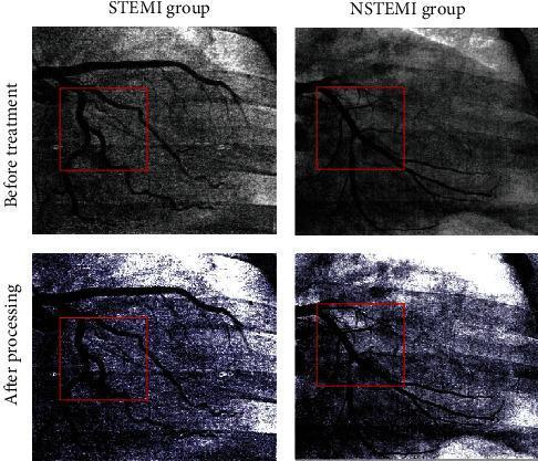
A comparison panel of Digital Subtraction Angiography (DSA) images showcasing coronary artery visualization in STEMI (ST-Elevation Myocardial Infarction) and NSTEMI (Non-ST-Elevation Myocardial Infarction) cases. The top row, labeled 'Before treatment,' shows raw coronary angiograms (CAG) where the vascular anatomy exhibits lower contrast and higher background noise, particularly within the red boxes highlighting target lesions. The bottom row, labeled 'After processing,' demonstrates the same clinical cases following the application of a neural network image enhancement algorithm. These processed images exhibit significantly improved vessel clarity, sharper edge definition of the coronary branches, and enhanced contrast against the background. This comparison illustrates the role of advanced image processing in clinical imaging to better visualize coronary stenosis and lesion morphology, facilitating diagnostic accuracy in interventional cardiology.
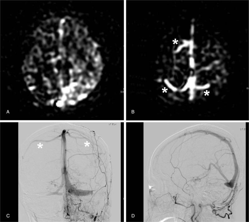
A multi-panel neuroradiological figure comparing Arterial Spin-Labeling Perfusion-Weighted Imaging (ASL-PWI) and Digital Subtraction Angiography (DSA) for the evaluation of a dural arteriovenous fistula (DAVF). Panels A and B display ASL-PWI axial slices; Panel A shows mottled high signal intensity in the parenchyma, while Panel B demonstrates a 'bright vessel sign' with hyperintense signal in the venous sinuses and cortical vessels (marked by asterisks) against a suppressed dark background, indicating abnormal arterialized flow. Panels C and D are DSA images in frontal and lateral projections, respectively, providing the gold standard comparison. These angiograms reveal the vascular architecture of the DAVF, showing early opacification of the left sigmoid sinus and associated venous structures. The collection illustrates the clinical utility of non-contrast MR perfusion techniques like ASL in detecting hemodynamically significant shunts by identifying hyperintense venous signals that represent shunted arterial blood, relevant for neuroradiology education and vascular pathology diagnosis.
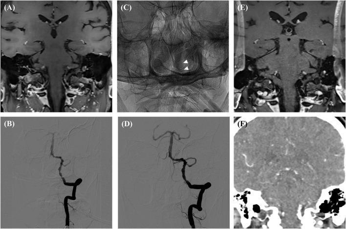
This composite figure illustrates a neurointerventional clinical case across multiple imaging modalities, including High-Resolution MRI (HR-MR), Digital Subtraction Angiography (DSA), and CT Angiography (CTA). Panels A and B display preprocedural states; Panel A shows a coronal HR-MR section of the skull base, while Panel B is a DSA revealing focal intracranial arterial stenosis. Panels C and D show the immediate post-procedural results, with Panel C (unsubtracted) highlighting a deployed Apollo stent (white arrows) and Panel D (subtracted) demonstrating restored luminal diameter and altered flow dynamics. Panel E represents a follow-up HR-MR, which exhibits a distinct signal drop (susceptibility artifact) at the vessel site due to the metallic properties of the stainless-steel stent, complicating luminal evaluation. Panel F provides a CTA for cross-sectional correlation. The collection serves to teach the radiographic appearance of intracranial stenting, the diagnostic utility of DSA in assessing stenosis, and the limitations of MRI follow-up due to metallic artifacts from certain vascular devices.
MR Angiography vs CT Angiography vs DSA (Digital Subtraction Angiography)
1. INTRODUCTION
- MR Angiography (MRA) - magnetic resonance-based, no ionising radiation
- CT Angiography (CTA) - computed tomography with iodinated contrast
- Digital Subtraction Angiography (DSA) - conventional catheter-based, the historic gold standard
2. PRINCIPLES / TECHNICAL BASIS
2.1 MR Angiography (MRA)
- Most common form in clinical practice
- Uses gradient-echo (GE) sequences with rapid radiofrequency (RF) pulses of short TR to saturate (darken) stationary background tissue
- Unsaturated spins from blood flowing into the imaging field of view (FOV) create "flow-related enhancement" - appearing bright without any contrast injection
- Can be acquired as 2D or 3D data sets
- Best for intracranial arterial imaging (Circle of Willis, MCA, ACA, PCA, vertebrobasilar)
- Limitation: overestimates stenosis at turbulent flow areas due to signal loss; in-plane flow saturation (vessels running parallel to slice give no signal)
- Based on detection of phase shifts generated by a flow-encoding gradient
- Velocity-encoding (VENC) must be set according to expected vessel velocity
- Independent of direction of flow but motion susceptible and time-consuming
- More sensitive to slow flow (useful for cerebral veins - MR venography/MRV)
- Used for dural venous sinus thrombosis, venous stenoses
- Can quantify direction of blood flow
- Does not suffer from T1-contamination artefact
- Generally inferior to 3D TOF for arteries
- Relies on IV gadolinium (Gd)-based contrast agents (GBCAs)
- Uses coronal 3D fast multiplanar gradient-echo breath-hold sequence after a timing bolus of contrast
- Reduces artefact from turbulent or slow flow (major advantage over TOF)
- Excellent for large FOV imaging - carotid and vertebral arteries in the neck
- Gadolinium now contraindicated in renal failure/severe dysfunction (eGFR <30) due to risk of Nephrogenic Systemic Fibrosis (NSF)
- Has shown benefit with intracranial 3D TOF MRA for AVMs and intracranial stenosis
2.2 CT Angiography (CTA)
- Contrast injection at high flow rates (typically 4-6 mL/s) with bolus tracking to time acquisition at peak arterial enhancement
- Sub-millimetre collimation (0.5-0.75 mm) allows reconstruction in any plane
- Post-processing: MPR, MIP, and VRT reconstructions routinely generated
- Can image aortic arch, extracranial carotids, intracranial vessels, and circle of Willis in a single acquisition (see Fig. 53.41 - CT angiogram of neck and circle of Willis, Grainger & Allison)
- CT perfusion (CTP) can be added in stroke protocol: measures CBF, CBV, MTT, and Tmax maps to identify ischaemic penumbra vs infarct core
- Excellent soft tissue, bony structure, and vascular imaging simultaneously - cannot be matched by DSA
2.3 Digital Subtraction Angiography (DSA)
- Arterial access (typically femoral artery via Seldinger technique)
- Catheter navigation to target vessel under fluoroscopic guidance
- Selective injection of iodinated contrast into desired vessel
- Digital subtraction of pre-contrast "mask" from contrast images
- Results in high-resolution, real-time 2D luminograms of vessels
- Rotational DSA and 3D reconstruction now possible with modern flat-panel detectors
3. COMPARISON TABLE
| Parameter | MRA | CTA | DSA |
|---|---|---|---|
| Modality | Magnetic resonance | X-ray CT | Fluoroscopy/catheter |
| Radiation | None | Yes (moderate) | Yes (significant) |
| Contrast | None (TOF/PC) or Gadolinium (CE-MRA) | Iodinated contrast IV | Iodinated contrast intra-arterial |
| Invasiveness | Non-invasive | Non-invasive | Invasive (arterial puncture) |
| Spatial resolution | Moderate (~1mm) | High (sub-mm isotropic) | Highest (conventional gold standard) |
| Temporal resolution | Low-moderate | Moderate | Highest (real-time flow) |
| Speed | Slow (minutes) | Fast (seconds) | Moderate (dependent on procedure) |
| Cost | High | Moderate | Highest |
| 3D reconstruction | Yes (MIP, VRT) | Yes (MIP, VRT, MPR) | Limited (rotational DSA) |
| Intervention | Not possible | Not possible | Possible (therapeutic/diagnostic) |
| Renal impairment | Gadolinium CI if eGFR <30 (risk of NSF) | Iodinated contrast nephrotoxic | Iodinated contrast nephrotoxic |
| Metal implants | Contraindicated (most) | Safe | Safe |
| Calcification | Not well shown | Excellent | Poorly shown (subtracted out) |
| Soft tissue detail | Excellent | Good | Poor |
| Plaque morphology | Good (fibrous cap, lipid core, intraplaque haemorrhage) | Good (plaque calcium, morphology) | Only lumen (no plaque info) |
| Flow dynamics | PC-MRA gives quantitative flow | No direct flow data | Real-time flow dynamics visible |
| Access complications | None | None | Haematoma (4%), neurological (2.6%) |
4. CLINICAL INDICATIONS
4.1 MRA - Indications
- Intracranial aneurysm screening (TOF)
- Carotid and vertebral artery dissection
- Cerebral venous sinus thrombosis (MRV)
- Vasculitis (large and medium vessels)
- Reversible Cerebral Vasoconstriction Syndrome (RCVS)
- Renal artery stenosis (CE-MRA - preferred in younger patients to avoid radiation)
- AVM characterisation
- Post-CEA (carotid endarterectomy) follow-up - avoids radiation
- Patients allergic to iodinated contrast
- Plaque morphology / intraplaque haemorrhage monitoring
4.2 CTA - Indications
- Acute stroke protocol (non-contrast CT + CTP + CTA in one sitting)
- Pulmonary embolism (CT pulmonary angiography - CTPA)
- Aortic dissection / aneurysm
- Carotid stenosis assessment pre-CEA or pre-CAS (images arch to circle of Willis)
- Acute GI bleeding (CTA sensitivity 85%, specificity 92% for active bleed)
- Renal artery stenosis
- Peripheral arterial disease (runoff CTA)
- Trauma (polytrauma whole-body CTA)
- Pre-operative vascular mapping
- Follow-up of stent/graft patency
4.3 DSA - Indications
- Gold standard for degree of stenosis when CTA and duplex ultrasound give conflicting results
- Pre-interventional angiography (diagnostic + therapeutic in same session)
- Cerebral angiography for AVMs, aneurysms (confirms findings before clipping/coiling)
- Intra-arterial thrombolysis / mechanical thrombectomy (confirmed on DSA, treated immediately)
- Renal artery angioplasty and stenting
- Peripheral angioplasty
- Vasculitis work-up (when MRA/CTA normal but clinical suspicion high)
- Hepatic/splenic/renal artery embolisation
- Mesenteric ischaemia confirmation and intervention
- CNS vasculitis: DSA should be considered when brain MRI is normal but vasculitis still suspected
5. ADVANTAGES AND LIMITATIONS (Detailed)
MRA
- No ionising radiation (ideal for young patients, pregnant women, follow-up studies)
- No nephrotoxic iodinated contrast needed for TOF/PC techniques
- Superior soft tissue contrast
- Shows plaque composition (fibrous cap integrity, lipid core, intraplaque haemorrhage)
- MRV detects cerebral venous thrombosis
- CE-MRA preferred for large FOV vascular imaging (neck vessels)
- Time-consuming; patient cooperation and breath-hold required
- Contraindicated in patients with ferromagnetic implants, pacemakers, certain metallic foreign bodies
- Claustrophobia a significant practical limitation
- Overestimates stenosis (turbulent flow = signal void mimicking occlusion)
- Gadolinium contraindicated if eGFR <30 (NSF risk)
- Calcified plaques not well visualised
- Occluded vessels may be missed
- Cost and limited availability
- Cannot guide intervention
CTA
- Fast (seconds), widely available, 24/7 accessible
- Sub-millimetre spatial resolution with excellent 3D post-processing
- Simultaneously images soft tissue, bones, and vessels
- Evaluates aortic arch anatomy, identifies tandem lesions
- Can identify calcified and non-calcified plaque
- Guides surgical/interventional planning (arch anatomy, proximal CCA lesions)
- More reproducible and less operator-dependent than MRA
- Useful in patients with implants who cannot undergo MRI
- Ionising radiation (concern especially in younger patients and serial imaging)
- Nephrotoxic iodinated contrast (avoid in CKD, hydration required)
- Contraindicated in contrast allergy (pre-medication required)
- Less accurate than duplex ultrasound for screening asymptomatic patients (SoVS level 1B evidence)
- Cannot guide intervention
- Metallic stents cause beam hardening artefact, limiting in-stent evaluation
DSA
- Historically the gold standard (highest spatial and temporal resolution)
- Real-time imaging of blood flow dynamics
- Enables immediate intervention (diagnostic + therapeutic in one procedure)
- Best for subtle flow abnormalities (slow flow, early venous drainage in AVM)
- Rotational DSA provides 3D vessel imaging
- Confirms stenosis degree when non-invasive tests conflict
- Invasive - arterial access required (femoral, radial)
- Risk of neurological complications (~2.6%) and access-site haematoma (~4%) in large series
- Significant radiation dose to patient and operator
- Iodinated contrast - nephrotoxicity risk
- Requires specialised facility (angiography suite) and operator expertise
- Expensive, resource-intensive
- Only shows vessel lumen (no plaque, no parenchymal information)
- Post-procedure monitoring and recovery required
- No longer used for routine diagnostic purposes in most centres; has been replaced by CTA/MRA
6. SPECIFIC SCENARIOS / COMPARATIVE USE
Stroke / Acute Cerebral Ischaemia
- First-line: Non-contrast CT + CTA + CTP (fast, widely available)
- CTA confirms large vessel occlusion (M1 MCA, ICA, basilar) for thrombectomy decision
- DSA is performed in the angiography suite immediately before mechanical thrombectomy
- MRA: used for follow-up, dissection diagnosis, AVMs
Carotid Artery Disease
- Duplex ultrasound = first-line screening
- CTA: pre-CEA/CAS planning (arch to circle of Willis in one study)
- MRA: alternative when radiation/contrast must be avoided
- DSA: only if CTA and duplex give conflicting stenosis grades
Intracranial Aneurysm
- MRA (3D TOF): screening in high-risk populations (family history, PKD)
- CTA: excellent sensitivity/specificity, faster, preferred in acute SAH
- DSA: gold standard for pre-treatment planning (clip vs coil), confirms aneurysm morphology, detects small aneurysms CTA/MRA may miss
Pulmonary Embolism
- CTPA (CT pulmonary angiography) = gold standard non-invasive investigation (has replaced DSA and V/Q in most centres)
- MRA: option when CT contrast contraindicated but sensitivity lower
- DSA: now only used if catheter-directed thrombolysis planned
Renal Artery Stenosis
- CE-MRA or CTA: initial investigation
- DSA: definitive if angioplasty/stenting planned (Brenner & Rector's The Kidney)
7. IMAGE EXAMPLES
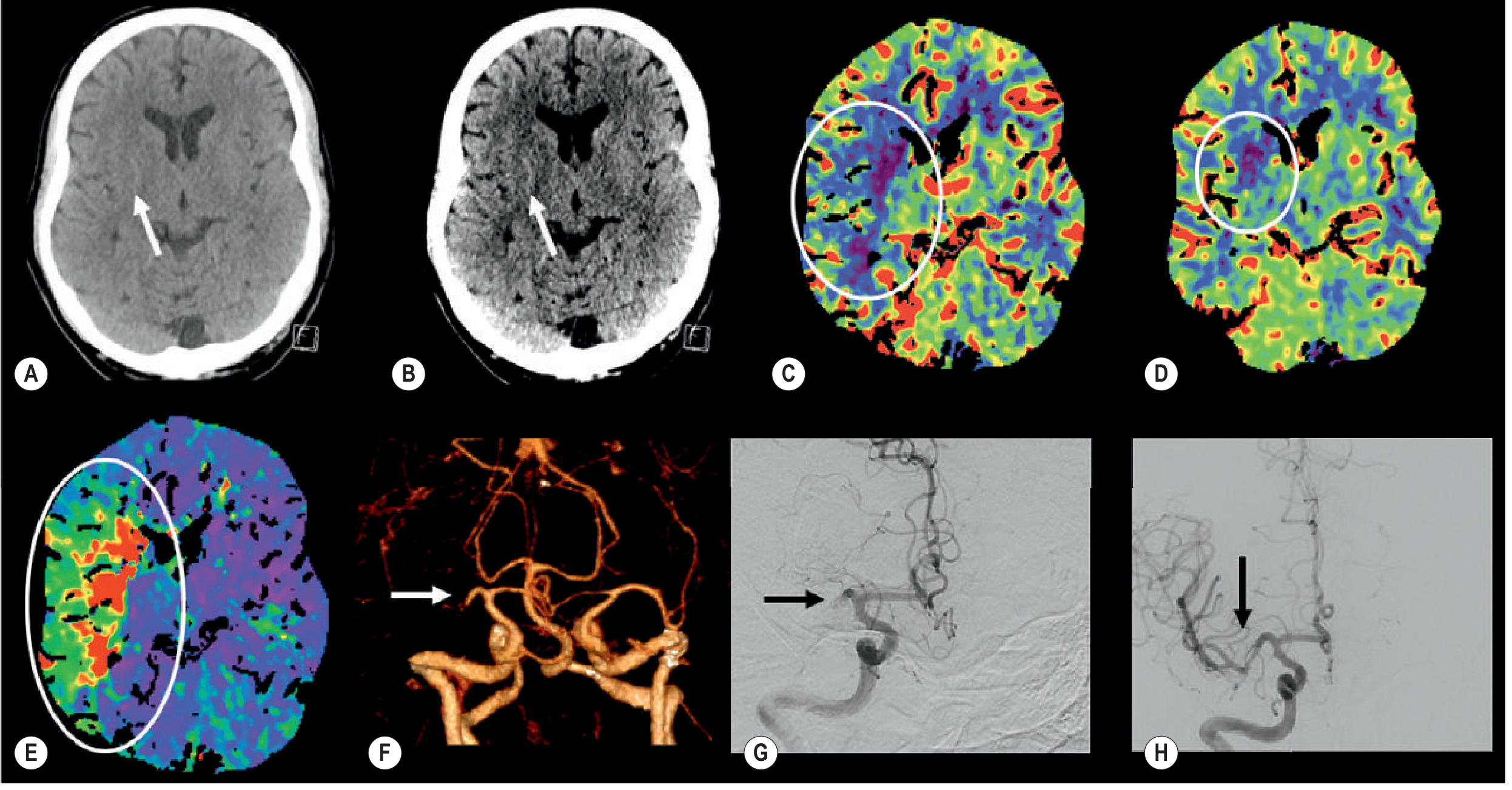
8. SUMMARY - KEY EXAM POINTS
| Point | Key Fact |
|---|---|
| Gold standard for stenosis | DSA (but being replaced by CTA/MRA) |
| Gold standard non-invasive | CTA (most organs); MRA (avoiding radiation/contrast) |
| No radiation modality | MRA |
| Gadolinium CI | eGFR <30 (NSF risk) |
| DSA complication rate | Neuro: 2.6%, haematoma: 4% |
| MRA overestimates stenosis | Due to turbulent flow signal loss |
| CTA advantage | Fast, 3D, simultaneous soft tissue + bone + vessel |
| DSA unique advantage | Real-time flow + immediate intervention |
| TOF MRA principle | Flow-related enhancement without contrast |
| PC MRA use | Slow flow (veins), MR venography |
| CE-MRA advantage | Large FOV, reduces turbulent flow artefact |
| CTA in acute stroke | CTA + CTP together for penumbra/core mapping |
- Grainger & Allison's Diagnostic Radiology (blocks 6, 10, 15)
- Sabiston Textbook of Surgery (block 24 - Carotid artery disease)
- Bradley & Daroff's Neurology in Clinical Practice (block 4)
- Rheumatology 2-Volume Set (Elsevier 2022) (block 22 - CNS vasculitis)
- Brenner & Rector's The Kidney (block 38)