
Thyroid physiology and anstomy full detail explain
Reading File
Searching sources...
Finding Sources
Finding Sources
Reading File
Reading File
Reading File
Reading File
Reading File
Reading File
Reading File
Finding Sources
Loading Image
Reading File
Reading File
Loading Image
Reading File
Reading File
Loading Image
Reading File
Loading Image
Reading File
Loading Image
I now have all the information needed to compile a comprehensive, well-illustrated answer. Let me deliver the full detailed response.
Thyroid Gland: Complete Anatomy and Physiology
1. Embryology
The thyroid is the first endocrine gland to develop, appearing at approximately day 24 of gestation. It arises from the median bud of the pharynx at the base of the tongue - the foramen caecum marks this site on the adult tongue (junction of anterior 2/3 and posterior 1/3).
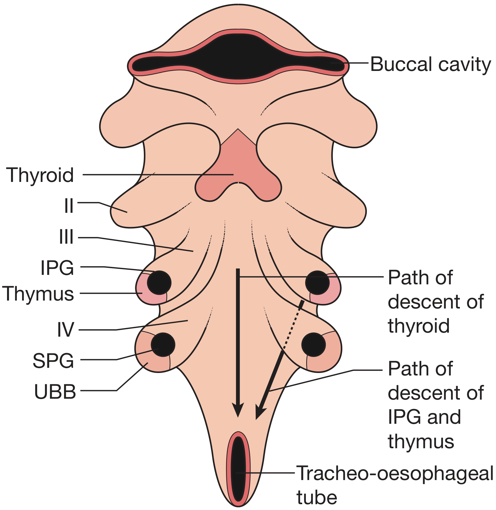
Key developmental events:
- The initially hollow thyroglossal duct migrates caudally, passing through or near the developing hyoid cartilage
- By week 7, the thyroid has assumed its definitive shape and position anterior to the trachea
- The thyroid lobes amalgamate with structures from the 4th pharyngeal pouch (superior parathyroid gland and ultimobranchial body)
- Parafollicular C cells (calcitonin-secreting) originate from neural crest cells and reach the thyroid via the ultimobranchial body
- Parathyroid glands develop from the 3rd (inferior) and 4th (superior) pharyngeal pouches
- The thyroglossal duct normally involutes; its remnant may persist as a pyramidal lobe (~50% of individuals) or cause pathology (thyroglossal duct cysts)
Anomalies of descent:
- Lingual thyroid: failure to descend, leaving gland at base of tongue
- Thyroglossal duct cysts: cystic remnants along the descent path, lined by pseudostratified ciliated columnar epithelium; treated by Sistrunk operation (en bloc cystectomy + central hyoid bone excision)
2. Gross Anatomy
Position and Shape
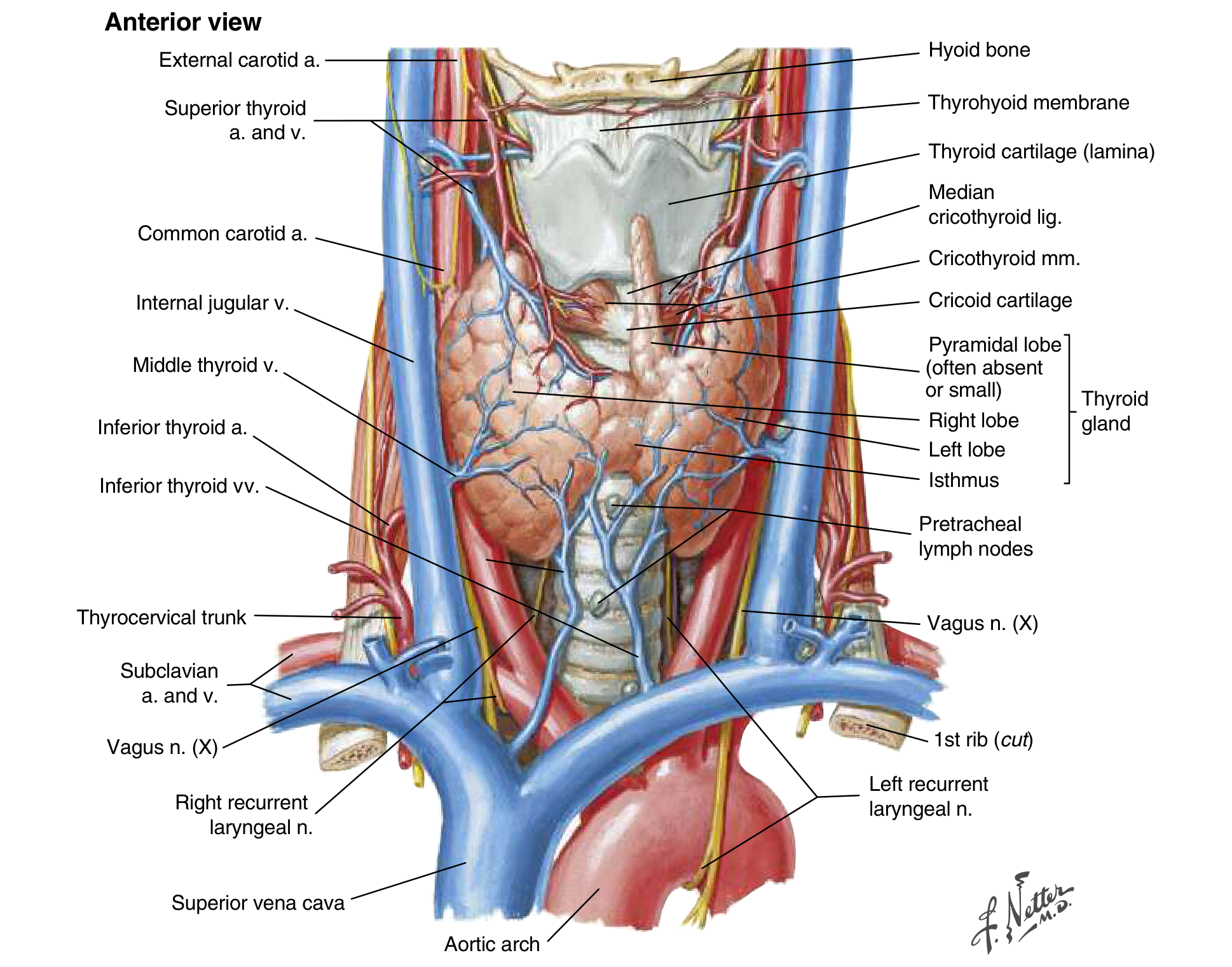
The normal adult thyroid is a brown, firm, butterfly-shaped gland weighing 20-25 g (varies with body weight and iodine intake). It lies posterior to the strap muscles and overlies the anterior trachea.
Lobes and Isthmus
- Two lobes (right and left), connected by an isthmus located just inferior to the cricoid cartilage
- Each lobe extends from the midthyroid cartilage superiorly, lying adjacent to the carotid sheaths and sternocleidomastoid muscles laterally
- Pyramidal lobe: present in ~50% of people, projects upward from the isthmus, just left or right of midline - becomes enlarged and palpable in Graves' disease or thyroiditis
Fascial Relations
- Enveloped by a loose fascia derived from the deep cervical fascia (anterior and posterior divisions)
- The true capsule is a thin, densely adherent fibrous layer that sends septa inward, forming pseudolobules
- The capsule condenses posteriorly into Berry's ligament (posterior suspensory ligament) near the cricoid and upper tracheal rings - this ligament anchors the thyroid to the trachea, causing the gland to move with swallowing
Strap Muscles (anterior)
Sternohyoid, sternothyroid, and superior belly of omohyoid - all innervated by the ansa cervicalis (ansa hypoglossi)
3. Blood Supply
Arteries
| Vessel | Origin | Supplies |
|---|---|---|
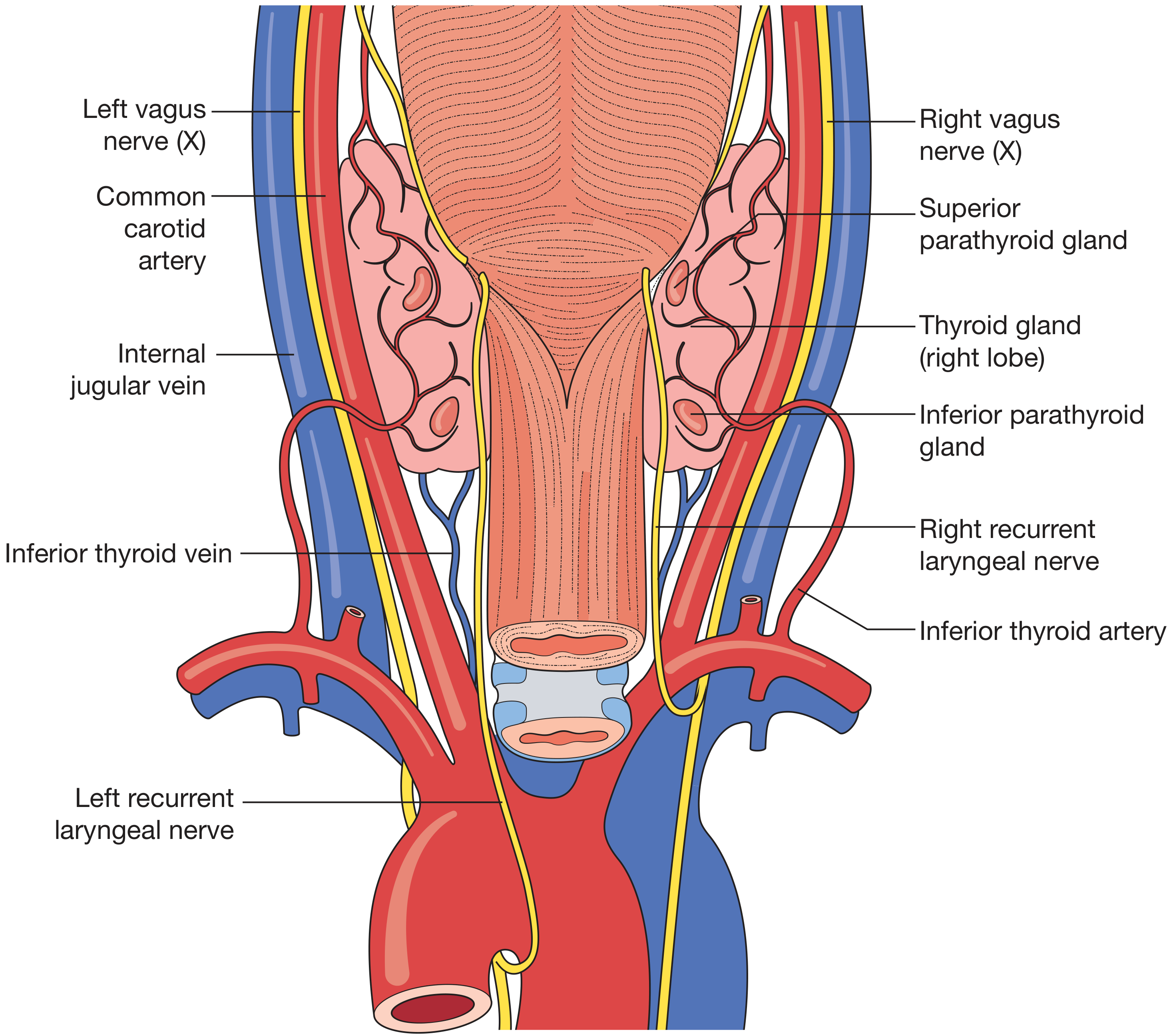
| Superior thyroid artery (×2) | External carotid artery | Apices of thyroid lobes; divides into anterior and posterior branches |
| Inferior thyroid artery (×2) | Thyrocervical trunk → subclavian artery | Lower poles; travels posterior to the carotid sheath |
| Thyroidea ima artery | Directly from aorta or innominate (~10%) | Isthmus (when present) |
Extensive anastomoses exist between these arteries and branches of the tracheal and oesophageal arteries.
Veins
- Superior thyroid veins - drain into the internal jugular vein
- Middle thyroid veins - drain into the internal jugular vein
- Inferior thyroid veins - drain into the brachiocephalic veins (right and left)
4. Nerve Supply
Recurrent Laryngeal Nerve (RLN)
This is the most surgically important nerve relation of the thyroid.
- A branch of the vagus nerve (CN X)
- Left RLN: recurs around the arch of the aorta - has more distance to travel, runs in a more medial plane in the tracheo-oesophageal groove
- Right RLN: recurs around the subclavian artery - has less distance, runs more obliquely to reach the groove
- ~2% of right nerves are non-recurrent (enter larynx from above) - associated with an aberrant right subclavian artery
- The nerve runs posterior to the thyroid and enters the larynx at the cricothyroid joint
- Critical relation: the RLN crosses the inferior thyroid artery; the superior parathyroid is characteristically dorsal to the nerve plane, the inferior parathyroid is ventral
- Injury → hoarseness (unilateral) or respiratory distress (bilateral)
Superior Laryngeal Nerve (SLN)
- Also a branch of the vagus
- External branch runs near the superior thyroid vessels and controls the cricothyroid muscle (voice pitch)
- Injury during superior pole dissection → monotone voice, difficulty singing high notes
5. Lymphatic Drainage
The thyroid has an extensive lymphatic network:
- Subcapsular plexus drains principally to:
- Central compartment (level VI): juxtathyroid "Delphian" nodes, paratracheal nodes, and nodes on superior and inferior thyroid veins
- Then onwards to:
- Deep cervical nodes (levels II, III, IV, V)
- Mediastinal nodes (level VII)
6. Histology (Microscopic Anatomy)
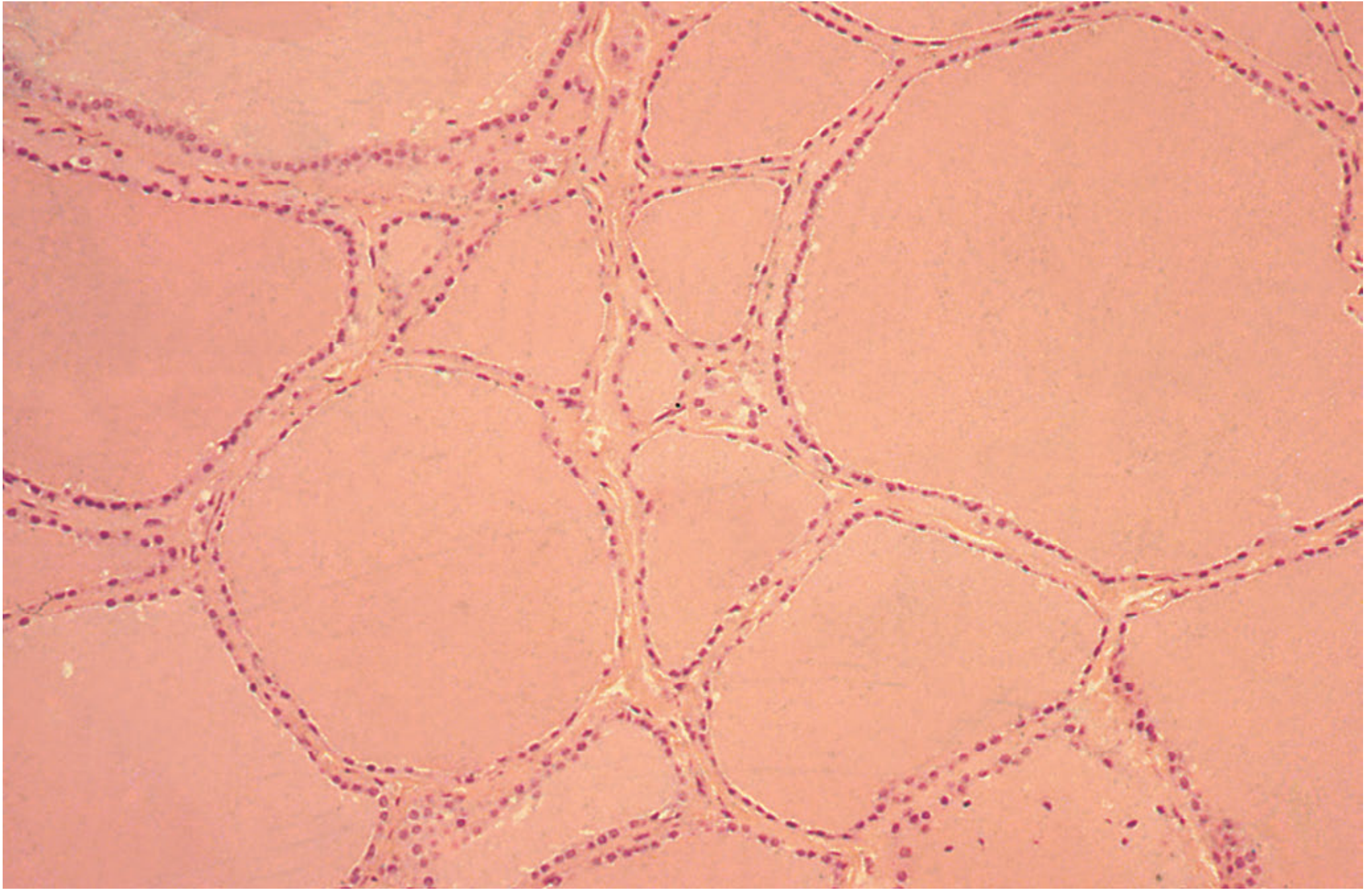
The functional unit of the thyroid is the follicle:
- Each lobule (supplied by a single arteriole) contains 24-40 follicles
- Each follicle measures 200-300 µm in diameter
- Lined by a single layer of cuboidal follicular epithelial cells (become columnar when active, flat when inactive)
- The cells have a basal membrane facing the blood and an apical membrane facing the follicular lumen
- The lumen contains colloid - a viscous material composed of thyroglobulin (TG) with attached thyroid hormones
- Between follicles lie parafollicular cells (C cells) - larger, pale cells that secrete calcitonin (lowers blood calcium)
7. Thyroid Hormone Synthesis (Step-by-Step)
The synthesis is uniquely complex - partially intracellular, partially extracellular, with storage in the follicular lumen.
The 8 Steps
Step 1 - Thyroglobulin synthesis:
TG (a large glycoprotein rich in tyrosine residues) is synthesized on the rough ER and Golgi apparatus of follicular cells, then extruded across the apical membrane into the follicular lumen.
Step 2 - Iodide trapping (Na⁺-I⁻ cotransport):
Iodide (I⁻) is actively transported from blood into follicular cells against both chemical and electrical gradients by the Na⁺/I⁻ symporter (NIS) at the basal membrane. Intracellular iodine concentration is many times higher than plasma. This pump is stimulated by TSH and by low iodine states.
- Inhibited by: perchlorate, thiocyanate
Step 3 - Oxidation of I⁻ → I₂:
At the apical membrane, I⁻ is oxidized to molecular iodine (I₂) by thyroid peroxidase (TPO).
- Inhibited by: propylthiouracil (PTU), methimazole
Step 4 - Organification (iodination of tyrosine):
I₂ combines with tyrosine residues on TG at the apical membrane (catalyzed by TPO):
- 1 iodine + tyrosine = monoiodotyrosine (MIT)
- 2 iodines + tyrosine = diiodotyrosine (DIT)
- High iodine levels block this step = Wolff-Chaikoff effect
Step 5 - Coupling reaction (still on TG in follicular lumen):
MIT and DIT undergo coupling (catalyzed by TPO):
- DIT + DIT = T₄ (thyroxine/tetraiodothyronine) - 4 iodine atoms
- DIT + MIT = T₃ (triiodothyronine) - 3 iodine atoms
- MIT + MIT = T₂ (inactive)
The major secretory product is T₄ (~80-90% of output). T₃ is more biologically active but only ~20% is directly secreted.
Step 6 - Endocytosis of TG:
When stimulated by TSH, follicular cells engulf TG-containing colloid via endocytosis at the apical membrane.
Step 7 - Hydrolysis and secretion:
Intracellular lysosomes contain proteases that cleave T₄ and T₃ from TG. The free hormones then enter the circulation via the basolateral membrane.
Step 8 - Deiodination of residual MIT/DIT:
Residual MIT and DIT are deiodinated intracellularly by thyroid deiodinase - the freed iodide is recycled back for new hormone synthesis (iodine conservation). Deficiency of this enzyme mimics dietary iodine deficiency.
| Step | Event | Site | Enzyme | Inhibitor |
|---|---|---|---|---|
| 1 | TG synthesis + extrusion | Rough ER, Golgi | - | - |
| 2 | Na⁺-I⁻ cotransport | Basal membrane | NIS | Perchlorate, thiocyanate |
| 3 | I⁻ → I₂ oxidation | Apical membrane | Peroxidase | PTU |
| 4 | Organification (MIT, DIT) | Apical membrane | Peroxidase | PTU |
| 5 | Coupling (T₃, T₄) | Apical membrane | Peroxidase | PTU |
| 6 | Endocytosis of TG | Apical membrane | - | - |
| 7 | Hydrolysis; T₃/T₄ released | Lysosomes | Proteases | - |
| 8 | Deiodination of MIT/DIT | Intracellular | Deiodinase | - |
8. Transport in Blood
- >99% of circulating thyroid hormones are bound to plasma proteins:
- Thyroxine-binding globulin (TBG) - primary carrier
- Transthyretin (T₄-binding prealbumin)
- Albumin
- <1% circulates as free (unbound) hormone - this is the biologically active fraction
- The bound pool serves as a large reservoir that buffers free hormone levels
- T₄ has a plasma half-life of ~7 days; T₃ ~1 day
9. Peripheral Conversion
- T₄ is the dominant secretory product but is the pro-hormone
- Target tissues (liver, kidney, brain, skeletal muscle, gut) convert T₄ → T₃ by 5'-deiodinase (removes one iodine) - this accounts for ~80% of circulating T₃
- Reverse T₃ (rT₃) - inactive isomer formed by deiodination at a different position; increases in illness/starvation (sick euthyroid syndrome)
10. Regulation: The HPT Axis
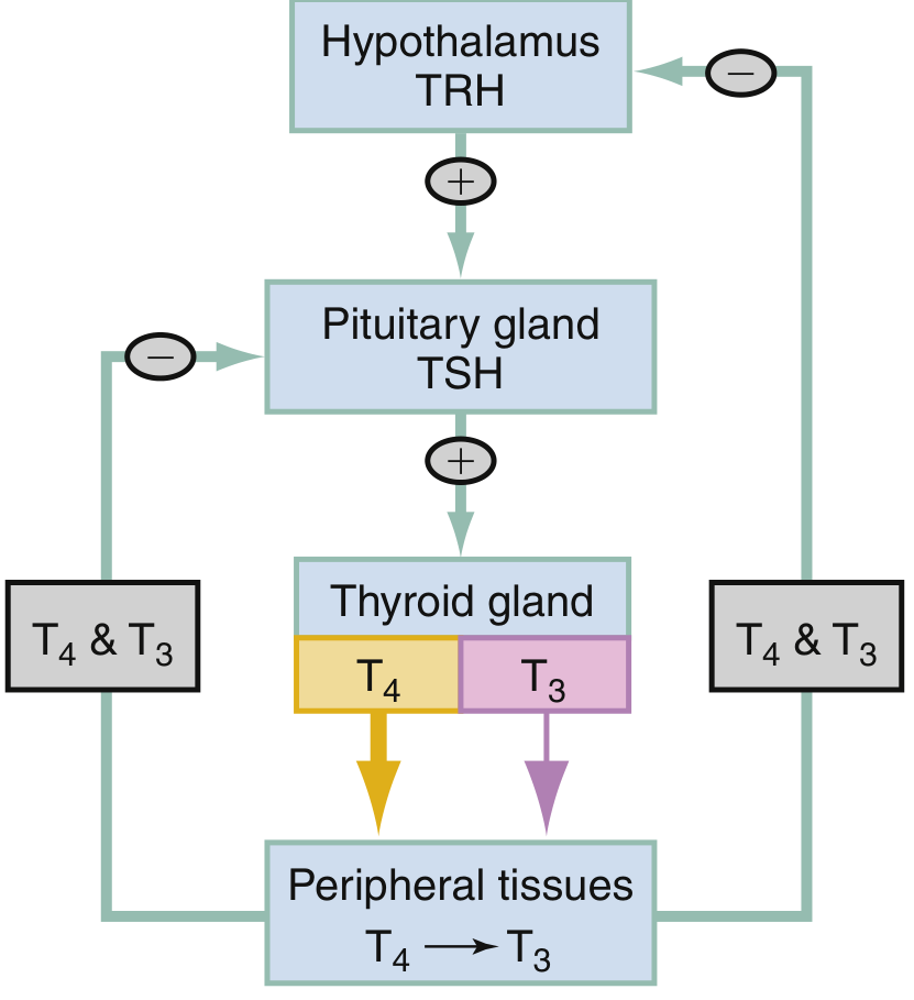
The Axis
- Hypothalamus releases TRH (thyrotropin-releasing hormone, a tripeptide) → stimulates anterior pituitary
- Anterior pituitary thyrotroph cells release TSH (thyroid-stimulating hormone, a glycoprotein) → binds receptors on follicular cells
- TSH actions on thyroid:
- Increases iodide uptake
- Stimulates all steps of hormone synthesis
- Stimulates endocytosis and secretion of T₃/T₄
- Promotes thyroid cell growth (hypertrophy and hyperplasia)
- Negative feedback: elevated T₃/T₄ suppresses both TRH (hypothalamus) and TSH (pituitary), completing the loop
Other regulators
- Iodine excess directly suppresses thyroid hormone synthesis (Wolff-Chaikoff effect) - used in preoperative preparation (Lugol's iodine)
- Dopamine and glucocorticoids suppress TSH secretion
11. Actions of Thyroid Hormones
T₃ enters the cell, translocates to the nucleus, and binds thyroid hormone receptors (TR) - nuclear receptors that regulate gene transcription.
Metabolic Actions
- Primary driver of basal metabolic rate (BMR)
- Stimulates mitochondrial ATP production, oxygen consumption, and glucose oxidation (calorigenic effect - generates heat)
- Increases lipid and carbohydrate metabolism
- Stimulates gluconeogenesis and lipolysis
Cardiovascular
- Increases cardiac output, heart rate, and stroke volume
- Causes peripheral vasodilation
- Synergistic with catecholamines via β-adrenergic receptors (explains why propranolol reduces symptoms of hyperthyroidism)
Growth and Development
- Essential for normal in utero and childhood growth (bone maturation, linear growth)
- Critical for CNS maturation in the perinatal period - neonatal hypothyroidism causes irreversible cretinism (mental retardation, growth failure) if untreated → mandates newborn screening
- In adults, hypothyroidism causes slowed cognition, lethargy, somnolence
Reproductive Function
- Normal reproductive function depends on adequate thyroid hormone
- Both hypo- and hyperthyroidism cause menstrual irregularities and subfertility
12. Calcitonin (C Cell Function)
- Secreted by parafollicular (C) cells in response to hypercalcemia
- Lowers blood calcium by inhibiting osteoclast activity and promoting renal calcium excretion
- Less important in adult calcium homeostasis than PTH
- Clinically significant as a tumor marker for medullary thyroid carcinoma (MTC)
13. Quick Pathophysiology Summary
| Parameter | Hyperthyroidism | Hypothyroidism |
|---|---|---|
| BMR | Increased | Decreased |
| Weight | Loss | Gain |
| Heart rate | Tachycardia, palpitations | Bradycardia |
| Heat | Heat intolerance, sweating | Cold intolerance |
| CNS | Tremor, anxiety, hyperreflexia | Lethargy, slowed cognition |
| Skin | Warm, moist | Dry, myxedematous |
| TSH | Low (usually) | High (primary) |
| Treatment | PTU, carbimazole, ¹³¹I, surgery | Levothyroxine (T₄) replacement |
Sources: Schwartz's Principles of Surgery (11th Ed.), Bailey & Love's Short Practice of Surgery (28th Ed.), Costanzo Physiology (7th Ed.), Sabiston Textbook of Surgery, Rosen's Emergency Medicine - all via the medical textbook library.
This is a shared conversation. Sign in to Orris to start your own chat.