# GROSS ANATOMY OF THE LIVER, SPLEEN, AND PANCREAS
---
# PART 1: THE LIVER (HEPAR)
---
## INTRODUCTION
The liver is the **largest solid organ** in the body, the **largest gland**, and the **largest accessory digestive gland**. It is a vital organ with over 500 functions, including metabolism, detoxification, bile production, and storage.
---
## GENERAL CHARACTERISTICS
| Feature | Detail |
|---|---|
| **Weight** | ~1500 g (1.5 kg) in males; ~1300 g in females |
| **Color** | Reddish-brown (due to rich vascularity) |
| **Consistency** | Soft and friable (easily torn/lacerated) |
| **Shape** | Wedge-shaped; likened to a "cap fitting over the right upper abdominal viscera" |
| **Percentage of body weight** | ~2–3% in adults; ~5% in neonates |
| **Blood content** | Contains ~13% of total blood volume at any time (~450 mL) |
---
## LOCATION AND EXTENT
- Occupies the **right hypochondrium**, extending into the **epigastrium** and reaching into the **left hypochondrium**
- Lies mostly under cover of the **right costal margin** and the **right dome of the diaphragm**
- Extends from the **right 5th intercostal space** (in the midclavicular line) superiorly to the **right costal margin** inferiorly
- It is almost entirely covered by the **rib cage** — only a small part projects below the costal margin in the epigastrium
- **In infants**, the liver is proportionally larger and extends 1–2 cm below the costal margin normally
### **Clinical: Hepatomegaly**
An enlarged liver (hepatomegaly) becomes palpable below the right costal margin. Causes include:
- **Congestive cardiac failure** (nutmeg liver — chronic passive venous congestion)
- **Cirrhosis** (early stages — later the liver shrinks)
- **Hepatitis** (viral, alcoholic)
- **Malignancy** (primary hepatocellular carcinoma, metastatic deposits — the liver is the **most common site for blood-borne metastases** from GI tract cancers via the portal vein)
- **Amyloidosis**
- **Fatty liver** (steatosis)
- **Tropical splenomegaly syndrome** with hepatomegaly
A cirrhotic liver becomes hard, nodular, and shrunken. Palpation reveals a firm, irregular edge.
---
## SURFACES AND BORDERS
The liver has **two surfaces** and **two borders** (some texts describe it as having five surfaces — superior, anterior, right, posterior, and inferior):
### 1. DIAPHRAGMATIC SURFACE
- **Smooth and convex**
- Faces superiorly, anteriorly, and to the right
- Related to the undersurface of the **diaphragm**, which separates it from:
- Right and left **pleural recesses**
- **Base (inferior surface) of the right lung**
- **Heart** and **pericardium** (via the central tendon of the diaphragm)
- Subdivided by the attachment of the **falciform ligament** into a larger **right part** and a smaller **left part**
- The **bare area** is on the posterosuperior aspect — the area not covered by peritoneum, bounded by the layers of the coronary ligament
#### Impressions on the Diaphragmatic Surface
- **Cardiac impression** — a shallow depression on the superior surface of the left lobe, produced by the heart through the diaphragm
### 2. VISCERAL (INFERIOR) SURFACE
- Faces **posteroinferiorly and to the left**
- **Irregular and concave** — molded by the underlying viscera
- Covered by peritoneum except at the **porta hepatis** and the **fossa for the gallbladder**
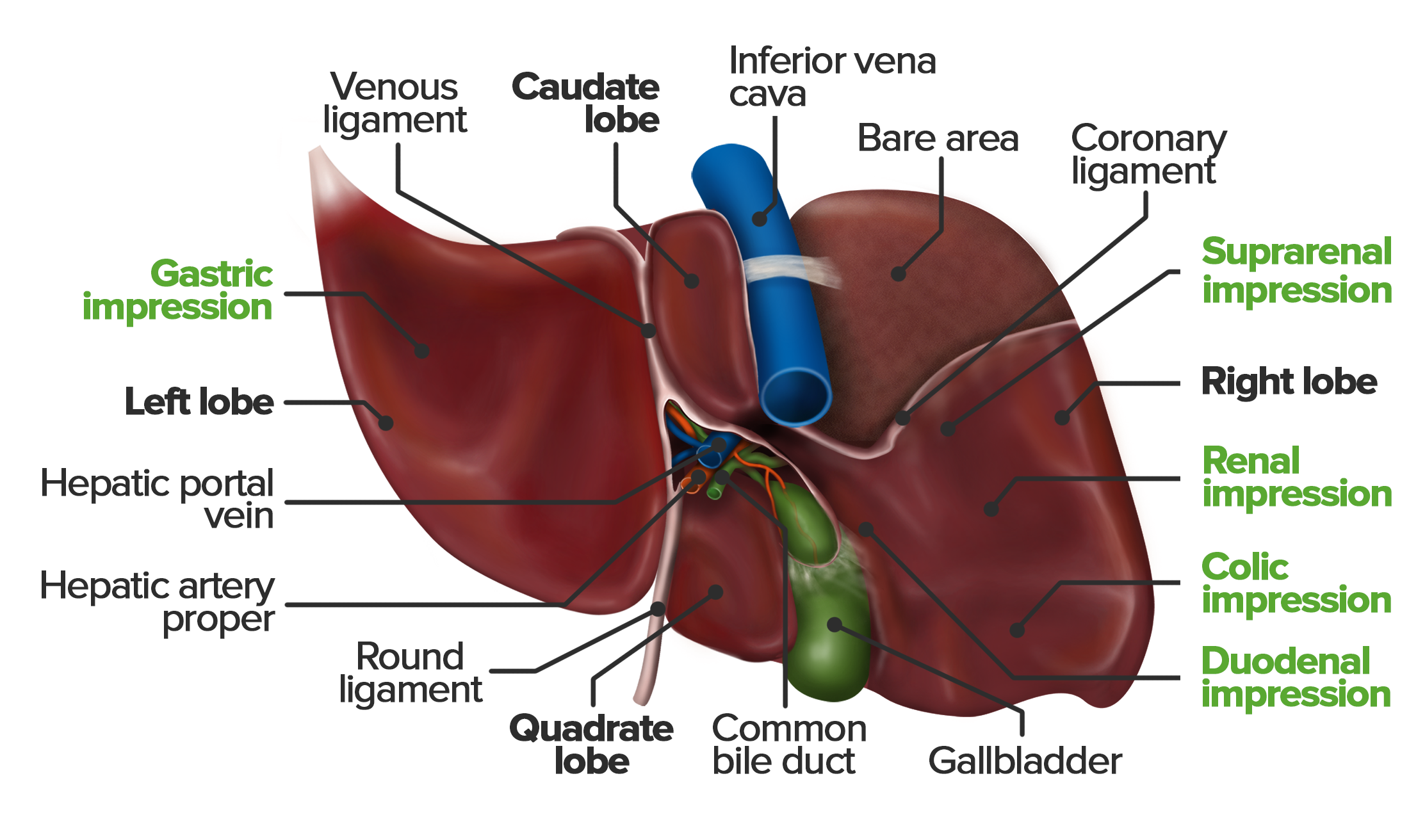
- Bears the **porta hepatis** (hilum of the liver)
- Has several **impressions** named after the organs that produce them
#### Impressions on the Visceral Surface
**On the Right Lobe:**
| Impression | Organ |
|---|---|
| **Colic impression** | Right (hepatic) flexure of the colon |
| **Renal impression** | Right kidney |
| **Duodenal impression** | First part of the duodenum (D1) |
| **Suprarenal impression** | Right suprarenal gland (on bare area posteriorly) |
**On the Left Lobe:**
| Impression | Organ |
|---|---|
| **Gastric impression** | Anterior surface of the stomach |
| **Oesophageal impression** | Abdominal oesophagus |
**On the Caudate Lobe:**
- Related to the **inferior vena cava** and **crura of the diaphragm**
**On the Quadrate Lobe:**
- Related to the **pylorus of the stomach** and **first part of the duodenum**
---
## BORDERS
### 1. Inferior (Anteroinferior) Border
- **Sharp** where the diaphragmatic and visceral surfaces meet anteriorly
- Has two **notches**:
- **Notch for the falciform ligament** (ligamentum teres) — where the round ligament (ligamentum teres hepatis) reaches the inferior border
- **Notch for the fundus of the gallbladder** (cystic notch) — at the tip of the right 9th costal cartilage
- The inferior border follows the right costal margin and crosses the epigastrium
### Clinical: Murphy's Sign and the Fundus of the Gallbladder
The **fundus of the gallbladder** projects at the junction of the right **linea semilunaris** (lateral border of the rectus abdominis) with the **right costal margin** (tip of the 9th costal cartilage). This is the surface marking used to locate the gallbladder.
**Murphy's sign**: The examiner places fingers at this point and asks the patient to take a deep breath. In **acute cholecystitis**, the inflamed gallbladder descends with the diaphragm and contacts the examiner's fingers, causing the patient to **catch their breath** (inspiratory arrest). This sign is **positive in acute cholecystitis**.
### 2. Posterior (Posterosuperior) Border
- **Rounded and blunt**
- Related to the **bare area** of the liver
---
## LOBES OF THE LIVER
The liver is divided into lobes by **anatomical (morphological)** and **functional (surgical)** criteria.
### A. ANATOMICAL LOBES (Four Lobes)
#### 1. Right Lobe
- The **largest lobe** — constitutes about 5/6 of the liver
- Separated from the left lobe on the **diaphragmatic surface** by the **falciform ligament**
- Separated from the left lobe on the **visceral surface** by the **left sagittal fissure** (containing the ligamentum teres anteriorly and ligamentum venosum posteriorly)
#### 2. Left Lobe
- Smaller, flatter, and thinner
- Occupies the epigastrium and left hypochondrium
- Has the **gastric impression** and **oesophageal groove** on its visceral surface
#### 3. Caudate Lobe (Spigelian Lobe)
- Located on the **posterosuperior aspect of the visceral surface**
- **Boundaries:**
- Left: **Fissure for the ligamentum venosum**
- Right: **Groove for the inferior vena cava**
- Inferior: **Porta hepatis**
- Has a **caudate process** (projects to the right, connecting it to the right lobe) and a **papillary process** (projects into the porta hepatis)
- **Unique feature**: Receives blood supply from **both** the right and left hepatic arteries and portal vein branches, and drains directly into the **IVC** via **small hepatic veins** (not via the main right, middle, or left hepatic veins)
#### Clinical: Budd-Chiari Syndrome and Caudate Lobe Hypertrophy
In **Budd-Chiari syndrome** (thrombosis of the hepatic veins), the main hepatic veins are occluded, but the caudate lobe's venous drainage directly into the IVC is preserved. This leads to **compensatory hypertrophy of the caudate lobe** while the rest of the liver is congested and swollen. This is a **classic radiological finding**.
#### 4. Quadrate Lobe
- Located on the **anteroinferior part of the visceral surface**
- **Boundaries:**
- Left: **Fissure for the ligamentum teres**
- Right: **Fossa for the gallbladder**
- Superior (posterior): **Porta hepatis**
- Functionally, the quadrate lobe belongs to the **left lobe** (it receives left hepatic artery and left portal vein branches, and bile drains into the left hepatic duct)
### B. FUNCTIONAL (SURGICAL/COUINAUD) LOBES
Based on the distribution of the **hepatic artery**, **portal vein**, and **hepatic duct** (the portal triad), the liver is divided into:
- **Right functional lobe** — supplied by the right hepatic artery, right portal vein, drained by the right hepatic duct
- **Left functional lobe** — supplied by the left hepatic artery, left portal vein, drained by the left hepatic duct
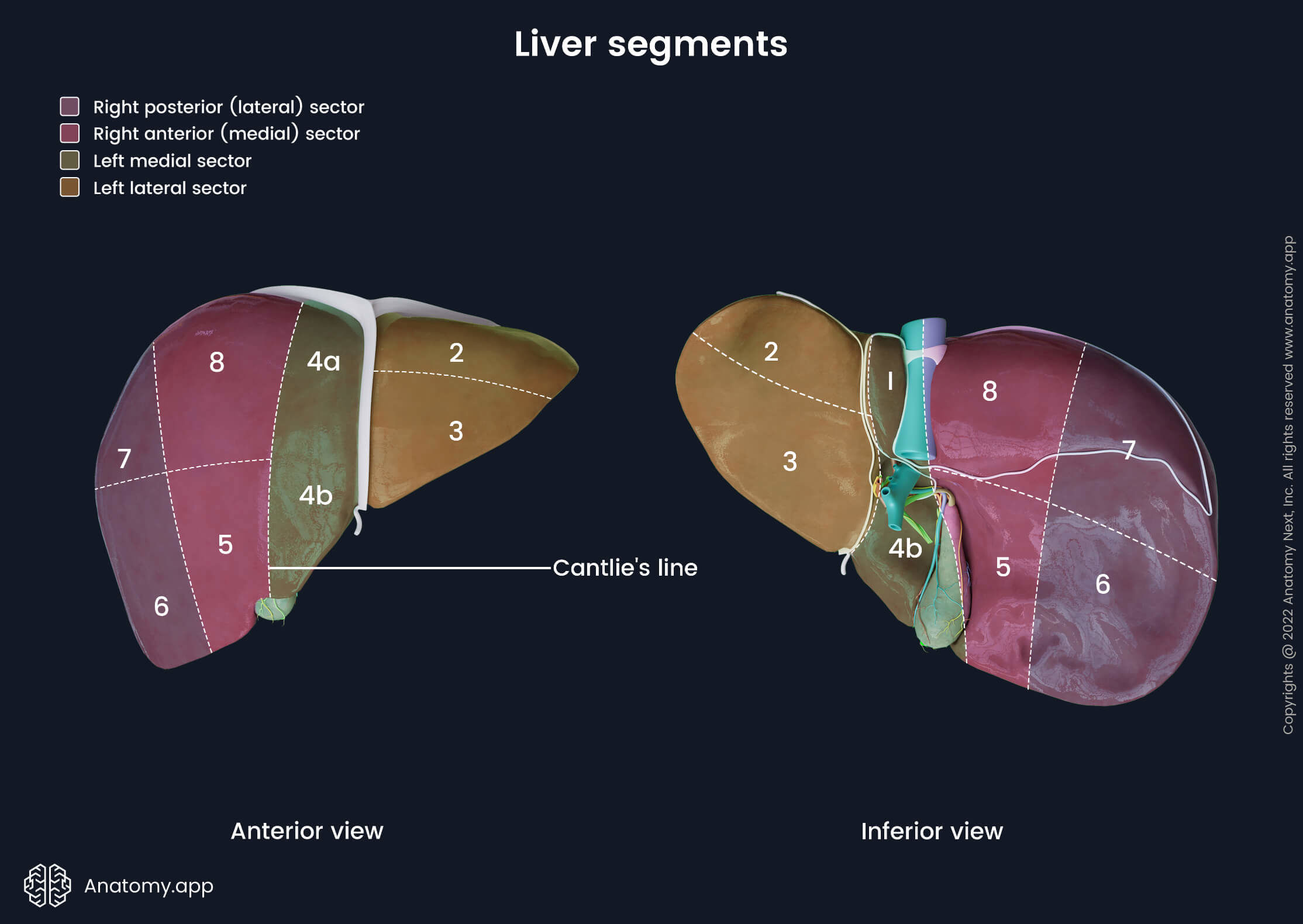
The **functional division** (Cantlie's line or Rex-Cantlie line) runs from the **gallbladder fossa** anteroinferiorly to the **IVC groove** posterosuperiorly. This line roughly corresponds to the course of the **middle hepatic vein**.
Thus, functionally:
- The **quadrate lobe** belongs to the **left functional lobe**
- The **caudate lobe** is considered **autonomous** (receives dual supply)
### C. COUINAUD'S SEGMENTAL ANATOMY (1957)
**Claude Couinaud** described **8 functionally independent segments**, each with its own **portal pedicle** (portal vein, hepatic artery, bile duct) and **hepatic venous drainage**.
| Segment | Location |
|---|---|
| **Segment I** | Caudate lobe |
| **Segment II** | Left lateral superior |
| **Segment III** | Left lateral inferior |
| **Segment IV** | Left medial (quadrate lobe) — divided into IVa (superior) and IVb (inferior) |
| **Segment V** | Right anterior inferior |
| **Segment VI** | Right posterior inferior |
| **Segment VII** | Right posterior superior |
| **Segment VIII** | Right anterior superior |
**Segments II and III** = Left lateral section
**Segment IV** = Left medial section
**Segments V and VIII** = Right anterior section
**Segments VI and VII** = Right posterior section
### Clinical: Couinaud Segments in Hepatic Surgery
Couinaud's segmental anatomy is the **basis of modern hepatic surgery**. Because each segment is functionally independent, individual segments can be resected (**segmentectomy**) without compromising the vascular supply or biliary drainage of the remaining liver.
**Types of hepatic resection:**
- **Right hepatectomy**: Segments V, VI, VII, VIII
- **Left hepatectomy**: Segments II, III, IV
- **Right lobectomy (extended right hepatectomy/right trisectionectomy)**: Segments IV, V, VI, VII, VIII
- **Left lateral sectionectomy**: Segments II, III (most common resection for living-donor liver transplant for pediatric recipients)
---
## THE "H"-SHAPED ARRANGEMENT ON THE VISCERAL SURFACE
On the visceral surface, two **sagittal fissures** and one **transverse fissure** form an **"H"**:
### Left Sagittal Fissure
Contains:
- **Anteriorly**: Fissure for the **ligamentum teres** (round ligament of the liver — obliterated left umbilical vein)
- **Posteriorly**: Fissure for the **ligamentum venosum** (obliterated ductus venosus)
### Right Sagittal Fissure
Contains:
- **Anteriorly**: **Fossa for the gallbladder**
- **Posteriorly**: **Groove for the IVC**
### Transverse Fissure (Porta Hepatis)
- The **hilum of the liver**
- Approximately **5 cm long**
- Located on the visceral surface between the caudate and quadrate lobes
- Contains the structures entering and leaving the liver (see below)
---
## PORTA HEPATIS (HILUM)
The porta hepatis is the transverse fissure on the visceral surface through which all major structures enter and exit the liver (except the hepatic veins and IVC).
### Contents (from anterior to posterior):
**Mnemonic: "DAV" — Duct Anteriorly, Artery in the middle, Vein posteriorly**
| Structure | Position |
|---|---|
| **Hepatic ducts** (right and left) | Most anterior |
| **Hepatic arteries** (right and left) | Intermediate |
| **Portal vein** (right and left branches) | Most posterior |
| **Hepatic nerve plexus** | Along the arteries |
| **Lymphatic vessels** | Accompanying the vessels |
Also:
- The hepatic duct lies to the **right**
- The hepatic artery lies to the **left**
- The portal vein lies **posterior and between** the two
### Clinical: Pringle Maneuver
The **Pringle maneuver** involves clamping the **hepatoduodenal ligament** (containing the portal triad — common bile duct, proper hepatic artery, and portal vein) between the thumb and index finger through the **epiploic foramen (foramen of Winslow)**. This is used to control **hemorrhage from the liver** during surgery or trauma. It interrupts the arterial and portal venous inflow to the liver. The liver can tolerate ischemia for about **15–20 minutes** at normothermia (longer with intermittent clamping or hypothermia).
If bleeding continues despite the Pringle maneuver, the source is likely the **hepatic veins or the IVC** (retrohepatic injury), which is a very serious injury with high mortality.
---
## PERITONEAL RELATIONS AND LIGAMENTS
The liver is almost completely covered by **peritoneum** except at:
1. The **bare area** (posterosuperior surface)
2. The **porta hepatis**
3. The **fossa for the gallbladder**
4. The **groove for the IVC**
5. A small area where the **falciform ligament** is attached
### Ligaments of the Liver
#### 1. Falciform Ligament
- A **sickle-shaped** fold of peritoneum
- Extends from the **anterior abdominal wall** (from the umbilicus to the diaphragm) to the **liver**
- Divides the **diaphragmatic surface** of the liver into anatomical right and left lobes
- Contains in its **free inferior border** the **ligamentum teres** (round ligament) — the obliterated **left umbilical vein**
- Also contains small **paraumbilical veins** (of Sappey) in its layers
#### Clinical: Caput Medusae
In **portal hypertension**, the paraumbilical veins within the falciform ligament may recanalize as a portosystemic collateral pathway. Blood flows from the portal system via the paraumbilical veins to the veins of the anterior abdominal wall (superior and inferior epigastric veins), producing **caput medusae** — dilated, tortuous, radiating periumbilical veins resembling the head of Medusa.
- Flow is **away from the umbilicus** in portal hypertension (distinguishing it from **IVC obstruction** where flow is **upward** toward the SVC)
The **Cruveilhier-Baumgarten syndrome** involves a patent umbilical vein with portal hypertension, producing a venous hum (bruit) heard over the umbilicus.
#### 2. Coronary Ligament
- The **most important ligament** in terms of area
- Consists of **anterior (superior) and posterior (inferior) layers** of peritoneum that reflect from the liver to the diaphragm
- The area between the two layers on the posterosuperior surface is the **bare area** of the liver
- The anterior layer is continuous with the right layer of the falciform ligament
- The posterior layer reflects onto the right kidney as the **hepatorenal ligament**
#### 3. Triangular Ligaments (Right and Left)
- Formed where the anterior and posterior layers of the coronary ligament meet laterally
- **Right triangular ligament**: Short, at the right extremity of the bare area
- **Left triangular ligament**: Longer, extends from the left end of the bare area; contains the **obliterated appendix fibrosa** (a fibrous remnant) in some individuals
#### 4. Ligamentum Teres Hepatis (Round Ligament)
- Obliterated **left umbilical vein**
- Runs from the **umbilicus** in the free border of the falciform ligament to the **left branch of the portal vein** at the umbilical fissure on the visceral surface
- The left portal vein is located at the attachment point of the ligamentum teres (Rex recessus)
#### Clinical: Transjugular Intrahepatic Portosystemic Shunt (TIPS) and Recanalization
In portal hypertension, the **round ligament (obliterated umbilical vein)** may be recanalized surgically or spontaneously. This is used in rare cases for **portal decompression**.
#### 5. Ligamentum Venosum
- Obliterated **ductus venosus** (which in fetal life shunted oxygenated blood from the left umbilical vein directly to the IVC, bypassing the liver sinusoids)
- Located in the **fissure for the ligamentum venosum** on the visceral surface
- Connects the left branch of the portal vein to the left hepatic vein/IVC
#### 6. Hepatoduodenal Ligament
- The **free right border** of the lesser omentum
- Extends from the porta hepatis to the **first part of the duodenum** and **lesser curvature of the stomach**
- Contains the **portal triad:**
- **Common bile duct** (CBD) — right and anterior
- **Proper hepatic artery** — left and anterior
- **Portal vein** — posterior
- Also: hepatic nerve plexus, lymphatics, and lymph nodes (hepatic/cystic nodes)
- Forms the **anterior boundary** of the **epiploic foramen (of Winslow)**
#### 7. Hepatogastric Ligament
- The thin, translucent part of the lesser omentum
- Extends from the liver (fissure for the ligamentum venosum) to the lesser curvature of the stomach
- Contains the **right and left gastric vessels**, the **hepatic branches of the vagus nerve**, and lymphatics
- May contain an **accessory/replaced left hepatic artery** from the left gastric artery (present in ~25% of people)
### Clinical: Variant Hepatic Arterial Anatomy
Variations in the hepatic arterial supply are **extremely common** (~40-45% of people have variant anatomy). The surgeon **must** be aware of these variations during hepatic, biliary, gastric, and pancreatic surgery.
**Common variations:**
| Variant | Incidence | Details |
|---|---|---|
| **Replaced right hepatic artery** from SMA | ~11-21% | Runs posterior to the portal vein and CBD, through the portal triad |
| **Replaced left hepatic artery** from left gastric artery | ~10-12% | Runs in the hepatogastric ligament |
| **Accessory right hepatic artery** from SMA | ~6% | In addition to normal right hepatic artery |
| **Accessory left hepatic artery** from left gastric artery | ~8% | In addition to normal left hepatic artery |
| **Common hepatic artery** from SMA | ~2.5% | No origin from celiac trunk |
**Surgical significance**: A replaced left hepatic artery from the left gastric artery runs in the hepatogastric ligament — it can be inadvertently ligated during gastrectomy, causing ischemia of the left lobe of the liver. Similarly, a replaced right hepatic artery from the SMA courses posterior to the head of the pancreas and can be injured during pancreaticoduodenectomy (Whipple's procedure).
---
## THE BARE AREA OF THE LIVER
- Located on the **posterosuperior surface**
- An area **devoid of peritoneum** where the liver is in direct contact with the diaphragm
- **Bounded by:**
- Superiorly: **Anterior (superior) layer of the coronary ligament**
- Inferiorly: **Posterior (inferior) layer of the coronary ligament**
- Right: **Right triangular ligament**
- Left: **Left triangular ligament/upper end of the lesser omentum**
- Contains the **groove for the IVC** and the **right suprarenal gland impression**
- The IVC is partially embedded in the bare area and is held in place by **hepatic veins** and connective tissue
### Clinical: Bare Area and Liver Abscess
- Because the bare area has no peritoneal covering, an **amoebic liver abscess** in the posterosuperior aspect of the right lobe can **rupture through the diaphragm** into the **right pleural cavity** or the **right lower lobe of the lung**, causing an **empyema** or a **hepatobronchial fistula** (patient coughs "anchovy sauce" sputum — chocolate-brown, odorless pus)
- In contrast, an abscess on the visceral surface would more likely rupture into the **peritoneal cavity**
- The bare area is also a route of direct spread of infection between the liver and the subphrenic/subhepatic spaces
---
## BLOOD SUPPLY
The liver has a **dual blood supply** — this is one of its most important characteristics:
### 1. Portal Vein (75-80% of hepatic blood flow)
- Formed behind the **neck of the pancreas** by the union of the **superior mesenteric vein (SMV)** and the **splenic vein**
- Length: ~8 cm; diameter: ~1.2 cm
- Carries **deoxygenated but nutrient-rich** blood from the GI tract, spleen, pancreas, and gallbladder
- Ascends in the **hepatoduodenal ligament** (posterior to the CBD and hepatic artery)
- At the porta hepatis, divides into **right and left branches**
- Right branch: Short, enters the right lobe directly
- Left branch: Longer, more horizontal, gives off branches to the caudate lobe, quadrate lobe, and left lobe; gives attachment to the ligamentum teres and ligamentum venosum
### 2. Hepatic Artery (20-25% of hepatic blood flow, but ~50% of oxygen supply)
- The **common hepatic artery** arises from the **celiac trunk**
- Courses to the right along the upper border of the pancreas
- At the upper border of D1, gives off the **gastroduodenal artery** and continues as the **proper hepatic artery**
- The proper hepatic artery ascends in the hepatoduodenal ligament (to the left of the CBD, anterior to the portal vein)
- Near the porta hepatis, divides into **right and left hepatic arteries**
- The **right hepatic artery** typically crosses **posterior** to the **common hepatic duct** (but may cross anteriorly in ~13-20% of cases — important surgical variation)
- Gives off the **cystic artery** (usually from the right hepatic artery, within **Calot's triangle**)
### Clinical: Calot's Triangle (Cystohepatic Triangle)
**Boundaries:**
- **Medially**: Common hepatic duct
- **Inferiorly**: Cystic duct
- **Superiorly**: Inferior surface of the liver (segment V/right lobe)
**Contents:**
- **Cystic artery** (most important content)
- Right hepatic artery (may pass through)
- Cystic lymph node (of Lund/Calot's node)
- Connective tissue, lymphatics, sometimes an accessory hepatic duct
**Surgical importance**: Calot's triangle is the key area of dissection during **cholecystectomy** (especially laparoscopic cholecystectomy). The surgeon must achieve the **Critical View of Safety (CVS)** — clearing Calot's triangle to positively identify the cystic duct and cystic artery before clipping and dividing them. Failure to do so can result in **bile duct injury** (inadvertent clipping/transection of the common bile duct or right hepatic duct) — one of the most feared complications of cholecystectomy.
### 3. Hepatic Veins (Drainage)
- **Three major hepatic veins**: Right, Middle (intermediate), and Left
- Drain directly into the **IVC** just below the **diaphragm**
- The **right hepatic vein** is the largest and drains the right posterior section
- The **middle hepatic vein** runs in **Cantlie's line** (along the principal plane of the liver) — drains segments IV, V, VIII
- The **left hepatic vein** drains segments II and III
- **Middle and left hepatic veins** often form a **common trunk** before entering the IVC
- Additionally, multiple **small hepatic veins (accessory hepatic veins)** drain the **caudate lobe** and parts of the right lobe directly into the IVC
### Clinical: Hepatic Vein Injury
- The hepatic veins have **no extrahepatic valves** and are embedded in the liver substance with short extrahepatic portions
- Injury to hepatic veins during liver surgery or trauma is dangerous — causes massive hemorrhage and **air embolism** (because of the negative intrathoracic pressure that can suck air into open veins)
- **Retrohepatic IVC injuries** (between the hepatic veins and the diaphragm) are among the **most lethal abdominal injuries** with mortality >80%
---
## LYMPHATIC DRAINAGE
The liver produces a **large proportion** of the body's lymph (about 25-50% of all thoracic duct lymph).
### Superficial Lymphatics
- From the **diaphragmatic surface**: drain to **diaphragmatic nodes** → **posterior mediastinal nodes** → right lymphatic duct or thoracic duct
- Some pass through the **bare area** to reach nodes along the IVC
- From the **anterior diaphragmatic surface**: follow the falciform ligament to **parasternal nodes**
### Deep Lymphatics
- Follow the portal triad to the **hepatic nodes** (in the hepatoduodenal ligament) → **celiac nodes** → **cisterna chyli** → **thoracic duct**
- Some drain directly through the diaphragm to **posterior mediastinal nodes**
### Clinical: Lymphatic Spread in Liver Disease
- Hepatocellular carcinoma and metastatic liver disease can spread via lymphatics to **hepatic hilar nodes**, **celiac nodes**, and even **mediastinal nodes**
- In **portal hypertension**, hepatic lymph production increases dramatically (up to 20-fold), and transudation of lymph from the liver surface contributes to **ascites formation**
---
## NERVE SUPPLY
### 1. Sympathetic
- From the **celiac plexus** (T7-T9 preganglionic fibers → celiac ganglion → postganglionic fibers)
- Travel along the hepatic artery as the **hepatic plexus**
- Function: vasoconstriction, glycogenolysis, gluconeogenesis regulation
### 2. Parasympathetic
- From the **vagus nerve** (mainly left vagus/anterior vagal trunk)
- The **hepatic branch** of the anterior vagal trunk passes through the hepatogastric ligament
- Function: promotes glycogen synthesis, bile secretion
### 3. Sensory (Phrenic Nerve)
- The liver parenchyma itself is insensitive to pain
- Pain fibers come from the **peritoneal covering** (visceral peritoneum) and the **liver capsule (Glisson's capsule)**
- Pain from stretching of the capsule is referred to the **right shoulder** and **right scapular region** via the **right phrenic nerve** (C3, C4, C5) — because the diaphragm and its peritoneum (which covers the superior liver) are innervated by the phrenic nerve
- This is called **Kehr's sign** — referred pain to the right shoulder
### Clinical: Referred Pain
- **Liver abscess** or **hepatic distension** causes dull aching pain in the right hypochondrium and may produce referred pain to the **right shoulder tip** (via phrenic nerve irritation of the diaphragm overlying the liver)
- **Acute cholecystitis** can also cause right shoulder pain by similar mechanism
- **Subphrenic abscess** classically causes referred shoulder tip pain
---
## HEPATIC SEGMENTS AND INTRAHEPATIC ANATOMY
### Glisson's Capsule (Capsula Fibrosa)
- A thin but tough **fibrous capsule** covering the entire liver
- At the porta hepatis, it forms sheaths around the portal triad structures as they enter the liver (forming the **periportal sheath** or Glisson's sheath)
- This sheath extends into the liver substance along the portal tracts, providing internal support
- The capsule is innervated — stretching causes pain
### Clinical: Subcapsular Hematoma
- Trauma to the liver may cause a **subcapsular hematoma** — blood collects between Glisson's capsule and the liver parenchyma
- This is dangerous because:
- The hematoma may expand and eventually **rupture**, causing sudden massive hemoperitoneum (delayed splenic rupture equivalent for the liver)
- It may be missed on initial assessment if the capsule is intact
- **CT scan** is the investigation of choice
---
## PERITONEAL RECESSES AROUND THE LIVER
### 1. Subphrenic Spaces
- Between the diaphragm and the liver
- Divided by the falciform ligament into:
- **Right anterior subphrenic space**
- **Left anterior subphrenic space**
### 2. Subhepatic Spaces
- Between the visceral surface of the liver and the underlying viscera
- **Right subhepatic space** — includes the **hepatorenal recess (Morison's pouch)**
- This is the **deepest part of the peritoneal cavity** in the supine position
- Bounded by the visceral surface of the right lobe superiorly, the right kidney and suprarenal gland posteriorly, and the right colic flexure and duodenum inferiorly
- Fluid (blood, bile, pus) tends to collect here in supine patients
### Clinical: Morison's Pouch
- In **abdominal trauma**, the **FAST scan** (Focused Assessment with Sonography for Trauma) looks for free fluid in Morison's pouch as the first and most sensitive sign of hemoperitoneum
- In **peritonitis**, abscess formation commonly occurs in Morison's pouch and the subphrenic spaces
- **Subphrenic abscess** is a complication of perforated peptic ulcer, perforated appendicitis, or post-surgical infection
### 3. Left Subhepatic Space
- Overlies the lesser omentum and the stomach
- Communicates with the **lesser sac (omental bursa)** through the epiploic foramen
---
## DEVELOPMENT OF THE LIVER (Brief Anatomical Overview)
- Develops from the **hepatic diverticulum** (liver bud) — an outgrowth of the **foregut endoderm** at the junction of the foregut and midgut during **week 3-4** of development
- The liver bud grows into the **septum transversum** (a mesodermal mass between the pericardial cavity and the yolk stalk)
- The septum transversum forms the **connective tissue stroma**, **Glisson's capsule**, and the **hematopoietic tissue** of the fetal liver
- The septum transversum contributes to the **central tendon of the diaphragm** — this is why the liver is closely related to the diaphragm
- The **bare area** represents the area where the liver was in direct contact with the septum transversum
### Clinical: Congenital Anomalies
- **Accessory lobes (Riedel's lobe)**: A tongue-like downward projection of the right lobe, more common in women, may be mistaken for hepatomegaly or a right-sided abdominal mass
- **Agenesis of the left lobe**: Rare; the right lobe hypertrophies compensatorily
- **Ectopic liver tissue**: Rare; may be found in the gallbladder wall, adrenal gland, or elsewhere
---
## SURFACE MARKING (CLINICAL ANATOMY)
### Upper Border (Diaphragmatic Surface)
- Right side: **Right 5th rib** in the midclavicular line (5th intercostal space)
- Center: Crosses the sternum at the level of the **xiphisternal joint**
- Left side: **Left 5th intercostal space** in the midclavicular line
### Lower Border
- Starts at the **right 10th rib** in the midaxillary line
- Crosses the **right costal margin** at the tip of the **9th costal cartilage** (gallbladder fundus)
- Crosses the midline at the **transpyloric plane** (L1 level)
- Reaches the **left 5th intercostal space** in the midclavicular line
### Clinical: Percussion of the Liver
- **Liver span**: Normally 6-12 cm in the right midclavicular line by percussion
- Decreased liver span may indicate:
- **Cirrhosis** (shrunken liver)
- **Perforation of hollow viscus** (free air under the diaphragm obliterates liver dullness — **loss of liver dullness** is a sign of pneumoperitoneum)
- The upper border of liver dullness coincides with the lower border of the right lung
---
## CLINICAL CONDITIONS OF THE LIVER — COMPREHENSIVE OVERVIEW
### 1. Cirrhosis
- Irreversible fibrosis and nodular regeneration
- Causes: Alcohol, hepatitis B/C, NAFLD, autoimmune, Wilson's disease, hemochromatosis, alpha-1 antitrypsin deficiency, primary biliary cholangitis
- Complications: Portal hypertension, ascites, variceal bleeding, hepatorenal syndrome, hepatic encephalopathy, hepatocellular carcinoma
### 2. Portal Hypertension
- Portal venous pressure >10 mmHg (normal ~5-8 mmHg)
- Causes: Prehepatic (portal vein thrombosis), hepatic (cirrhosis — most common), posthepatic (Budd-Chiari, constrictive pericarditis)
- **Portosystemic anastomoses** (sites where portal and systemic venous systems communicate):
| Site | Portal Tributary | Systemic Tributary | Clinical Manifestation |
|---|---|---|---|
| **Lower esophagus** | Left gastric vein | Esophageal veins → azygos vein | **Esophageal varices** (life-threatening bleeding) |
| **Anorectal junction** | Superior rectal vein | Middle and inferior rectal veins | **Anorectal varices** (not to be confused with hemorrhoids) |
| **Periumbilical** | Paraumbilical veins | Superficial epigastric veins | **Caput medusae** |
| **Bare area of liver** | Hepatic veins (portal branches) | Phrenic veins → IVC | Retroperitoneal collaterals |
| **Retroperitoneal** | Colic veins | Retroperitoneal veins (renal, gonadal, lumbar veins) | **Veins of Retzius** |
### 3. Liver Transplantation
- Anatomical considerations:
- The **suprahepatic IVC** and **infrahepatic IVC** must be dissected or the "piggyback" technique (preserving recipient's IVC) may be used
- **Hepatic artery**, **portal vein**, and **bile duct** anastomoses are performed
- Duct-to-duct anastomosis (choledochocholedochostomy) or Roux-en-Y hepaticojejunostomy for biliary reconstruction
- Knowledge of arterial variants is critical
### 4. Liver Trauma
- The liver is the **most commonly injured abdominal organ** in penetrating trauma and the **second most commonly injured** (after the spleen) in blunt trauma
- Classification: **AAST grading** (I-VI) based on depth and extent of laceration/hematoma
- Management: Hemodynamically stable → non-operative (CT monitoring); unstable → operative (damage control surgery, packing, Pringle maneuver)
---
# PART 2: THE SPLEEN (LIEN/SPLEN)
---
## INTRODUCTION
The spleen is the **largest lymphoid organ** in the body. It is NOT a vital organ (life is compatible without it), but it plays critical roles in **immune function** and **blood filtration**.
---
## GENERAL CHARACTERISTICS
| Feature | Detail |
|---|---|
| **Weight** | ~150 g (80-300 g range; average 7 oz) |
| **Dimensions** | ~12 cm × 7 cm × 3 cm (1 × 3 × 5 × 7 × 9 × 11 rule — see below) |
| **Color** | Dark purplish-red |
| **Consistency** | Soft, highly vascular, friable (easily ruptured) |
| **Shape** | Ovoid; like a "coffee bean" or "clenched fist" |
| **Location** | Left hypochondrium |
| **Vertebral level** | 9th to 11th ribs (long axis along the 10th rib) |
### The "1, 3, 5, 7, 9, 11" Rule (Mnemonic)
- **1** × **3** × **5** inches (dimensions)
- **7** ounces (weight)
- Lies deep to ribs **9, 10, 11**
- **1** — single organ
- **3** — surfaces
- **5** — ligaments (some texts; varies)
---
## LOCATION AND RELATIONS
- Lies in the **left hypochondrium**, between the **gastric fundus** and the **left hemidiaphragm**
- Long axis corresponds to the **10th rib**
- Does not extend beyond the **midaxillary line** normally (important for clinical examination)
- Completely covered by the **rib cage** and not normally palpable
### Clinical: Splenomegaly
The spleen **must enlarge to about 2-3 times its normal size** before it becomes palpable below the left costal margin. When palpable, it descends **medially and inferiorly** toward the **right iliac fossa** (along its long axis/10th rib direction).
**Characteristics of a palpable spleen** (to distinguish from left kidney):
1. **Moves with respiration** (descends on inspiration — pushed down by the diaphragm)
2. Has a **notched anterior border** (pathognomonic)
3. **Cannot get above it** (hand cannot be insinuated between the spleen and the costal margin)
4. Extends toward the **right iliac fossa** (not into the loin)
5. **Dull to percussion** over the 9th, 10th, and 11th ribs in the left midaxillary line (Castell's point — normally resonant; dullness suggests splenomegaly)
6. Not bimanually palpable (unlike a kidney)
**Massive splenomegaly (crossing the umbilicus) — Causes:**
- Chronic myeloid leukemia (CML) — **most common cause of massive splenomegaly**
- Myelofibrosis
- Malaria (chronic/hyperreactive malarial splenomegaly)
- Kala-azar (visceral leishmaniasis)
- Gaucher's disease
- Thalassemia major
- Polycythemia vera
- Lymphoma
---
## SURFACES
The spleen has **two surfaces** and **two borders**:
### 1. Diaphragmatic Surface
- **Smooth and convex**
- Related to the undersurface of the **left dome of the diaphragm**
- Through the diaphragm, related to:
- **Left lung** (base/costophrenic recess)
- **Left pleural recess** (costodiaphragmatic recess)
- **Ribs 9, 10, 11**
### Clinical: Rib Fractures and Splenic Injury
Fractures of the **left lower ribs (9th, 10th, 11th)** should always raise suspicion of **splenic injury**, which is the **most commonly injured organ in blunt abdominal trauma**. Always examine for:
- Left upper quadrant tenderness and guarding
- Signs of peritonism
- **Kehr's sign** — referred pain to the **left shoulder tip** due to diaphragmatic irritation by blood pooling in the left subphrenic space (blood irritates the phrenic nerve → referred pain to C3, C4, C5 dermatomes at the shoulder)
- **Balance's sign** — dullness to percussion in the left flank that doesn't shift with position change (due to clotted blood around the spleen), with shifting dullness on the right
### 2. Visceral Surface
- **Concave and irregular**
- Related to adjacent viscera
- Bears the **hilum** (hilus) of the spleen, through which the splenic vessels enter and leave
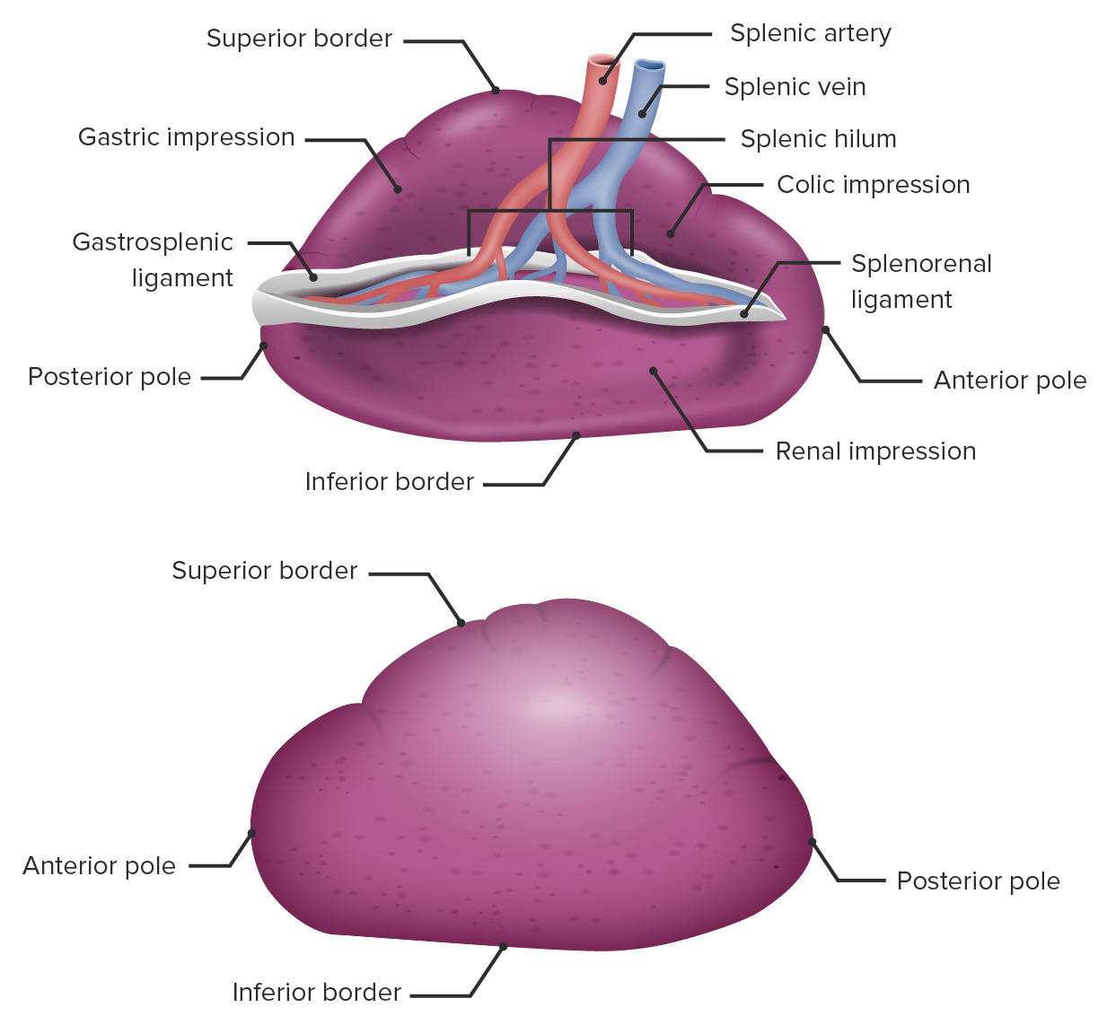
#### Impressions on the Visceral Surface
| Impression | Related Organ | Location |
|---|---|---|
| **Gastric impression** | Fundus of the stomach | Large, anterior and superior |
| **Renal impression** | Left kidney (upper pole) | Posterior and inferior |
| **Colic impression** | Left colic (splenic) flexure | Inferior |
| **Pancreatic impression** | Tail of the pancreas | Near the hilum, between gastric and renal impressions |
---
## BORDERS
### 1. Superior (Anterior) Border
- **Thin and sharp**
- Separates the gastric impression from the diaphragmatic surface
- Has characteristic **notches** (1-3 notches) — remnants of the lobulated fetal spleen
- These notches are **palpable** when the spleen is enlarged — this is a **diagnostic feature** of splenomegaly
### 2. Inferior (Posterior) Border
- **Rounded and blunt**
- Separates the renal impression from the diaphragmatic surface
- Rests on the **phrenicocolic ligament** (sustentaculum lienis — the "shelf" that supports the spleen)
### 3. Medial End (Anterior/Superior Extremity)
- Pointed, directed anteromedially
### 4. Lateral End (Posterior/Inferior Extremity)
- Rounded, directed posterolaterally
---
## POLES
- **Superior (anterior) pole**: Pointed, close to the greater curvature of the stomach
- **Inferior (posterior) pole**: Rounded, rests on the splenic flexure of the colon and the phrenicocolic ligament
---
## HILUM OF THE SPLEEN
- Located on the **visceral surface** between the gastric and renal impressions
- A long fissure through which the **splenic vessels** and **nerves** enter and leave
- The **tail of the pancreas** is closely related to the hilum (extends to within 1-2 cm of the hilum in 75% of cases — directly contacts the hilum in ~30%)
### Structures at the Hilum
- **Splenic artery** (branches into multiple terminal branches → enters hilum)
- **Splenic vein** (formed by union of tributaries leaving the hilum)
- **Lymphatics**
- **Splenic nerve plexus**
### Clinical: Tail of Pancreas and Splenic Hilum
The close relationship between the **tail of the pancreas** and the splenic hilum has important surgical implications:
- During **splenectomy**, the tail of the pancreas may be inadvertently injured, leading to **pancreatic fistula** or **pancreatitis**
- During **distal pancreatectomy** for pancreatic tail tumors, the spleen is usually removed en bloc (**distal pancreatectomy with splenectomy**), though spleen-preserving distal pancreatectomy (Kimura or Warshaw technique) can be attempted
---
## PERITONEAL RELATIONS AND LIGAMENTS
The spleen is **entirely covered by peritoneum** except at the hilum. It is an **intraperitoneal organ** and is relatively mobile.
### Ligaments (Peritoneal Folds)
#### 1. Gastrosplenic Ligament (Gastrolienal Ligament)
- Connects the **hilum of the spleen** to the **greater curvature of the stomach**
- Contains:
- **Short gastric arteries** (4-5 branches from the splenic artery)
- **Short gastric veins**
- **Left gastroepiploic (gastroomental) artery and vein**
#### 2. Splenorenal Ligament (Lienorenal Ligament)
- Connects the **hilum of the spleen** to the **left kidney** (anterior surface)
- Contains:
- **Splenic artery** (terminal branches)
- **Splenic vein**
- **Tail of the pancreas** (runs within this ligament)
#### 3. Phrenicocolic Ligament (Sustentaculum Lienis)
- Extends from the **left colic flexure** to the **diaphragm**
- Acts as a **shelf** supporting the inferior pole of the spleen
- Does NOT contain vessels — it is an avascular fold
- Important during spleen mobilization in surgery
#### 4. Phrenosplenic (Splenophrenic) Ligament
- Some texts describe a separate fold connecting the **superior pole of the spleen** to the **diaphragm**
- May contain polar vessels
#### Clinical: Ligaments and Splenectomy
During **splenectomy**, the surgeon must systematically divide all the ligaments:
1. **Splenocolic ligament** (if present) — inferior attachments
2. **Gastrosplenic ligament** — ligate short gastric and left gastroepiploic vessels
3. **Splenorenal ligament** — ligate splenic artery and vein (protect the tail of the pancreas!)
4. **Phrenosplenic ligament** — divide superior attachments
In **laparoscopic splenectomy** (now the gold standard for elective splenectomy), the lateral decubitus position is used to allow gravity to retract the spleen medially.
---
## BLOOD SUPPLY
### Arterial Supply: Splenic Artery
- The **largest branch** of the **celiac trunk**
- One of the most **tortuous arteries** in the body (becomes increasingly tortuous with age — possibly to accommodate the changes in stomach volume and splenic size)
- Course:
1. Arises from the celiac trunk (behind the upper border of the pancreas)
2. Runs to the left along the **superior border of the pancreas** (in a tortuous course)
3. Enters the **splenorenal ligament** with the tail of the pancreas
4. Near the hilum, divides into **terminal branches** (2 primary branches — superior and inferior — corresponding to the vascular segments of the spleen)
#### Branches of the Splenic Artery
| Branch | Details |
|---|---|
| **Pancreatic branches** | Multiple small branches to the body and tail of the pancreas |
| **Dorsal pancreatic artery** | Arises near the origin of the splenic artery (or from celiac/SMA/common hepatic); descends behind the pancreas |
| **Greater pancreatic artery (arteria pancreatica magna)** | Largest pancreatic branch; supplies body of pancreas |
| **Short gastric arteries** (4-5) | Arise near the hilum; pass in the gastrosplenic ligament to the fundus of the stomach |
| **Left gastroepiploic (gastroomental) artery** | Arises near the hilum; passes in the gastrosplenic ligament along the greater curvature of the stomach |
| **Polar arteries** | Terminal branches to the upper and lower poles of the spleen |
| **Terminal (segmental) branches** | 2-3 branches entering the hilum |
### Clinical: Splenic Artery Aneurysm
- **Most common visceral artery aneurysm** (60% of all visceral aneurysms)
- More common in **women** (4:1 F:M ratio), especially multiparous women
- Risk factors: Pregnancy (increased blood volume and hormonal effects on vessel wall), portal hypertension, atherosclerosis, pancreatitis
- **Rupture risk** increases during pregnancy (especially 3rd trimester) — can be catastrophic with maternal and fetal mortality
- May present as an incidental finding (calcified ring on abdominal X-ray — "signet ring" sign)
### Venous Drainage: Splenic Vein
- Formed by the union of **5-6 tributaries** emerging from the hilum
- Exits the hilum through the splenorenal ligament
- Courses to the right, **behind the body and tail of the pancreas** (posterior to the pancreas — important!)
- Unites with the **superior mesenteric vein** behind the **neck of the pancreas** to form the **portal vein**
- **Tributaries:**
- Short gastric veins
- Left gastroepiploic vein
- Pancreatic veins
- **Inferior mesenteric vein (IMV)** — usually drains into the splenic vein near its junction with the SMV (but may drain into the SMV or the SMV-splenic vein junction)
### Clinical: Splenic Vein Thrombosis
- The splenic vein runs **posterior to the pancreas** — diseases of the pancreas (especially **chronic pancreatitis** and **pancreatic cancer**) can cause **splenic vein thrombosis**
- This leads to **sinistral (left-sided) portal hypertension** (also called **segmental portal hypertension**) with:
- **Isolated gastric varices** (especially fundal varices) — because blood from the spleen must find alternative routes: via short gastric veins → gastric fundal veins → left gastric vein → portal vein
- Splenomegaly
- Normal liver function
- Treatment: **Splenectomy** (curative! — removes the source of increased venous pressure)
---
## VASCULAR SEGMENTS OF THE SPLEEN
- The splenic artery typically divides into **2 terminal branches** (superior and inferior) — corresponding to **2 vascular segments**
- Some divisions give 3 or more segments
- These segments are **functionally independent** — no anastomoses between segmental arteries
- This is the basis for **partial (segmental) splenectomy** when preservation of splenic tissue is desired
### Avascular Plane
- The line between the upper and lower vascular segments is relatively avascular — the **avascular plane of the spleen**
- Can be used for partial splenectomy
---
## LYMPHATIC DRAINAGE
- Drains to **splenic hilar nodes** (pancreaticosplenic nodes) → **celiac nodes** → **cisterna chyli** → **thoracic duct**
---
## NERVE SUPPLY
- **Sympathetic**: From the **celiac plexus** via the **splenic plexus** (travels along the splenic artery)
- T6-T8 segments
- Vasomotor function (contraction of splenic capsule and trabeculae → expulsion of stored blood)
- **Parasympathetic**: Vagal fibers via the celiac plexus (functional significance debated)
- The spleen has **no sensory innervation** of its parenchyma (splenic disease is usually painless unless the capsule is stretched or peritoneum is irritated)
---
## DEVELOPMENT
- Develops from **mesenchymal condensation** in the **dorsal mesogastrium** during **week 5** of intrauterine life
- **Mesodermal** in origin (unlike lymph nodes, which develop from mesenchyme; the spleen is NOT a lymph node — it has no afferent lymphatics)
- Initially **lobulated** (the notches on the adult superior border are remnants of this lobulation)
- The rotation of the stomach during development carries the spleen to the **left hypochondrium**
### Clinical: Accessory Spleens (Splenunculi)
- Present in **10-30% of individuals**
- Usually found near the **hilum of the spleen** (most common site — ~75%), in the **gastrosplenic ligament**, **splenorenal ligament**, **greater omentum**, **splenic pedicle**, or even in the **pelvis** (wandering spleen) or **scrotum**
- Usually 1-2 cm in diameter; resemble lymph nodes
- **Clinical significance:**
- During **splenectomy for hematological disorders** (e.g., ITP — immune thrombocytopenic purpura, hereditary spherocytosis), **ALL accessory spleens must be identified and removed**, otherwise the hematological condition may recur or persist
- Can be detected preoperatively with **technetium-99m sulfur colloid scan** or **heat-damaged RBC scan**
- May be mistaken for **lymphadenopathy** or a **mass** on imaging
### Clinical: Asplenia and Hyposplenia
**Post-splenectomy or functional asplenia** (e.g., sickle cell disease — autosplenectomy):
- Increased susceptibility to **encapsulated organisms**: **Streptococcus pneumoniae** (most dangerous), **Haemophilus influenzae type b**, **Neisseria meningitidis** — leading to **overwhelming post-splenectomy infection (OPSI)**
- OPSI has a mortality rate of **50-70%** if untreated
- Risk is lifelong but greatest in the **first 2 years** after splenectomy and in **children under 5 years**
- **Prevention:**
- **Vaccinations** (ideally 2 weeks BEFORE elective splenectomy): Pneumococcal, Meningococcal, Haemophilus influenzae type b, Influenza
- **Lifelong prophylactic antibiotics** (penicillin V or amoxicillin) — at least for 2 years; some guidelines recommend lifelong prophylaxis in children
- Patient education: seek immediate medical attention for any febrile illness
- Medic-Alert bracelet
**Blood film findings after splenectomy:**
- **Howell-Jolly bodies** (nuclear remnants in RBCs — normally removed by the spleen; their presence indicates absent/nonfunctional splenic tissue)
- **Target cells**
- **Pappenheimer bodies** (iron-containing granules)
- **Acanthocytes**
- **Thrombocytosis** (initially transient; may persist)
- **Lymphocytosis** and **monocytosis**
- **Heinz bodies** may be seen
---
## SURFACE MARKING
- Long axis along the **left 10th rib**
- Upper border: **Left 9th rib** along the midaxillary line
- Lower border: **Left 11th rib** along the midaxillary line
- Does not extend **anterior to the midaxillary line** (if it does, it is enlarged)
- Medial end reaches to within **4 cm of the midline** at the level of **T10 vertebral spine**
---
## CLINICAL CONDITIONS OF THE SPLEEN
### 1. Splenic Rupture/Trauma
- **Most commonly injured abdominal organ in blunt trauma**
- Causes: Motor vehicle accidents, falls, sports injuries, left lower rib fractures
- **Types:**
- **Immediate rupture**: Direct trauma → capsular tear → hemoperitoneum
- **Delayed rupture**: Initial subcapsular hematoma → expansion over hours to days (up to 2 weeks) → rupture → sudden hemoperitoneum
- Patient initially appears stable ("lucid interval") then suddenly deteriorates
- **Occult/subcapsular rupture**: Hematoma contained within the capsule; may resolve or rupture
- **Diagnosis**: FAST ultrasound (free fluid in the left upper quadrant/splenorenal recess), CT scan with contrast (gold standard for stable patients)
- **Management**:
- Hemodynamically stable with low-grade injury: **Non-operative management** (observation, bed rest, serial imaging, serial hemoglobin) — successful in ~60-90% of cases
- Hemodynamically unstable or high-grade injury: **Operative management** — splenectomy or splenorrhaphy (splenic repair) or partial splenectomy (to preserve immune function, especially in children)
- **Angioembolization** for intermediate-grade injuries
- Trend toward **splenic preservation** when possible, especially in children
### 2. Wandering (Ectopic/Ptotic) Spleen
- Occurs when the splenic ligaments are elongated or absent (congenital or acquired — e.g., in multiparous women)
- The spleen "wanders" and may be found in the pelvis or elsewhere
- Risk of **splenic torsion** → vascular compromise → splenic infarction
- May present as an acute abdomen
- Treatment: **Splenopexy** (surgical fixation) or splenectomy if infarcted
### 3. Splenic Infarction
- Occurs due to occlusion of the splenic artery or its branches
- Causes: Emboli (atrial fibrillation, infective endocarditis), sickle cell disease, myeloproliferative disorders, splenic vein thrombosis, torsion of a wandering spleen
- Presents with sudden left upper quadrant pain, fever, leukocytosis
### 4. Splenosis vs. Accessory Spleen
- **Splenosis**: Autotransplantation of splenic tissue after splenic rupture or splenectomy — implants on peritoneal surfaces throughout the abdomen
- **Accessory spleen**: Congenital; present from birth
- Both may be found incidentally on imaging
---
# PART 3: THE PANCREAS
---
## INTRODUCTION
The pancreas is a **retroperitoneal, mixed (both endocrine and exocrine) gland** that lies transversely across the posterior abdominal wall. It is the **second largest gland** associated with the GI tract (after the liver).
---
## GENERAL CHARACTERISTICS
| Feature | Detail |
|---|---|
| **Weight** | ~80-90 g (some sources say 70-110 g) |
| **Length** | ~15-20 cm (6-8 inches) |
| **Color** | Yellowish-pink (lobulated appearance) |
| **Shape** | Elongated, flattened, resembling a "hockey stick" or a "tadpole" |
| **Location** | Retroperitoneal, across the upper abdomen at the level of **L1-L2** vertebrae |
| **Consistency** | Soft, lobulated |
| **Peritoneal status** | **Retroperitoneal** (secondarily — most of it; the tail is intraperitoneal within the splenorenal ligament) |
---
## PARTS OF THE PANCREAS
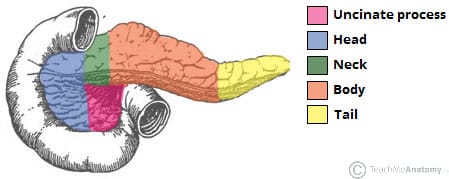
The pancreas has **four parts**: Head, Neck, Body, and Tail (some add the **Uncinate Process** as a separate part).
### 1. HEAD OF THE PANCREAS
- The **broadest and thickest part**
- Lies within the **concavity of the C-loop of the duodenum** (D1, D2, D3)
- Flattened anteroposteriorly
- **Relations:**
| Relation | Structure |
|---|---|
| **Anterior** | Transverse colon and transverse mesocolon; root of small bowel mesentery; gastroduodenal artery |
| **Posterior** | IVC, right renal vein, right crus of diaphragm, common bile duct (embedded in posterior surface or groove), aorta |
| **Medial (left)** | Superior mesenteric vessels (SMV/SMA emerge from between the head/uncinate and the neck) |
| **Lateral (right)** | Duodenum (D1, D2, D3) |
| **Superior** | First part of duodenum (D1), portal vein (formed behind the neck) |
| **Inferior** | Third part of duodenum (D3) |
#### UNCINATE PROCESS
- A hook-like projection extending from the **lower and left part of the head**
- Projects **behind the superior mesenteric vessels** (SMA and SMV)
- Derived from the **ventral pancreatic bud** embryologically
**Relations of the uncinate process:**
- Anterior: SMA and SMV
- Posterior: Aorta, IVC
- Superior: Splenic vein (as it joins the SMV to form the portal vein)
- Inferior: Third part of duodenum (D3)
### Clinical: Pancreatic Head Tumors and Obstructive Jaundice
- Tumors of the **head of the pancreas** (most commonly **pancreatic ductal adenocarcinoma**) cause compression/obstruction of the **intrapancreatic portion of the common bile duct**, leading to:
- **Painless, progressive, obstructive jaundice** (conjugated hyperbilirubinemia)
- **Courvoisier's law**: "In the presence of jaundice, if the gallbladder is palpable, the jaundice is unlikely to be due to gallstones." (Because gallstones cause chronic inflammation → fibrosis → non-distensible gallbladder, whereas pancreatic head tumors cause gradual obstruction → the gallbladder distends progressively)
- Dark urine (cola-colored), pale/clay-colored stools (acholic stools), pruritus
- Steatorrhea (fat malabsorption due to lack of bile in the intestine)
- **Whipple's procedure (pancreaticoduodenectomy)**: The classic operation for resectable pancreatic head tumors — removes the head of the pancreas, duodenum, distal common bile duct, gallbladder, and often the distal stomach (in classic Whipple; pylorus-preserving Whipple preserves the stomach)
- **Reconstructions (3 anastomoses):**
1. **Pancreaticojejunostomy** (or pancreaticogastrostomy) — connecting the remnant pancreas to the jejunum
2. **Hepaticojejunostomy** — connecting the common hepatic duct to the jejunum
3. **Gastrojejunostomy** (or duodenojejunostomy in pylorus-preserving variant)
### 2. NECK OF THE PANCREAS
- A short (about 2 cm), **constricted portion** connecting the head to the body
- Lies **anterior to** the **superior mesenteric vessels** (SMA and SMV) and the **formation of the portal vein**
- The **portal vein** is formed behind the neck by the union of the SMV and splenic vein
**Relations:**
| Relation | Structure |
|---|---|
| **Anterior** | Pylorus of the stomach (transpyloric plane — L1); lesser sac (omental bursa) |
| **Posterior** | Formation of the portal vein; beginning of the portal vein; SMV; SMA origin |
| **Superior** | Celiac trunk origin (from aorta at T12/L1) |
### Clinical: Portal Vein Formation and Pancreatic Cancer
Because the portal vein is formed immediately behind the neck of the pancreas, tumors of the pancreatic head/neck may invade or compress the portal vein and/or the SMV. **Involvement of the SMA (>180° encasement) is a criterion for unresectability** in pancreatic cancer staging.
However, involvement of the SMV/portal vein alone may still be resectable with **vascular resection and reconstruction** in experienced centers (borderline resectable pancreatic cancer).
### 3. BODY OF THE PANCREAS
- The longest part
- Elongated and triangular in cross-section
- Lies to the left of the neck, across the **aorta** and **L2 vertebra**
**Surfaces (three):**
| Surface | Relations |
|---|---|
| **Anterior** | Stomach (separated by the lesser sac/omental bursa); transverse mesocolon attached to its anterior inferior margin |
| **Posterior** | Aorta, left crus of diaphragm, left suprarenal gland, left kidney, left renal vessels, splenic vein (runs along posterior surface), SMA origin |
| **Inferior** | Duodenojejunal flexure, loops of jejunum, left colic flexure |
**Borders (three):**
| Border | Details |
|---|---|
| **Superior** | Splenic artery runs along this border (tortuous course); also related to celiac trunk, celiac lymph nodes |
| **Anterior** | Attachment of the transverse mesocolon |
| **Inferior** | Inferior mesenteric vein runs behind/along this border |
### Clinical: The Omental Bursa (Lesser Sac) and Pancreatic Disease
The **lesser sac** lies directly anterior to the body of the pancreas, separated only by the peritoneum. Therefore:
- **Acute pancreatitis** causes inflammation that spreads into the lesser sac, producing **fluid collections** and potentially **pancreatic pseudocysts** within the lesser sac
- A **pancreatic pseudocyst** in the lesser sac can be drained surgically by **cystogastrostomy** (creating a window between the pseudocyst and the posterior wall of the stomach — both of which abut the lesser sac)
### 4. TAIL OF THE PANCREAS
- The narrow, tapered, **left end** of the pancreas
- Relatively mobile (the only part that is somewhat intraperitoneal — it lies within the **splenorenal ligament**)
- Extends to the left and passes between the two layers of the **splenorenal ligament** to the **hilum of the spleen**
- The tail comes within **1-2 cm of the splenic hilum** in most individuals (directly contacts the hilum in ~30%)
**Relations:**
| Relation | Structure |
|---|---|
| **Anterior** | Stomach (fundus) |
| **Posterior** | Left kidney (upper pole), left suprarenal gland |
| **Lateral** | Hilum of the spleen |
### Clinical: Tail of Pancreas and Splenectomy
- During **splenectomy**, the tail of the pancreas is closely applied to the splenic hilum → risk of **pancreatic injury** → **pancreatic fistula** (leakage of pancreatic juice) or **pancreatitis**
- Conversely, during **distal pancreatectomy** (for tumors of the body/tail), the spleen is usually removed because its blood supply (splenic artery and vein) passes along the pancreas
---
## PANCREATIC DUCT SYSTEM
### 1. Main Pancreatic Duct (Duct of Wirsung)
- Begins in the **tail** and runs through the body toward the head
- Receives smaller tributary ducts along its length (herringbone pattern)
- In the **head**, it turns inferiorly and posteriorly, merging with the **common bile duct (CBD)** to form the **hepatopancreatic ampulla (ampulla of Vater)**
- The ampulla opens into the **second part of the duodenum (D2)** at the **major duodenal papilla** (papilla of Vater), located on the **posteromedial wall** of D2, about **8-10 cm from the pylorus**
- The ampulla is surrounded by the **sphincter of Oddi** (hepatopancreatic sphincter), which has three components:
1. Sphincter of the bile duct (choledochal sphincter)
2. Sphincter of the pancreatic duct
3. Sphincter of the ampulla
### 2. Accessory Pancreatic Duct (Duct of Santorini)
- A smaller duct draining the **upper part of the head** (and sometimes part of the body)
- Opens into D2 at the **minor duodenal papilla**, located about **2 cm proximal (superior)** to the major papilla
- Communicates with the main duct in most people
- Embryologically drains the **dorsal pancreatic bud**
### Clinical: Pancreas Divisum
- The **most common congenital anomaly** of the pancreas (occurs in ~5-10% of the population)
- Results from **failure of fusion** of the ventral and dorsal pancreatic buds during development
- Consequence: The **major part** of the pancreas (body and tail — from the dorsal bud) drains through the **accessory duct (of Santorini)** via the **minor papilla** (which is smaller)
- The ventral bud (uncinate process and inferior head) drains through the main duct via the major papilla
- Most individuals are **asymptomatic**
- In some cases, the small size of the minor papilla creates relative **drainage obstruction** → recurrent acute pancreatitis or chronic pancreatitis
- Treatment (if symptomatic): **Minor papilla sphincterotomy** (endoscopic or surgical) or stenting
### Clinical: Annular Pancreas
- A ring of pancreatic tissue surrounds the **second part of the duodenum**
- Caused by failure of the ventral bud to rotate properly during development
- Can cause **duodenal obstruction** — presents in neonates with **bilious vomiting** and the **"double bubble" sign** on X-ray (if the annulus is distal to the ampulla; proximal to the ampulla → non-bilious)
- May also present in adults with duodenal obstruction, pancreatitis, or peptic ulceration
- Treatment: **Duodenoduodenostomy** or **duodenojejunostomy** (bypass) — NOT resection of the annular tissue (risk of pancreatic fistula and injury to the common bile duct)
### Clinical: Gallstone Pancreatitis
- A gallstone migrating through the CBD can impact at the **ampulla of Vater** → obstruction of the pancreatic duct → reflux of bile into the pancreatic duct → activation of pancreatic enzymes → **acute pancreatitis**
- This is the **most common cause of acute pancreatitis** (along with alcohol)
- **Gallstone pancreatitis management**: ERCP with sphincterotomy and stone extraction (if CBD stone is present) + cholecystectomy (ideally during the same admission to prevent recurrence)
---
## BLOOD SUPPLY
The pancreas has a **dual blood supply** reflecting its dual embryological origin (ventral and dorsal buds):
### Arterial Supply
#### Head of the Pancreas
The head is supplied by two **arterial arcades** (anterior and posterior pancreaticoduodenal arcades):
**Anterior Pancreaticoduodenal Arcade:**
| Artery | Origin |
|---|---|
| **Anterior superior pancreaticoduodenal artery** | Gastroduodenal artery (from common hepatic artery/celiac trunk) |
| **Anterior inferior pancreaticoduodenal artery** | Inferior pancreaticoduodenal artery (from SMA) |
**Posterior Pancreaticoduodenal Arcade:**
| Artery | Origin |
|---|---|
| **Posterior superior pancreaticoduodenal artery** | Gastroduodenal artery |
| **Posterior inferior pancreaticoduodenal artery** | Inferior pancreaticoduodenal artery (from SMA) or directly from SMA |
This dual supply from the **celiac trunk** (via gastroduodenal artery) and the **SMA** makes the head of the pancreas and the duodenum a **"watershed" zone** between the two arterial territories.
### Clinical: Why the Duodenum and Pancreatic Head Are Resected Together
The duodenum and the head of the pancreas share a **common blood supply** (the pancreaticoduodenal arcades). Therefore:
- You **cannot resect the head of the pancreas without resecting the duodenum** (and vice versa) — because the shared blood supply would leave one ischemic
- This is the anatomical basis for the **Whipple procedure** (pancreaticoduodenectomy)
- Similarly, in **duodenal injuries** or tumors, the pancreatic head must be considered
#### Body and Tail of the Pancreas
| Artery | Origin |
|---|---|
| **Dorsal pancreatic artery** | Splenic artery, celiac trunk, SMA, or common hepatic artery |
| **Greater pancreatic artery (arteria pancreatica magna)** | Splenic artery (largest branch to the pancreatic body) |
| **Transverse (inferior) pancreatic artery** | Branch of dorsal pancreatic artery; runs along the inferior border |
| **Caudal pancreatic artery** | Splenic artery or left gastroepiploic artery; supplies the tail |
| **Multiple small branches** | From the splenic artery along the superior border of the body |
### Venous Drainage
- Veins correspond to the arteries
- **Head**: Anterior and posterior pancreaticoduodenal veins → drain into the **SMV** (anterior) and **portal vein** (posterior)
- **Body and tail**: Drain into the **splenic vein** (which runs along the posterior surface of the body)
- Ultimately all drain into the **portal vein**
### Clinical: Pancreatic Cancer and Vascular Involvement
Pancreatic cancer (especially of the head/uncinate) frequently involves the:
- **SMV/Portal vein**: May be resected and reconstructed in borderline resectable cases
- **SMA**: Encasement of the SMA >180° is considered **locally advanced/unresectable** in most classification systems
- **Celiac trunk**: Involvement may preclude standard resection; however, the **Appleby procedure** (modified distal pancreatectomy with celiac axis resection) can be performed for body tumors encasing the celiac trunk, relying on retrograde flow through the pancreaticoduodenal arcades from the SMA to supply the liver (via the common hepatic artery)
---
## LYMPHATIC DRAINAGE
The lymphatic drainage of the pancreas is extensive and follows the arterial supply:
### Head
- Drains to **pancreaticoduodenal nodes** (anterior and posterior) → **hepatic nodes** → **celiac nodes** → **cisterna chyli**
- Also drains to **superior mesenteric nodes**
- May also drain to **pyloric nodes**
### Body and Tail
- Drains to **pancreaticosplenic nodes** (along the splenic artery and at the splenic hilum) → **celiac nodes** → **cisterna chyli**
- Also drains to **superior mesenteric nodes** and **para-aortic (lumbar) nodes**
### Clinical: Lymph Node Involvement in Pancreatic Cancer
Pancreatic cancer has a propensity for **early lymphatic spread** — at the time of diagnosis, lymph node metastases are present in **>70% of cases**. This contributes to the poor prognosis of pancreatic cancer (5-year survival ~10% overall; ~20% even after curative resection).
Extended lymphadenectomy during pancreaticoduodenectomy has been studied but has NOT shown survival benefit in randomized trials compared to standard lymphadenectomy.
---
## NERVE SUPPLY
### Sympathetic
- From the **celiac plexus** and **superior mesenteric plexus** (T5-T9 through the greater splanchnic nerve, and T10-T11 through the lesser splanchnic nerve)
- **Pain fibers** travel with the sympathetic nerves
- Pancreatic pain is typically **epigastric**, radiating through to the **back** (because the pancreas is retroperitoneal and lies against the vertebral column/aorta)
### Parasympathetic
- From the **vagus nerve** (posterior vagal trunk)
- Stimulates exocrine secretion (enzyme and bicarbonate secretion)
### Clinical: Celiac Plexus Block
- Severe, intractable pain from **pancreatic cancer** (especially body/tail tumors) can be managed by **celiac plexus neurolysis** — injection of alcohol or phenol into the celiac plexus to destroy the pain fibers
- Performed percutaneously (under CT or fluoroscopic guidance), endoscopically (EUS-guided), or surgically
- Provides significant pain relief in 70-90% of patients with pancreatic cancer
### Clinical: Pancreatic Pain Characteristics
- Pain from the **head** of the pancreas: Referred to the **right side of the epigastrium** and the **right paravertebral area**
- Pain from the **body** of the pancreas: Referred to the **epigastrium** and the **mid-back**
- Pain from the **tail** of the pancreas: Referred to the **left side of the epigastrium** and the **left paravertebral area**
- Pancreatic pain is often **worsened by lying supine** (pancreas compressed against the vertebral column) and **relieved by leaning forward** or sitting in a **"jack-knife" position** — this is a classic feature of chronic pancreatitis and pancreatic cancer
---
## RELATIONS OF THE PANCREAS — COMPREHENSIVE SUMMARY
### Anterior Relations (from right to left)
1. Transverse colon and mesocolon (attached to the anterior surface at the anterior border)
2. Lesser sac (omental bursa) — separates the pancreas from the stomach
3. Stomach (body — through the lesser sac)
### Posterior Relations (from right to left)
1. **Head**: IVC, right renal vein, right crus of diaphragm, CBD (in a groove or tunnel)
2. **Uncinate process**: Aorta, IVC
3. **Neck**: Portal vein formation (SMV + splenic vein), SMV, SMA
4. **Body**: Aorta, SMA origin, left crus of diaphragm, left suprarenal gland, left kidney, left renal vein, splenic vein
5. **Tail**: Left kidney (upper pole)
### Superior Relations
1. Splenic artery (runs along the superior border of the body)
2. Celiac trunk
3. Common hepatic artery (along the head/neck — superior border)
4. Portal vein (behind the neck)
### Inferior Relations
1. Third and fourth parts of the duodenum (D3, D4)
2. Duodenojejunal flexure (ligament of Treitz)
3. Superior mesenteric vessels (crossing anterior to D3)
### Clinical: SMA Syndrome (Wilkie's Syndrome)
The **third part of the duodenum (D3)** passes between the **aorta** posteriorly and the **SMA** anteriorly. The angle between the aorta and SMA is normally 38-65° with the mesenteric fat pad cushioning D3.
In conditions of **extreme weight loss** (anorexia nervosa, severe burns, prolonged bed rest, body casting for scoliosis), the **mesenteric fat pad** is lost → the angle narrows → **compression of D3** between the SMA and aorta → **duodenal obstruction**.
Symptoms: Postprandial epigastric pain, nausea, bilious vomiting, weight loss
Diagnosis: CT scan showing reduced aortomesenteric angle (<25°) and distance (<8 mm)
Treatment: Nutritional rehabilitation (to restore mesenteric fat), positional changes (left lateral decubitus or prone position), surgery (duodenojejunostomy or Strong's procedure) if conservative management fails
---
## DEVELOPMENT OF THE PANCREAS
The pancreas develops from **two buds** that arise from the **foregut endoderm**:
### 1. Ventral Pancreatic Bud (Smaller)
- Arises from the **hepatic diverticulum** (same origin as the liver and gallbladder)
- Initially lies ventral to the duodenum (D2)
- **Rotates clockwise** (from the ventral to the dorsal side) during the rotation of the duodenum
- Forms the **uncinate process** and the **inferior part of the head**
- Its duct becomes the **main pancreatic duct (duct of Wirsung)** — the distal/terminal portion
### 2. Dorsal Pancreatic Bud (Larger)
- Arises directly from the **dorsal wall of the duodenum** (D2)
- Forms the **body, tail, and the superior part of the head**
- Its duct becomes the **proximal portion of the main pancreatic duct** (in the body and tail) and the **accessory duct (duct of Santorini)**
### Fusion
- The two buds normally fuse during **week 6-7** of development
- The duct systems also fuse — the proximal part of the dorsal duct joins the ventral duct to form the **main pancreatic duct**
- The proximal part of the dorsal bud's duct (from the dorsal bud to the duodenum) persists as the **accessory duct (of Santorini)** opening at the minor papilla
### Clinical: Ectopic Pancreatic Tissue (Pancreatic Heterotopia)
- Pancreatic tissue found outside the pancreas — most commonly in the:
- **Stomach** (antrum/prepyloric region — most common site)
- **Duodenum** (second most common)
- **Jejunum**
- **Meckel's diverticulum**
- **Ileum**
- Usually asymptomatic; found incidentally
- May cause **ulceration**, **bleeding**, **obstruction**, or rarely **malignant transformation**
- On upper GI endoscopy/barium study: appears as a **submucosal nodule** with a **central umbilication** (the ductal opening)
---
## PERITONEAL RELATIONS
- The pancreas is **secondarily retroperitoneal** (originally intraperitoneal; became retroperitoneal during fetal development when the dorsal mesogastrium fused with the posterior body wall)
- The **anterior surface of the body** is covered by peritoneum (forming the posterior wall of the lesser sac)
- The **transverse mesocolon** is attached to the **anterior surface** of the pancreas along a line from the inferior border of the body
- The **tail** is relatively mobile and lies within the **splenorenal ligament** (somewhat intraperitoneal)
- The **head** is largely covered by peritoneum anteriorly but is retroperitoneal
---
## IMPORTANT CLINICAL CONDITIONS OF THE PANCREAS
### 1. Acute Pancreatitis
- Causes (mnemonic: **I GET SMASHED**):
- **I** — Idiopathic
- **G** — Gallstones (most common cause with alcohol)
- **E** — Ethanol (alcohol — chronic use)
- **T** — Trauma
- **S** — Steroids
- **M** — Mumps (and other infections)
- **A** — Autoimmune
- **S** — Scorpion sting (Tityus trinitatis), surgery (post-ERCP)
- **H** — Hyperlipidemia, hypothermia, hypercalcemia, hyperparathyroidism
- **E** — ERCP
- **D** — Drugs (azathioprine, valproic acid, thiazides, sulfonamides)
- **Anatomical basis of clinical features:**
- **Grey Turner's sign**: Bluish discoloration of the **flanks** — retroperitoneal hemorrhage tracking along the fascial planes to the lateral abdominal wall
- **Cullen's sign**: Bluish discoloration of the **periumbilical region** — hemorrhage tracking along the falciform ligament to the umbilicus
- **Fox's sign**: Ecchymosis of the **inguinal ligament** area
- These signs indicate **hemorrhagic (necrotizing) pancreatitis** — severe disease with high mortality
- Pain radiating to the back (retroperitoneal location)
- Pain relieved by leaning forward
### 2. Chronic Pancreatitis
- Progressive inflammatory disease with irreversible fibrosis
- Most common cause: **Alcohol** (70-80%)
- Leads to:
- **Exocrine insufficiency**: Steatorrhea (fat malabsorption), weight loss (occurs when >90% of exocrine function is lost)
- **Endocrine insufficiency**: Diabetes mellitus (type 3c — pancreatogenic diabetes)
- **Chronic pain**: Due to neural inflammation and increased intraductal pressure
- **Pancreatic calcifications**: Visible on X-ray/CT — pathognomonic
- **CBD obstruction**: Fibrosis of the head can compress the intrapancreatic CBD → obstructive jaundice
- **Duodenal obstruction**: Severe fibrosis of the head (rare — "groove pancreatitis")
- **Splenic vein thrombosis**: Inflammation of the body/tail → sinistral portal hypertension → gastric varices
- **Pseudocyst formation**: Collection of pancreatic secretions contained by a wall of fibrous tissue (no epithelial lining — distinguishing it from a true cyst)
### 3. Pancreatic Cancer
- **Pancreatic ductal adenocarcinoma** (PDAC) is the most common type (~85-90%)
- **Location**: 60-70% in the head, 15-20% in the body, 5-10% in the tail
- **Risk factors**: Smoking, chronic pancreatitis, family history, obesity, diabetes mellitus, BRCA2 mutation
- **Prognosis**: One of the most lethal cancers — overall 5-year survival ~10%
- **Presentation varies by location:**
- **Head**: Painless obstructive jaundice (60-70%), weight loss, Courvoisier gallbladder, clay-colored stools, dark urine
- **Body/Tail**: Insidious, often presents late with pain, weight loss, and sometimes **Trousseau's sign** (migratory thrombophlebitis — paraneoplastic hypercoagulable state)
- **New-onset diabetes** in a thin elderly patient may be the first sign
- **Staging**: CT scan with pancreas protocol (triple-phase contrast-enhanced CT) — assesses vascular involvement (SMA, celiac, SMV/portal vein)
- **CA 19-9**: Tumor marker (not specific; elevated in biliary obstruction of any cause)
### 4. Pancreatic Pseudocyst
- Collection of pancreatic fluid (rich in amylase/lipase) enclosed by a wall of **fibrous tissue and granulation tissue** (NO epithelial lining)
- Usually occurs 4-6 weeks after acute pancreatitis or in chronic pancreatitis
- Usually located in the **lesser sac** (posterior to the stomach)
- If >6 cm or symptomatic → drainage:
- **Endoscopic**: EUS-guided cystogastrostomy or cystoduodenostomy
- **Surgical**: Cystogastrostomy (if pseudocyst abuts the posterior wall of the stomach), cystojejunostomy (Roux-en-Y)
- **Percutaneous drainage**: Reserved for infected pseudocysts or when endoscopic/surgical drainage is not possible
### 5. Pancreatic Neuroendocrine Tumors (PNETs)
- Arise from the **islets of Langerhans** (endocrine pancreas)
- Types:
| Tumor | Hormone | Syndrome | Location (most common) |
|---|---|---|---|
| **Insulinoma** | Insulin | Hypoglycemia (Whipple's triad) | Body/tail; usually benign (90%) |
| **Gastrinoma** | Gastrin | Zollinger-Ellison syndrome (peptic ulcers, diarrhea) | **"Gastrinoma triangle"** (junction of cystic duct-CBD, junction of D2-D3, junction of neck-body of pancreas); 60% malignant |
| **Glucagonoma** | Glucagon | Diabetes, necrolytic migratory erythema, weight loss | Body/tail; usually malignant |
| **VIPoma** | VIP | WDHA syndrome (watery diarrhea, hypokalemia, achlorhydria) | Body/tail |
| **Somatostatinoma** | Somatostatin | Diabetes, gallstones, steatorrhea | Head; usually malignant |
---
## SURFACE MARKING OF THE PANCREAS
- The pancreas lies along the **transpyloric plane** (halfway between the jugular notch and the pubic symphysis — at the level of **L1 vertebra**)
- The head lies to the right of the midline, within the C-loop of the duodenum
- The body extends to the left across the midline
- The tail extends toward the **left 10th rib** in the midaxillary line (toward the splenic hilum)
---
## SUMMARY TABLE: COMPARATIVE ANATOMY
| Feature | Liver | Spleen | Pancreas |
|---|---|---|---|
| **Weight** | ~1500 g | ~150 g | ~80-90 g |
| **Location** | Right hypochondrium | Left hypochondrium | Retroperitoneal, epigastrium |
| **Peritoneal status** | Mostly intraperitoneal (except bare area) | Completely intraperitoneal | Retroperitoneal (tail partially intraperitoneal) |
| **Arterial supply** | Hepatic artery (celiac trunk) + Portal vein | Splenic artery (celiac trunk) | Celiac trunk + SMA branches |
| **Venous drainage** | Hepatic veins → IVC | Splenic vein → Portal vein | Into splenic vein and SMV → Portal vein |
| **Lymphatic drainage** | Hepatic nodes → Celiac nodes | Pancreaticosplenic nodes → Celiac nodes | Pancreaticoduodenal/pancreaticosplenic nodes → Celiac nodes |
| **Nerve supply** | Celiac plexus (sympathetic) + Vagus (parasympathetic) | Celiac/splenic plexus | Celiac/superior mesenteric plexus + Vagus |
| **Development** | Foregut (hepatic diverticulum) + Septum transversum | Dorsal mesogastrium (mesoderm) | Foregut (ventral + dorsal pancreatic buds) |
| **Most common associated cancer** | Hepatocellular carcinoma; metastases (most common malignancy) | Lymphoma (most common primary splenic malignancy) | Ductal adenocarcinoma |
---
## ADDITIONAL CLINICAL PEARLS
### Trousseau's Sign of Malignancy
- Migratory superficial thrombophlebitis
- Strongly associated with **pancreatic cancer** (body/tail) and other mucin-secreting adenocarcinomas
- Due to paraneoplastic hypercoagulable state (tumor produces procoagulant factors)
### Virchow's Node (Troisier's Sign)
- Enlarged left supraclavicular lymph node
- May be a sign of metastatic **gastric**, **pancreatic**, or other abdominal malignancy
- Located at the junction of the thoracic duct with the left subclavian vein
### Sister Mary Joseph's Nodule
- Metastatic deposit at the **umbilicus** (via peritoneal spread or through lymphatics in the falciform ligament/round ligament)
- Associated with **gastric**, **pancreatic**, **ovarian**, and other intra-abdominal cancers
### Krukenberg Tumor
- Metastatic tumor of the **ovary** — transcoelomic spread from **gastric** or other GI cancers (including pancreatic)
- Signet ring cells with mucin
---
This comprehensive review covers the **gross anatomy**, **relations**, **blood supply**, **lymphatic drainage**, **nerve supply**, **development**, **peritoneal relations**, **surface markings**, and **all major clinical correlations** for the liver, spleen, and pancreas. Each section integrates anatomical details with their surgical and clinical significance, providing a thorough understanding of these vital abdominal organs.
Rewrite the content as it is except the last ending paragraph that says this covers that that. Plus add images along the content from your side