
Cytitis in 5 year old
cystitis urinary tract infection children
Cystitis (Urinary Tract Infection) in a 5-Year-Old Child
Epidemiology & Risk Factors
- Approximately 1% of boys and 3% of girls are diagnosed with a UTI before puberty
- Females have 3x higher risk than males; uncircumcised males have 4-20x higher risk than circumcised males
- African American children have half the risk compared to non-black children
- History of a previous UTI doubles the risk
- Fever >39°C and duration >48 hours in girls also increases risk
Causative Organisms
- Escherichia coli is by far the most common cause in all pediatric age groups (accounts for ~80%)
- Other organisms: Klebsiella, Proteus mirabilis (especially in boys; associated with urolithiasis), Enterococcus, Staphylococcus saprophyticus (adolescent girls)
- Adenovirus (types 11, 21) can cause hemorrhagic (viral) cystitis - typically self-limiting
Clinical Presentation in a 5-Year-Old
| Symptom | Notes |
|---|---|
| Dysuria (painful/burning urination) | Most common complaint |
| Urinary frequency / urgency | Hallmark of bladder irritation |
| Suprapubic pain/tenderness | Dysuria + suprapubic tenderness = classic cystitis constellation |
| Malodorous or cloudy urine | Common finding |
| New enuresis / incontinence | Can be presenting sign |
| Low-grade fever | May or may not be present; high fever more suggestive of upper tract |
Diagnosis
- In toilet-trained children (age 5), a midstream clean-catch urine is acceptable
- Avoid bag specimens for culture (high contamination rate)
| Test | Sensitivity | Specificity | Notes |
|---|---|---|---|
| Leukocyte esterase | 83% | 78% | Indicates pyuria |
| Nitrite | 53% | 98% | High specificity; positive = very likely UTI |
| Both positive | ~99% | 70% | Best combined screen |
- Pyuria (>5 WBC/hpf) + bacteriuria = strong presumptive diagnosis
- Nitrite may be falsely negative if urine has not been in bladder long enough
- A single midstream specimen with ≥10^5 CFU/mL confirms UTI
- Always obtain culture before starting antibiotics - it sterilizes the urine within hours and guides definitive therapy
Treatment
- A 2-4 day course is acceptable for afebrile cystitis per some guidelines
| Antibiotic | Dose | Notes |
|---|---|---|
| Trimethoprim-sulfamethoxazole (TMP-SMX) | 6-12 mg/kg/day TMP component PO in 2 divided doses | Check local resistance - many E. coli strains now resistant |
| Cephalexin (1st gen cephalosporin) | 25-50 mg/kg/day PO in 3-4 divided doses | Good first-line option |
| Cefixime (3rd gen cephalosporin) | 8 mg/kg/day PO once daily | Excellent coverage |
| Nitrofurantoin | 5-7 mg/kg/day in 4 divided doses | Only for lower tract (cystitis) - NOT for febrile UTI/pyelonephritis; avoid if renal involvement suspected |
| Amoxicillin-clavulanate | 25-45 mg/kg/day in 2-3 divided doses | Broad coverage but high resistance rates for plain amoxicillin |
Important: Fluoroquinolones should only be used in children if sensitivities indicate no other options, due to cartilage toxicity concerns. Local/regional antibiograms should guide empirical choice due to evolving resistance patterns.
When to Admit / Use IV Antibiotics
- High fever (febrile UTI suggesting pyelonephritis)
- Vomiting / unable to tolerate oral medications
- Toxic appearance, dehydration, sepsis
- Underlying immunocompromise or known urological anomaly
Imaging & Follow-up
- First febrile UTI: Renal-bladder ultrasound is recommended to rule out structural anomalies (hydronephrosis, obstructive uropathy, renal abscess)
- Voiding cystourethrogram (VCUG): Recommended if ultrasound is abnormal or if recurrent UTIs - to rule out vesicoureteral reflux (VUR)
- For a simple, first episode of afebrile cystitis, extensive imaging is generally not needed but follow-up with urine culture in 2-3 days is advisable
Complications & Sequelae
- 10-30% of children develop at least one recurrent UTI
- Treatment delay of >48 hours significantly increases risk of renal scarring
- Renal scarring is linked to hypertension, proteinuria, preeclampsia risk, and renal insufficiency long-term
- Children with significant bilateral renal scars warrant long-term follow-up
Special Considerations in a 5-Year-Old
- Bowel and bladder dysfunction (BBD): Constipation and voiding dysfunction are very common in this age group and strongly predispose to recurrent UTIs. Must be evaluated and addressed.
- Vesicoureteral reflux (VUR): Should be considered in recurrent or febrile UTIs - antibiotic prophylaxis is recommended in girls with dilating (high-grade) reflux
- Vulvovaginitis / pinworm infestation: Common in this age group and can mimic cystitis symptoms - consider as differential diagnosis
- Asymptomatic bacteriuria (ASB): In preschool girls without VUR, ASB does not require antibiotics as it carries low risk of renal damage
Mesentric adenitis
mesenteric adenitis children diagnosis
mesenteric adenitis lymph nodes ultrasound
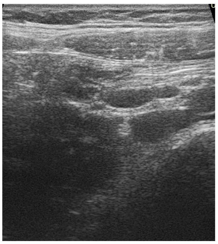
This abdominal ultrasound image demonstrates findings consistent with mesenteric adenitis. The primary diagnostic focus is on multiple enlarged lymph nodes located within the mesentery. These nodes appear as oval to rounded hypoechoic structures with relatively well-defined borders. Surrounding the clustered lymphadenopathy, the mesenteric fat exhibits increased echogenicity (hyperechoic appearance) and a somewhat ill-defined texture, which is a characteristic sonographic sign of localized inflammation or edema. There is a lack of significant fluid collection in the visualized field. The image illustrates the typical reactive appearance of mesenteric lymph nodes in a pediatric or adolescent clinical context, often associated with systemic inflammatory responses or localized gastrointestinal triggers. This diagnostic image serves as a clinical example of adenomesenteritis, showcasing the use of ultrasound in evaluating acute abdominal pain through the identification of lymph node morphology and mesenteric fat changes.
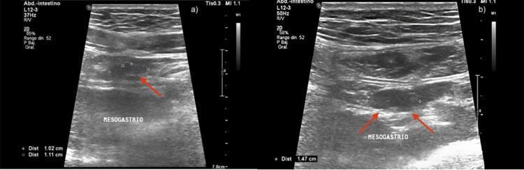
Two grayscale abdominal ultrasound images (a and b) demonstrating clinical findings consistent with mesenteric adenitis. The images focus on the mesogastrium region using a high-frequency linear transducer. Image (a) reveals multiple clustered, hypoechoic, oval-shaped structures representing enlarged mesenteric lymph nodes, with electronic calipers measuring approximately 1.02 cm and 1.11 cm. Red arrows highlight the largest nodes. Image (b) shows a prominent, swollen hypoechoic lymph node with a measurement of 1.47 cm. Trace anechoic free fluid is visible in the surrounding peritoneal space, particularly noted in the clinical context of the right iliac fossa. The lack of appendiceal wall thickening or surrounding fat stranding helps differentiate this from acute appendicitis. This diagnostic imaging serves as a pedagogical example for identifying pediatric or adolescent mesenteric lymphadenopathy and evaluating non-specific abdominal pain.
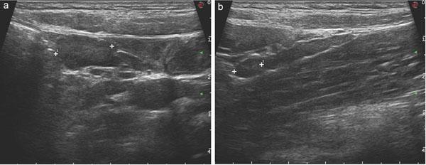
Two grayscale ultrasound images (labeled 'a' and 'b') of the abdominal cavity demonstrating findings consistent with mesenteric adenitis (adenomesenteritis), a common cause of acute abdomen in pediatric patients. In view 'a', multiple enlarged, hypoechoic lymph nodes are visible within the mesentery. These nodes are clustered and exhibit a characteristic chain-like arrangement, showing well-defined rounded to oval shapes with loss of the normal fatty hilum. View 'b' provides a detailed view of a specific enlarged mesenteric lymph node, delineated by caliper markers for measurement. The surrounding mesenteric fat appears slightly hyperechoic (inflamed). The imaging modality highlights the lack of radiation exposure, making it the preferred diagnostic tool for differentiating mesenteric lymphadenopathy from acute appendicitis in children. Key features illustrated include lymph node morphology, size, and spatial distribution along mesenteric vessels.
Mesenteric Adenitis (Mesenteric Lymphadenitis)
Intraoperative Appearance
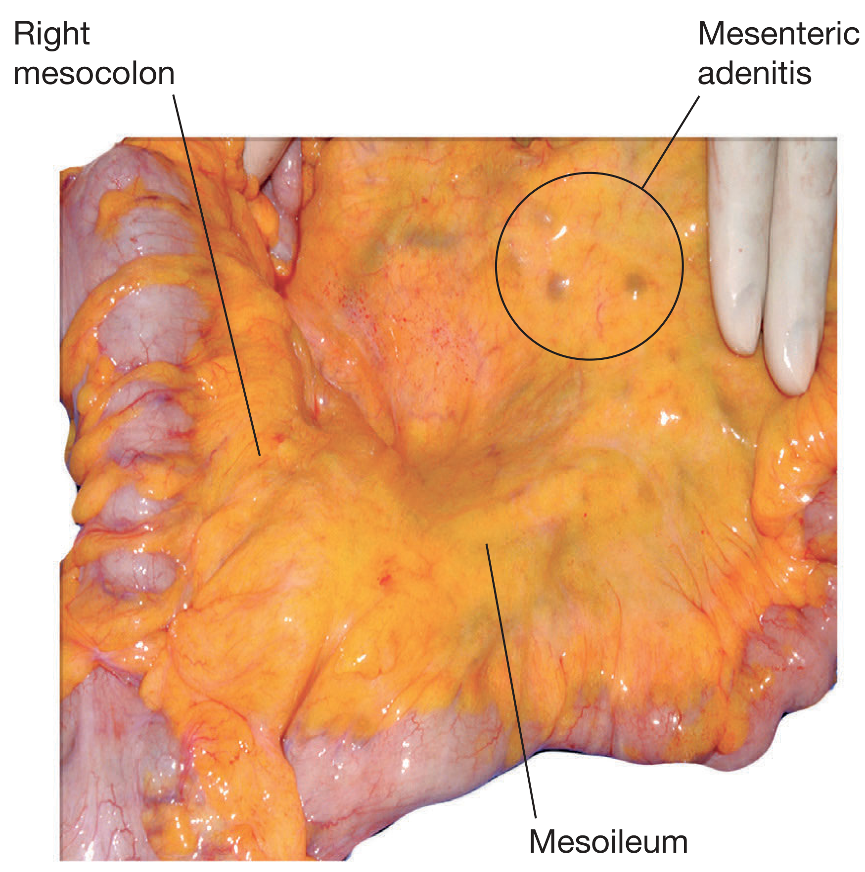
Anatomy & Pathophysiology
- The mesentery is rich in lymphatic tissue, and the ileocaecal region contains the highest concentration - hence adenitis predominates here
- Swelling of ileocaecal lymph nodes causes capsular stretch, producing somatic pain in the right iliac fossa (RIF)
- The nodes enlarge in response to infection draining from the terminal ileum, colon, or via systemic hematogenous spread
Causes
| Type | Causes |
|---|---|
| Non-specific viral (most common) | Adenovirus, EBV, enteroviruses; often follows URTI |
| Bacterial | Yersinia enterocolitica, Yersinia pseudotuberculosis, Campylobacter spp., group A Streptococcus, Mycobacterium tuberculosis |
- Yersinia pseudotuberculosis - produces acute appendicitis-like syndrome; the most classic bacterial cause. At laparotomy, the appendix is usually normal
- Yersinia enterocolitica - causes acute/chronic gastroenteritis AND mesenteric lymphadenitis; especially in infants and children; can produce reactive arthritis (via HLA-B27 molecular mimicry) and erythema nodosum
- SARS-CoV-2 - mesenteric adenitis is now recognized as not uncommon with COVID-19 infection
Clinical Features
| Feature | Detail |
|---|---|
| Age | School-age children and adolescents; classic pediatric condition |
| Pain | Short attacks of central/periumbilical abdominal pain, 10-30 minutes each; may localize to RIF |
| Vomiting | Common |
| Fever | Present in >50% of cases; may be markedly elevated |
| General appearance | Patient seldom looks ill (key differentiator from appendicitis) |
| Prior illness | Often preceded by sore throat / upper respiratory tract infection |
| Lymphadenopathy | Cervical, axillary, or inguinal nodes may be enlarged - always palpate |
- Abdominal tenderness is poorly localised
- Shifting tenderness (tenderness moves as position changes) - valuable sign distinguishing mesenteric adenitis from appendicitis
- No true peritoneal signs (no guarding, no rebound) - this is the critical differentiator from appendicitis
Differentiating from Acute Appendicitis
| Feature | Mesenteric Adenitis | Acute Appendicitis |
|---|---|---|
| Prior URTI | Common | Uncommon |
| Pain character | Colicky, migratory | Progressive, constant |
| Pain localization | Poorly localized, shifting | Progressively localizes to RIF |
| General appearance | Not ill | Increasingly unwell |
| Peritoneal signs | Absent | Present (guarding, rebound) |
| Cervical lymph nodes | Often palpable | Not enlarged |
| WBC (day 1) | Elevated 10,000-12,000/µL | Elevated, sustained |
| WBC (day 2) | Falls | Remains elevated / rises |
| Ultrasound | Enlarged lymph nodes, normal appendix | Distended, non-compressible appendix |
Investigations
- Leukocytosis of 10,000-12,000/µL on day 1 - importantly, falls by day 2 (helpful differentiating feature from appendicitis where it persists)
- CRP may be elevated
- Multiple enlarged (>10 mm short axis) hypoechoic, oval-to-rounded lymph nodes clustered in the mesentery
- Surrounding hyperechoic mesenteric fat (inflammation)
- Normal-calibre appendix (critical for excluding appendicitis)
- May show trace free fluid
- Reserved for equivocal cases where ultrasound is non-diagnostic
- Shows enlarged mesenteric nodes + normal appendix
- Sometimes required when appendicitis cannot be excluded clinically or radiologically
Microbiology Workup (for bacterial cases)
- Stool culture for Yersinia, Campylobacter, Salmonella
- Serology for Yersinia (especially if reactive arthritis or erythema nodosum present)
- Throat swab for Group A Streptococcus
- Monospot / EBV serology if glandular fever suspected
- Mantoux / IGRA if TB exposure risk
Treatment
- Supportive treatment only - analgesia, hydration, rest
- Resolves spontaneously but can recur
- No antibiotics needed
- Reassure parents and discharge with safety-net advice
- Symptoms can be severe (cramping pain, vomiting, diarrhoea)
- May require hospitalisation for IV fluids and supportive care
- Antibiotics directed at causative organism:
- Yersinia: ciprofloxacin or TMP-SMX (though most cases self-resolve)
- Campylobacter: azithromycin or ciprofloxacin
- Group A Streptococcus: amoxicillin or penicillin
- TB: standard anti-TB regimen
- Ileocaecal mesenteric adenitis is a feature of ileocolic Crohn's disease
- Mesentery is thickened, oedematous, shortened - "fat wrapping" / creeping fat is pathognomonic
- Managed as part of the underlying IBD
Complications
- Recurrence - viral adenitis commonly recurs
- Can be a presentation of underlying Crohn's disease (in recurrent cases)
- Yersinia complications: reactive arthritis, erythema nodosum, iritis (especially HLA-B27 positive patients)
- Tuberculous mesenteric lymphadenitis: calcified nodes visible on plain X-ray; may present with constitutional symptoms (fever, weight loss)
- Intussusception: enlarged mesenteric nodes can act as a lead point, especially in children
Key Summary Points
- Mesenteric adenitis is the #1 mimic of appendicitis - always consider it in a child with RIF pain
- The shifting tenderness and absent peritoneal signs are the most valuable clinical differentiators
- WBC that falls on day 2 supports mesenteric adenitis over appendicitis
- Ultrasound is the investigation of choice - look for multiple enlarged hypoechoic nodes with a normal appendix
- Treat viral form with supportive care only; bacterial forms may need antibiotics and occasionally hospitalisation
- Always check for Yersinia in persistent or recurrent cases with reactive arthritis/erythema nodosum
Precautions and food to be taken for both conditions
diet precautions cystitis UTI children 5 year old food to avoid
mesenteric adenitis diet food precautions children recovery
https://www.niddk.nih.gov/health-information/urologic-diseas…
Precautions & Dietary Guide for Both Conditions in a 5-Year-Old
PART 1 - CYSTITIS (Bladder Infection / UTI)
Diet - What TO Give
| Food / Drink | Why It Helps |
|---|---|
| Water (most important) | Flushes bacteria out of the urinary tract; prevents concentration of urine that bacteria thrive in |
| Berries (cranberries, blueberries, raspberries) | Contain proanthocyanidins - a compound that prevents bacteria (especially E. coli) from sticking to the bladder wall |
| High-fibre foods (whole grains, oats, lentils, fruits, vegetables) | Prevent constipation, which is a direct risk factor for UTI in children |
| Yogurt with live cultures (probiotics) | Supports healthy gut and perineal flora, reducing colonisation with uropathogenic bacteria |
| Cucumber, watermelon | High water content; naturally increases fluid intake |
| Diluted fresh fruit juices | Mild vitamin C content may acidify urine slightly, making it less hospitable to bacteria |
Diet - What to AVOID
| Food / Drink | Reason to Avoid |
|---|---|
| Sugary drinks, sweets, fruit concentrates | High sugar feeds bacterial growth and suppresses immune response |
| Spicy foods | Irritate the bladder lining and worsen dysuria |
| Carbonated drinks (cola, soda) | Acidic + caffeine content irritates the bladder |
| Caffeinated drinks (tea, chocolate milk in excess) | Diuretic effect + bladder irritant |
| Citrus fruits in excess (orange, lemon) | Can irritate a sensitive inflamed bladder |
| Processed/packaged snacks | High salt content concentrates urine |
Precautions for Cystitis
- Wipe front to back after every toilet visit - never back to front; this is the single most important hygiene measure to prevent faecal bacteria reaching the urethra
- Wash the perineal area with plain warm water daily; avoid bubble baths, scented soaps, and bath salts as these irritate the urethra
- Change underwear daily; use cotton underwear - avoid tight synthetic fabrics that trap moisture
- Teach the child to urinate regularly - every 2-3 hours; do not "hold" urine for long periods
- Ensure complete bladder emptying - instruct double voiding (urinate, wait a moment, try again)
- Never rush on the toilet; relaxed voiding posture (feet supported) helps complete emptying
- Urinate after swimming in pools (chlorine + prolonged moisture)
- Treat constipation promptly - a loaded bowel compresses the bladder and urethra, directly increasing UTI risk
- High-fibre diet + adequate fluids to maintain soft, regular stools
- If constipation is recurrent, discuss with the doctor (lactulose, stool softeners)
- Avoid tight jeans, leggings for prolonged periods during active infection
- Change out of wet swimwear promptly
- Complete the full antibiotic course even if the child feels better in 2-3 days
- Do not self-stop antibiotics - incomplete treatment leads to recurrence and resistance
- Return for urine culture follow-up in 2-3 days to confirm clearance
- Fever returns or worsens after starting antibiotics
- Child is vomiting and cannot take oral medication
- No improvement after 48 hours of antibiotics
- New loin/flank pain or back pain (suggests spread to kidney)
PART 2 - MESENTERIC ADENITIS
Diet - What TO Give
| Food / Drink | Why It Helps |
|---|---|
| Plain water and oral rehydration solution (ORS) | Restores fluids lost through vomiting/diarrhoea; prevents dehydration |
| Toast, plain crackers, rice cakes | Easy to digest; gentle on the stomach |
| Bananas | Soft, easy to digest; replenishes potassium lost through vomiting; naturally soothing |
| Boiled/steamed rice | Bland, low-residue; easy on inflamed bowel |
| Boiled potatoes (plain, no butter) | Gentle starch; easily tolerated |
| Steamed vegetables (carrots, zucchini, pumpkin) | Soft, low-fibre options that are easy to digest |
| Plain boiled chicken or fish | Light protein source; easy to digest |
| Clear soups / broths | Provides hydration, electrolytes, and mild nutrition |
| Yogurt (plain) | Probiotics support gut recovery, especially if antibiotics are being used for bacterial adenitis |
| Applesauce | The BRAT diet element; easy to digest, gentle |
Follow the BRAT diet principle during the acute phase: Bananas, Rice, Applesauce, Toast - these are the foundation for recovery
Diet - What to AVOID
| Food / Drink | Reason to Avoid |
|---|---|
| Fatty / greasy / fried foods | Slow digestion; worsen nausea and abdominal discomfort |
| Spicy foods | Irritate the bowel and worsen cramping |
| Milk and dairy products (temporarily) | Can upset the stomach and worsen nausea during the acute phase; reintroduce once recovered |
| Raw vegetables and salads | High insoluble fibre; harder to digest during active inflammation |
| Whole nuts, seeds, popcorn | Hard to digest; may worsen cramping |
| Sugary drinks, sweets, candy | Worsens diarrhoea (osmotic effect); feeds pathogens |
| Carbonated drinks | Causes bloating and gas that worsens abdominal pain |
| Caffeinated drinks | Dehydrating; irritates the gut |
| Heavy meals | Large portions stress the inflamed gut; prefer small, frequent meals |
Precautions for Mesenteric Adenitis
- Keep the child at home and rested during the acute phase - avoid school until fever-free for 24 hours and pain has settled
- Gentle activity is fine once the child feels better; no need for strict bed rest
- Give paracetamol (15 mg/kg every 4-6 hours) or ibuprofen (10 mg/kg every 6-8 hours with food) for pain and fever
- A warm compress or heat pad on the abdomen can soothe cramping
- Push fluids consistently throughout the day - small sips frequently if nauseous
- If the child is vomiting and not tolerating fluids, seek medical attention (IV fluids may be needed)
- Signs of dehydration to watch for: dry mouth, no tears when crying, no urine for >6-8 hours, sunken eyes
- During vomiting: withhold solid food for 1-2 hours after vomiting; then restart with small sips of ORS/water
- Once vomiting settles: start BRAT foods in small amounts every 2-3 hours
- Gradually reintroduce normal diet over 3-5 days as the child tolerates
- Pain not improving after 3 days
- Pain is getting significantly worse or becomes constant (not colicky/wave-like)
- High fever persisting beyond 3-4 days
- Child develops a rigid, board-like abdomen (peritoneal sign - must rule out appendicitis urgently)
- Vomiting is persistent and child cannot keep fluids down
- Child is unusually lethargic or looks unwell
Side-by-Side Quick Reference
| Cystitis | Mesenteric Adenitis | |
|---|---|---|
| Most important fluid | Lots of water (flush bacteria) | ORS + water (replace losses) |
| Best foods | Berries, high-fibre, yogurt | BRAT diet (banana, rice, toast), broth, boiled chicken |
| Avoid | Sugar, spicy foods, carbonated drinks | Fatty/fried food, dairy (acutely), spicy food, carbonated drinks |
| Key hygiene | Front-to-back wiping, cotton underwear | Handwashing before and after meals, after toilet |
| Activity | Normal (avoid tight clothing) | Rest during fever/pain |
| Key prevention | Avoid constipation, regular voiding | Handwashing, food hygiene to prevent viral/bacterial gut infections |
| Danger sign | Fever + flank pain (pyelonephritis) | Constant (not colicky) pain + rigid abdomen (appendicitis) |