
If a patient is completely normal and all the other blood work is normal but if SGPT is slightly decreased and Total protein is mildly increased in LFT what could be the diagnosis?
low SGPT ALT decreased total protein elevated LFT diagnosis causes
Slightly Decreased SGPT (ALT) + Mildly Increased Total Protein in LFT - in an Otherwise Asymptomatic, Normal Patient
1. Vitamin B6 (Pyridoxine) Deficiency - Most Common Cause of Low ALT
- ALT activity in the blood is reduced, giving a spuriously low or low-normal reading
- AST is also B6-dependent but to a lesser degree
- The liver itself is not diseased - it's a cofactor deficiency artifact
- Patients on isoniazid, hydralazine, or oral contraceptives (B6 antagonists)
- Chronic alcoholics (poor diet + B6 depletion)
- Patients on hemodialysis
- Elderly with poor nutritional intake
- Pregnant women (especially with hyperemesis)
2. Elevated Total Protein - What Does "Mildly Increased" Mean?
"Elevated protein levels in the absence of dehydration are usually related to increased globulin levels... Diffuse elevations in the γ region can occur with chronic infections, liver disease, autoimmune disease..."
| Cause | Mechanism | Clue |
|---|---|---|
| Chronic infection (TB, HIV, hepatitis C, endocarditis, malaria) | Polyclonal IgG/IgM rise | History, travel history |
| Autoimmune disease (SLE, rheumatoid arthritis, Sjogren's) | Polyclonal hyperglobulinemia | ANA, anti-dsDNA |
| Chronic liver disease (early cirrhosis) | Impaired clearance of gut antigens → globulin rise | May have low ALT if hepatocytes are "burnt out" |
| Multiple myeloma / MGUS | Monoclonal paraprotein (M-spike) | SPEP shows monoclonal band |
| Sarcoidosis | Polyclonal hypergammaglobulinemia | Raised ACE, chest X-ray |
| Dehydration | Hemoconcentration | Clinical context, raised hematocrit |
3. The Combined Pattern: Low ALT + High Total Protein Together
A. Early/Compensated Cirrhosis ⭐
- In end-stage fibrosis, there are so few functioning hepatocytes that ALT production is actually reduced - so ALT can be low-normal or even below normal
- Meanwhile, polyclonal hypergammaglobulinemia (from portosystemic shunting of gut antigens) raises the globulin fraction and thus total protein
- The patient may be entirely asymptomatic at this stage
- Key investigation: Albumin, PT/INR, bilirubin, platelet count, ultrasound abdomen, FibroScan
B. Vitamin B6 Deficiency + Concomitant Chronic Infection or Autoimmune Disease
C. MGUS (Monoclonal Gammopathy of Undetermined Significance)
- Monoclonal immunoglobulin raises total protein
- ALT is incidentally low (or within low-normal range)
- Patient is asymptomatic by definition
- Key investigation: Serum protein electrophoresis (SPEP) + immunofixation
D. Chronic Hepatitis C with Advanced Fibrosis
- Chronic HCV can cause polyclonal hypergammaglobulinemia raising total protein
- In late-stage disease, ALT paradoxically normalizes or decreases
- Patient may feel completely well
Recommended Workup
- Repeat LFT in 4-6 weeks (confirm it's not a lab artifact)
- Serum protein electrophoresis (SPEP) - to detect monoclonal or polyclonal hypergammaglobulinemia
- Serum albumin separately - to calculate A:G (albumin:globulin) ratio; a low ratio points to liver disease or chronic inflammation
- Vitamin B6 level (plasma pyridoxal phosphate) - especially if on medications that deplete B6
- Hepatitis B and C serology
- ANA, anti-dsDNA (if autoimmune features suspected)
- Ultrasound abdomen - to look for early cirrhotic changes
- CBC - thrombocytopenia would suggest portal hypertension
Summary
| Finding | Most likely explanation |
|---|---|
| Slightly decreased SGPT (ALT) | Vitamin B6 deficiency (most common) OR "burnt-out" hepatocyte loss in advanced liver disease |
| Mildly increased total protein | Hyperglobulinemia from chronic infection, autoimmune disease, cirrhosis, or MGUS |
| Both together, asymptomatic | Early/compensated cirrhosis, or MGUS, or incidental separate causes |
- Textbook of Family Medicine 9e, p. 840-844 (Total Protein section)
- StatPearls - Liver Function Tests
- WebMD - ALT/SGPT interpretation
The above patient has been having little blood with sputum for the past 3 months . What could be the correlation between the blood analysis and symptoms?
pulmonary tuberculosis chest X-ray cavitary lesion upper lobe
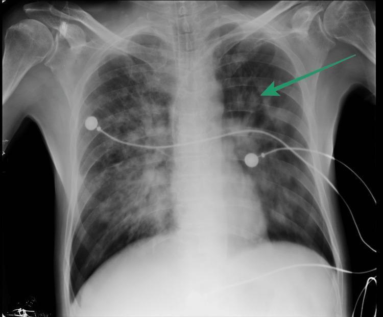
This diagnostic image is an anteroposterior (AP) chest X-ray demonstrating significant pulmonary pathology. The primary finding is a large, well-defined cavitary lesion in the left upper lobe, indicated by a green arrow, which shows increased lucency surrounded by thick, heterogeneous walls. Both lung fields exhibit diffuse, bilateral patchy opacities and infiltrates, more pronounced in the mid and upper zones, suggesting extensive consolidation or inflammatory process consistent with active pulmonary tuberculosis. Extrathoracic structures include radiopaque medical instrumentation: several ECG leads and circular electrode pads are superimposed across the chest cavity. The cardiomediastinal silhouette appears within normal limits for an AP view, though partially obscured by overlying pulmonary pathology. Skeletal structures, including the ribs, clavicles, and humeral heads, are visible. This radiographic presentation is highly characteristic of mycobacterial infection with cavitary disease, serving as a critical educational example of secondary tuberculosis findings in an adult patient.
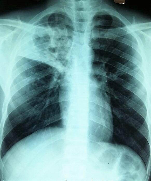
A posterior-anterior (PA) chest X-ray of an adult patient demonstrating significant pulmonary pathology. The most prominent finding is a large, poly-excavated (multi-cavitated) lesion in the right upper lobe. This cavity exhibits irregular, thickened walls with internal lucencies suggesting parenchymal destruction. The lesion appears to cross the midline or involve mediastinal structures, as evidenced by its contralateral extension. In the left lung, particularly within the hilar and axillary regions, there are visible micronodular and infiltrative opacities. These small, scattered densities suggest secondary spread or simultaneous involvement. The overall radiographic presentation is highly characteristic of advanced cavitary pulmonary tuberculosis or other chronic necrotizing infections. Key educational features include the demonstration of a 'destroyed lung' appearance and the distinction between large cavitary lesions and diffuse micronodular patterns (miliary or bronchogenic spread).
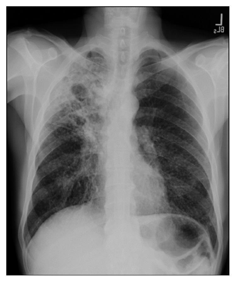
This diagnostic image is a posterior-anterior (PA) chest X-ray demonstrating significant pulmonary pathology. The primary finding is a large, prominent cavitary opacity localized in the right upper lobe. The lesion displays a thick, irregular wall with a central lucency, indicating an air-filled cavity within a region of parenchymal consolidation. Surrounding the main cavity, there are heterogeneous, patchy infiltrates and smaller areas of increased density throughout the right upper and middle lung fields. The left lung appears relatively clear in comparison, though subtle reticulonodular markings are present. The trachea is midline, and the cardiomediastinal silhouette appears within normal limits for this projection. This visual presentation is highly characteristic of secondary (reactivation) tuberculosis or a necrotizing fungal pneumonia. The image serves as a classic educational example of cavitary lung disease, illustrating the destruction of lung parenchyma and the formation of gas-filled spaces often associated with Mycobacterium tuberculosis infection.
Correlation: Blood-Streaked Sputum for 3 Months + Low SGPT + Elevated Total Protein
The Unifying Diagnosis: Pulmonary Tuberculosis
"Patients typically have nonspecific complaints of malaise, weight loss, cough, and night sweats. Sputum may be scant or bloody and purulent. Blood-streaked sputum (hemoptysis) is associated with tissue destruction (e.g., cavitary disease). One or both upper lobes of the lungs are usually involved."
How TB Explains the LFT Pattern
1. Low SGPT (ALT) - The Isoniazid / Vitamin B6 Link
"Isoniazid, a drug commonly used to treat tuberculosis, can induce a vitamin B6 deficiency by forming an inactive derivative with pyridoxal phosphate (PLP). Thus, B6 supplementation is essential for some patients to prevent peripheral neuropathy."
- INH forms a hydrazone complex with pyridoxal phosphate, depleting active B6
- Since ALT requires PLP as a cofactor for its enzymatic activity, B6 depletion directly causes a falsely low or suppressed ALT reading
- The liver is not actually damaged - it is a cofactor artifact
- Chronic TB leads to malnutrition, poor appetite, and wasting
- B6 stores become depleted through poor intake and the metabolic demands of chronic infection
- This similarly reduces ALT activity in blood
2. Elevated Total Protein - The Chronic Infection Immune Response
- Macrophages and T-cells are perpetually activated against Mycobacterium tuberculosis
- This triggers prolonged B-cell stimulation and polyclonal immunoglobulin production (polyclonal hypergammaglobulinemia)
- The IgG, IgM, and IgA fractions of serum globulins all rise
- Since Total Protein = Albumin + Globulins, a rise in globulins elevates total protein - even when albumin is normal
The Mechanistic Summary
Mycobacterium tuberculosis (Chronic Pulmonary Infection)
            |
            ├──► Cavitary lung destruction ──► Hemoptysis / Blood-streaked sputum
            |
            ├──► Chronic immune activation ──► Polyclonal hypergammaglobulinemia
            |         (B-cell stimulation)          ──► ↑ Total Protein
            |
            └──► Vitamin B6 depletion ──► ↓ PLP cofactor for ALT
                  (chronic illness / INH          ──► ↓ SGPT (ALT)
                   treatment)
Why the Patient Appears "Normal" Otherwise
- Early-to-moderate pulmonary TB is notoriously subclinical
- The cough and hemoptysis may be mild enough to be dismissed or attributed to other causes
- Bilirubin, ALP, albumin, and other LFT parameters remain normal because hepatocytes themselves are not being destroyed
- The immune system contains the infection in granulomas, preserving overall function while causing the chronic inflammatory perturbations seen in the blood work
Characteristic Chest X-Ray of Pulmonary TB
Recommended Investigations to Confirm
| Investigation | Expected Finding in TB |
|---|---|
| Sputum AFB smear x 3 | Acid-fast bacilli on Ziehl-Neelsen stain |
| Sputum culture (LJ medium) | M. tuberculosis growth (gold standard) |
| Sputum GeneXpert/CBNAAT | MTB detected, +/- rifampicin resistance |
| Chest X-ray / HRCT chest | Upper lobe cavitation, infiltrates, fibrosis |
| Mantoux (TST) / IGRA | Positive (though IGRA more specific) |
| Serum protein electrophoresis | Polyclonal rise in gamma region |
| Serum vitamin B6 level | Low pyridoxal phosphate |
| CBC | Lymphocytosis, anemia of chronic disease |
| ESR | Elevated (often >50 mm/hr) |
Key Teaching Points
- Low ALT in TB = vitamin B6 (PLP) depletion from either chronic illness or INH therapy, since ALT needs PLP as a cofactor
- Elevated total protein in TB = polyclonal hypergammaglobulinemia from chronic mycobacterial immune stimulation
- Hemoptysis in TB = cavitary disease eroding pulmonary vessels (Rasmussen's aneurysm in severe cases)
- "Normal" appearance is the hallmark of smoldering TB - the disease is insidious and systemic blood work changes precede overt symptoms
- Medical Microbiology 9e (Mycobacterium tuberculosis section, p. 553)
- Lippincott Biochemistry 8e (Vitamin B6 / Isoniazid, p. 1061)
- Harrison's Principles of Internal Medicine 22E (TB Regimens section)
- Textbook of Family Medicine 9e (Total Protein, p. 840-844)