
In ckd there is cmd difference do tell me what is cmd and how cmd difference cSuses in easy way
Reading File
In CKD, CMD stands for Chronic Kidney Disease - Mineral and Bone Disorder (CKD-MBD). Let me search the textbooks for detailed content.
Searching sources...
Finding Sources
Finding Sources
Reading File
Reading File
Reading File
Searching Images
CKD-MBD mineral bone disorder pathophysiology phosphorus calcium PTH vitamin D
I now have rich textbook content. Let me compile a clear, easy-to-understand answer.
CKD-MBD: What It Is and Why It Happens (Explained Simply)
What does CMD mean?
In CKD, CMD = CKD-MBD = Chronic Kidney Disease - Mineral and Bone Disorder
It is a systemic (whole-body) disorder of mineral and bone metabolism that happens because the kidneys are failing. It involves three interconnected problems:
- Abnormal levels of calcium, phosphorus, PTH (parathyroid hormone), or Vitamin D
- Bone abnormalities - weak bones, poor bone remodeling (called "renal osteodystrophy")
- Calcium deposits in blood vessels and soft tissues (vascular calcification)
(National Kidney Foundation Primer on Kidney Diseases, 8e)
Normal Situation First (What the Healthy Kidney Does)
In healthy people, three hormones work together to keep calcium and phosphorus in perfect balance:
| Hormone | Made by | Job |
|---|---|---|
| PTH | Parathyroid glands | Raises blood calcium; lowers phosphorus |
| Calcitriol (active Vitamin D) | Kidneys (activate it) | Absorbs calcium from the gut |
| FGF-23 | Bone cells | Lowers phosphorus; reduces Vitamin D activation |
The kidneys are the master controller of all three.
How CKD Causes MBD - Step by Step (Easy Way)
Think of it as a chain reaction that starts the moment kidneys begin to fail:
Step 1 - Phosphorus starts to build up
- Healthy kidneys excrete phosphorus in urine. When kidneys fail, phosphorus (Pi) accumulates in the blood.
- This rise in phosphorus is the trigger for everything else.
Step 2 - FGF-23 rises as the first alarm
- Bone cells sense high phosphorus and release FGF-23 to try to dump phosphorus in the urine.
- But FGF-23 also blocks Vitamin D activation in the kidneys.
- Result: Active Vitamin D (calcitriol) drops.
Step 3 - Calcitriol (Active Vitamin D) falls
- The failing kidney also loses the ability to activate Vitamin D on its own (independent of FGF-23).
- Less active Vitamin D means less calcium absorbed from the gut.
- Blood calcium begins to fall (hypocalcemia).
Step 4 - PTH skyrockets (Secondary Hyperparathyroidism)
- The parathyroid glands sense low calcium and low Vitamin D, and produce massively elevated PTH.
- High PTH tries to:
- Pull calcium OUT of bones (→ weakens bones)
- Make the kidneys hold onto calcium
- Dump more phosphorus in urine (but with CKD, kidneys can no longer do this well)
- The net result is chronic, uncontrolled PTH elevation = Secondary Hyperparathyroidism.
Step 5 - Bones suffer
- With PTH continuously pulling calcium from bones, bones become thin, weak, and deformed.
- This bone damage = Renal Osteodystrophy.
- Types include: high-turnover (osteitis fibrosa cystica), low-turnover (adynamic bone disease), mixed, and osteomalacia.
Step 6 - Blood vessels calcify
- High phosphorus + high calcium in the blood combine to form calcium-phosphate crystals.
- These deposit in artery walls and soft tissues.
- This causes vascular calcification, stiffening arteries, and dramatically increasing the risk of heart attack and stroke.
- This is a major reason why CKD patients die from cardiovascular disease.
Summary Diagram (Simple Chain)
Kidneys Failing
      ↓
Phosphorus builds up in blood
      ↓
FGF-23 rises → blocks Vitamin D activation
      ↓
Active Vitamin D ↓ → less calcium absorbed from gut
      ↓
Blood Calcium ↓ → Parathyroid glands work overtime
      ↓
PTH ↑↑↑ (Secondary Hyperparathyroidism)
      ↓                        ↓
Calcium pulled from bones    Phosphorus stays high
      ↓                        ↓
Weak bones (Renal          Calcium + Phosphorus
Osteodystrophy)            deposit in arteries
                            (Vascular Calcification)
When Does This Start?
These changes begin even at early CKD and become nearly universal once GFR falls below 30 mL/min (CKD Stage 3b-4). By dialysis (Stage 5D), almost every patient has some degree of CKD-MBD.
(Brenner and Rector's The Kidney, 2-Volume Set)
Why Does It Matter?
CKD-MBD leads to:
- Fractures - hip fractures in dialysis patients are more deadly than in the general population
- Cardiovascular disease - calcified arteries raise the risk of heart attacks
- Increased mortality
Key Lab Findings in CKD-MBD
| Lab | What Happens | Why |
|---|---|---|
| Phosphorus | High | Kidneys can't excrete it |
| Calcium | Low (early) | Gut absorption falls |
| Active Vitamin D | Low | Kidneys can't activate it |
| PTH | Very High | Compensating for low Ca and low Vit D |
| FGF-23 | Very High | Early marker, rises first |
| Alkaline Phosphatase | High | Bone turnover increased |
In short: CKD-MBD is what happens when the kidney, which is the master of calcium-phosphorus balance, stops working properly. The whole system - hormones, bones, and blood vessels - gets thrown off in a predictable cascade.
I saying about cortico medullary difference
Searching sources...
Finding Sources
Finding Sources
Reading File
Finding Sources
Reading File
Reading File
Searching Images
kidney ultrasound corticomedullary differentiation lost CKD chronic kidney disease
Got it! Now I have everything to explain this clearly.
Cortico-Medullary Differentiation (CMD) in CKD - Explained Simply
What is CMD?
CMD = Cortico-Medullary Differentiation
It refers to how clearly you can distinguish (tell apart) the cortex from the medulla of the kidney on ultrasound (USG).
Normal Kidney on Ultrasound - What You See
The kidney has two zones:
| Zone | Location | Appearance on USG |
|---|---|---|
| Cortex | Outer layer | Slightly darker (less echogenic) than liver/spleen |
| Medulla (pyramids) | Inner layer | Darker still (hypoechoic, triangular pyramids pointing inward) |
In a healthy kidney, these two zones look clearly different from each other. The cortex is slightly brighter than the dark medullary pyramids. The arcuate arteries at the corticomedullary junction create a visible bright line separating the two.
So in a normal kidney - CMD is preserved = you can clearly see the difference between cortex and medulla.
What Happens in CKD - CMD is LOST
In CKD, the kidney undergoes progressive damage:
- Interstitial fibrosis (scarring of tissue)
- Tubular atrophy (tubules waste away)
- Global glomerulosclerosis (glomeruli scar over)
All this scarring makes the cortex become brighter (more echogenic) on ultrasound.
The cortex gradually becomes as bright as or brighter than the medulla.
Now on ultrasound, you cannot tell the cortex and medulla apart - they look like the same grey zone.
This is called loss of CMD or CMD is obliterated/absent.
Simple Analogy
Imagine a white piece of paper (cortex) placed next to a light grey piece (medulla) - you can easily tell them apart.Now stain the white paper grey (fibrosis makes cortex echogenic) - suddenly both look the same shade and you can't distinguish them.That's loss of CMD.
Why Does CMD Get Lost in CKD? - The Mechanism
Kidneys damaged (CKD)
        ↓
Fibrosis + tubular atrophy + glomerulosclerosis in the CORTEX
        ↓
Cortex becomes fibrosed and dense
        ↓
Cortex looks BRIGHTER on ultrasound (↑ echogenicity)
        ↓
Cortex echogenicity = Medulla echogenicity
        ↓
Can't distinguish cortex from medulla
        ↓
CMD is LOST
(Brenner and Rector's The Kidney, 2-Volume Set)
How Severe is the Loss of CMD?
The worse the CKD, the more the CMD is lost. It directly correlates with:
- Degree of interstitial fibrosis
- Degree of tubular atrophy
- Number of sclerosed glomeruli
- Reduction in kidney size (small, shrunken kidneys)
CMD on Ultrasound - Quick Summary
| Finding | What it Means |
|---|---|
| CMD preserved (cortex and medulla clearly distinct) | Normal kidney |
| CMD reduced | Early CKD / some parenchymal damage |
| CMD lost / absent | Advanced CKD - significant fibrosis and scarring |
| Bright (echogenic) cortex + small kidney | Classic USG appearance of Chronic Kidney Disease |
Clinical Importance
- Loss of CMD on USG is a non-invasive clue that the kidney has chronic parenchymal damage
- It helps differentiate acute vs chronic kidney disease (though overlap exists)
- If CMD is lost + kidneys are small + bilateral = strongly suggests CKD
- Definitive diagnosis still requires kidney biopsy (USG alone is non-specific)
(Comprehensive Clinical Nephrology, 7th Edition)
In one line: In CKD, the scarring of the kidney cortex makes it appear equally bright as the medulla on ultrasound - so you can no longer tell them apart. This is called loss of cortico-medullary differentiation (CMD).
In detail how cmd change and what are its causes
Searching sources...
Finding Sources
Finding Sources
Finding Sources
Finding Sources
Reading File
Finding Sources
Finding Sources
Finding Sources
Finding Sources
Reading File
Excellent - I now have rich textbook content and an image. Let me compile a thorough, detailed answer.
CMD (Cortico-Medullary Differentiation) - Detailed Explanation
PART 1: Understanding the Normal Kidney Structure First
The kidney has two main zones:
The Cortex (Outer Layer)
- Contains: glomeruli, proximal tubules, distal tubules, peritubular capillaries
- Sits just beneath the renal capsule
- Rich in tubular cells - these cells are large, complex, full of mitochondria and protein machinery
- On ultrasound: appears dark grey (hypoechoic - less echogenic than liver/spleen)
The Medulla (Inner Layer)
- Contains: loops of Henle, collecting ducts, vasa recta
- Arranged as triangular pyramids (8-18 per kidney) pointing toward the renal pelvis
- Has very little protein or cellular complexity - mostly fluid-filled tubules
- On ultrasound: appears darker than cortex (more hypoechoic) because of high fluid content
The Key Separator - Arcuate Arteries
- At the corticomedullary junction, the arcuate arteries run horizontally
- These form a bright echogenic line on ultrasound
- This line is what makes the CMD visible - it is the border you see on USG
PART 2: Why Cortex Looks Different From Medulla on USG
On ultrasound, echogenicity depends on how much a tissue reflects sound waves:
| Tissue | Echogenicity | Why |
|---|---|---|
| Renal sinus (fat, pelvis) | Brightest (hyperechoic) | Fat reflects sound strongly |
| Renal cortex | Medium grey (hypoechoic) | Cellular but less dense than liver |
| Renal medullary pyramids | Darkest (most hypoechoic) | Mostly fluid-filled tubules, few reflectors |
So normal CMD = you can clearly see bright cortex vs. dark pyramids separated by the bright arcuate artery line.
(Comprehensive Clinical Nephrology, 7th Edition)
Here is a real USG comparison showing normal vs. reduced CMD in kidney disease:
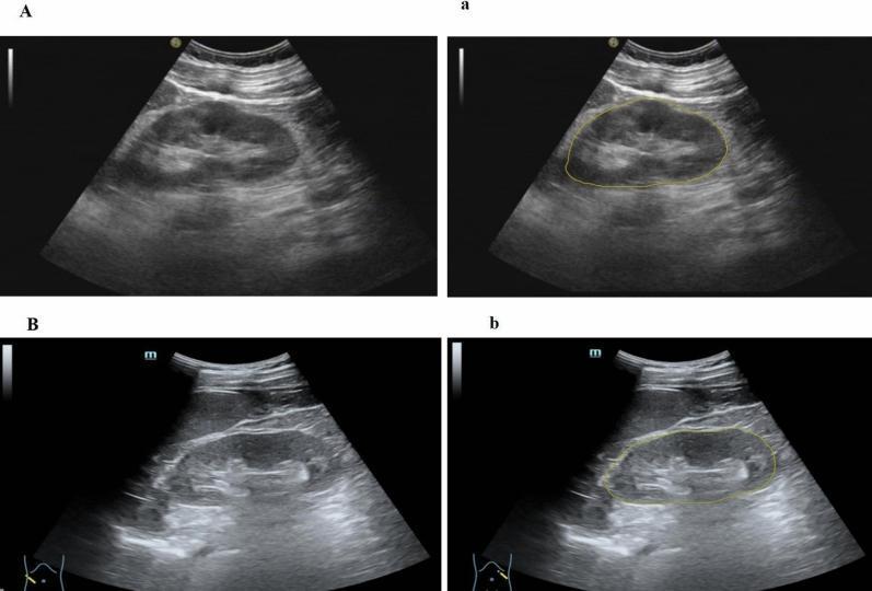
(Row A = diseased kidney with increased cortical echogenicity and reduced CMD. Row B = normal kidney with preserved CMD.)
PART 3: How CMD Changes in Disease - Step by Step
Step 1 - Normal CMD (Healthy Kidney)
[Capsule]
[CORTEX - grey, medium echogenicity]
[----arcuate arteries - bright line = CMD visible----]
[MEDULLA - dark pyramids, very hypoechoic]
[Renal Sinus - bright fat]
You can clearly see 3 distinct zones. CMD is present and sharp.
Step 2 - Cortex starts to scar (Early Disease)
When the kidney is repeatedly injured (by diabetes, hypertension, glomerulonephritis, etc.):
- Tubular cells in the cortex begin to die and shrink (tubular atrophy)
- The space left behind gets filled with collagen fibers (fibrosis)
- Collagen is a strong reflector of ultrasound waves
- So the cortex starts to become brighter on USG
The medulla is relatively less affected early, so it stays dark.
CMD is still visible but becoming less sharp.
Step 3 - Fibrosis progresses (Advanced CKD)
- More and more glomeruli get scarred over = glomerulosclerosis
- More tubules collapse = tubular atrophy
- More collagen deposited throughout = interstitial fibrosis
- Hyaline casts accumulate in tubular lumens (also reflect sound)
Now the entire cortex is full of collagen and scarred tissue - it becomes as bright or brighter than the medulla.
You can no longer tell cortex from medulla = CMD is LOST.
(Brenner and Rector's The Kidney)
The Four Microscopic Changes That Cause CMD Loss
| Pathological Change | What It Means | Why It Increases Echogenicity |
|---|---|---|
| Interstitial Fibrosis | Collagen replaces normal tissue | Collagen strongly reflects ultrasound |
| Tubular Atrophy | Tubular cells shrink/die | Fewer normal cells = more acoustic reflectors per unit area |
| Global Glomerulosclerosis | Whole glomeruli scar over | Scarred glomeruli = dense collagen deposits |
| Hyaline Casts in Tubules | Protein debris plugs tubules | Dense protein material reflects sound |
The degree of CMD loss directly correlates with the severity of all four changes above.
PART 4: Causes of Loss of CMD
CMD can be lost in many conditions - not just CKD. Here is a full classification:
A. Chronic Conditions (Gradual, Bilateral Loss)
| Cause | Mechanism |
|---|---|
| Chronic Kidney Disease (any cause) | Progressive fibrosis + tubular atrophy throughout cortex |
| Diabetic Nephropathy | Glomerulosclerosis + tubular damage from hyperglycemia |
| Hypertensive Nephrosclerosis | Ischemic scarring of cortex from reduced blood supply |
| Chronic Glomerulonephritis | Immune-mediated scarring of glomeruli spreading to interstitium |
| Reflux Nephropathy | Repeated urine reflux causes cortical scarring, especially at poles |
| Polycystic Kidney Disease | Progressive cyst replacement distorts normal architecture |
| Chronic Obstruction | Back-pressure atrophies cortex from inside out |
B. Acute Conditions (Sudden, Often Reversible CMD Loss)
| Cause | Mechanism |
|---|---|
| Acute Glomerulonephritis | Acute inflammation + edema + cellular infiltration of cortex |
| Lupus Nephritis | Immune complex deposition + inflammatory edema in cortex |
| Acute Interstitial Nephritis (AIN) | Inflammatory infiltrate fills interstitium (cells, edema) |
| Acute Tubular Necrosis (ATN) | Tubular cell death + edema increases cortical echogenicity |
| Renal Vein Thrombosis | Venous congestion → cortical edema → increased echogenicity |
| Pyelonephritis (severe) | Inflammatory edema alters cortical texture |
Important: In acute conditions, CMD loss may be reversible if the disease is treated. In CKD, the fibrosis is permanent and CMD loss is irreversible.
C. Infiltrative/Depositing Conditions
| Cause | Mechanism |
|---|---|
| Renal Amyloidosis | Amyloid protein deposits throughout cortex and medulla |
| Multiple Myeloma | Light chain deposition + cast nephropathy |
| Sarcoidosis of kidney | Granuloma formation throughout parenchyma |
PART 5: Grading CMD Change on Ultrasound
Radiologists often grade kidney echogenicity to describe how much CMD is lost:
| Grade | Appearance | Clinical Meaning |
|---|---|---|
| Grade 0 | Cortex less echogenic than liver | Normal |
| Grade 1 | Cortex equal to liver (isoechoic) | Mild parenchymal disease |
| Grade 2 | Cortex more echogenic than liver but less than sinus fat | Moderate disease |
| Grade 3 | Cortex as bright as sinus fat | Severe disease, CMD fully lost |
As grade increases, CMD is progressively lost.
PART 6: CKD vs Acute - Key CMD Differences
| Feature | CKD (Chronic) | Acute Disease |
|---|---|---|
| Kidney size | Small, shrunken | Normal or enlarged |
| CMD loss | Gradual, permanent | May be sudden, potentially reversible |
| Cortical echogenicity | Progressively increases | Acutely increased |
| Both kidneys affected | Yes, bilateral | May be bilateral or unilateral |
| Cause of echogenicity | Fibrosis + collagen | Edema + inflammation |
| Reversibility | No | Possibly, if treated early |
(Brenner and Rector's The Kidney, 2-Volume Set)
Summary in One Paragraph
In a healthy kidney, the cortex looks medium grey on ultrasound and the medullary pyramids look darker, creating a clear visual border called CMD. In disease, the cortex gets damaged by fibrosis (collagen deposition), tubular atrophy, glomerulosclerosis, and hyaline casts. Collagen and scar tissue reflect ultrasound strongly, making the cortex appear brighter and brighter until it matches the brightness of the medulla - at which point you can no longer distinguish the two layers and CMD is said to be lost. This happens progressively in CKD (due to any cause - diabetes, hypertension, chronic GN, obstruction) and can also happen acutely in conditions like lupus nephritis or AIN due to inflammation and edema. The more the CMD is lost, the more severe the underlying renal parenchymal damage.
How to differentiate between ckd and aki want diagonis to do and is cmd difference in ckd or in aki also
Searching sources...
Finding Sources
Finding Sources
Reading File
Finding Sources
Finding Sources
Finding Sources
Finding Sources
Finding Sources
Finding Sources
Searching Images
AKI vs CKD kidney ultrasound size comparison normal versus small shrunken
Reading File
Finding Sources
Reading File
Loading Image
I have all the material needed. Here is a comprehensive, detailed answer.
How to Differentiate AKI from CKD - Complete Guide
First, understand their relationship - AKI and CKD can overlap (a patient with CKD can get AKI on top - called AKI-on-CKD):
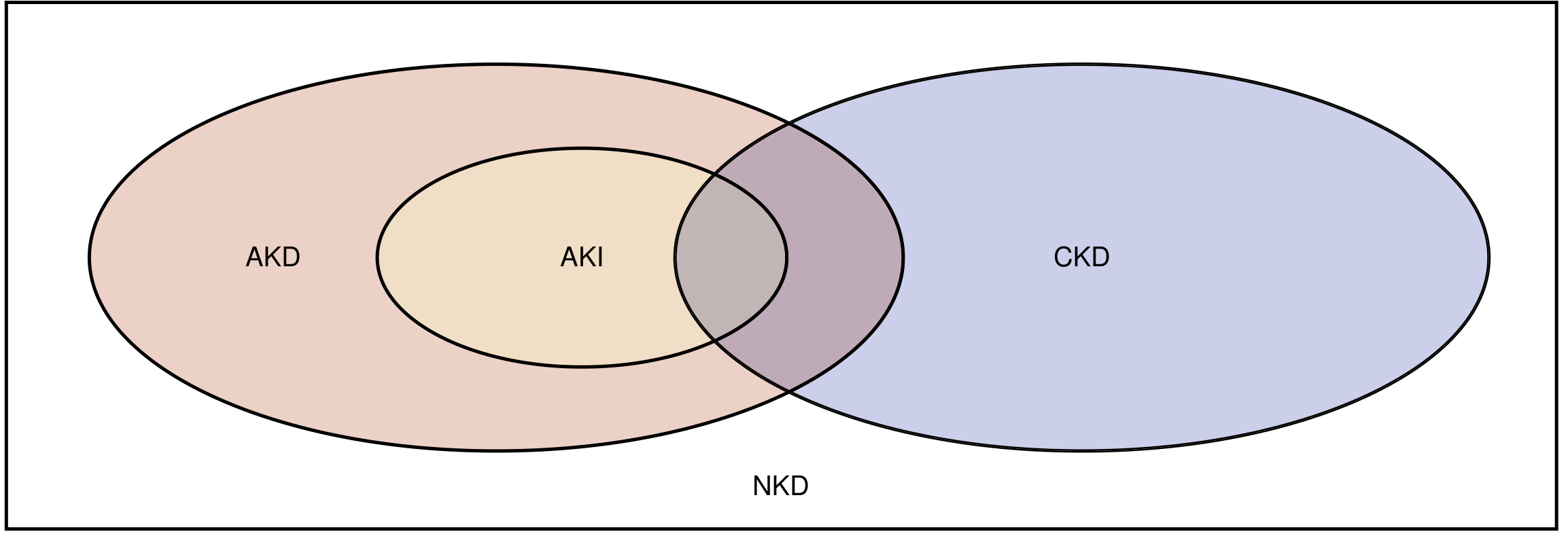
(National Kidney Foundation Primer on Kidney Diseases, 8e)
Definitions First
| AKI | CKD | |
|---|---|---|
| What it is | Sudden, rapid fall in kidney function | Permanent, irreversible kidney damage |
| Duration | Develops over hours to days | Present for ≥3 months |
| Reversible? | Often YES (if treated early) | NO - permanent damage |
| Creatinine rise | ≥0.3 mg/dL in 48 hrs OR 1.5× baseline in 7 days | Chronically elevated for >3 months |
(Rheumatology, 2-Volume Set, Elsevier 2022)
PART 1: HISTORY - Ask These Questions
| Question | AKI | CKD |
|---|---|---|
| How long has creatinine been high? | Days to weeks | Months to years |
| Any previous creatinine records? | Previously normal | Always been elevated |
| Sudden trigger? | YES - infection, surgery, drugs, dehydration, contrast | NO clear trigger - gradual |
| Known CKD before this? | No (unless AKI-on-CKD) | Yes - diabetes, HTN, GN for years |
| Symptoms duration | Sudden (days) | Months - fatigue, nocturia, poor appetite |
| Previous dialysis or kidney biopsy? | No | Possibly |
PART 2: CLINICAL EXAMINATION - Signs to Look For
Signs pointing to CKD (Chronicity):
| Sign | Explanation |
|---|---|
| Pallor (anaemia) | CKD causes chronic anaemia (low EPO) - takes months to develop. AKI does NOT cause anaemia quickly |
| Uraemic frost | White crystalline deposits on skin - only in very chronic disease |
| Peripheral neuropathy | Numbness/tingling in feet - develops over months in CKD |
| Muehrcke's lines / Leukonychia (white nails) | Nail changes from chronic hypoalbuminaemia in CKD |
| Renal osteodystrophy | Bone pain, deformities - only in CKD from chronic PTH excess |
| Growth retardation | In children - only chronic disease causes this |
| Hypertension (long-standing) | Chronic hypertension with LVH - suggests CKD |
| No oliguria or fluid overload early | CKD patients adapt over time and may still pass urine |
Signs pointing to AKI (Acuity):
| Sign | Explanation |
|---|---|
| Sudden oliguria/anuria | Abrupt drop in urine output |
| Fluid overload rapidly | Sudden pulmonary oedema, raised JVP |
| Normal-sized kidneys | No time for shrinkage yet |
| No anaemia (initially) | Anaemia has not had time to develop |
| Clear trigger | Sepsis, nephrotoxic drug, contrast, surgery, bleeding |
PART 3: INVESTIGATIONS - What to Do and What They Show
A. Blood Tests
| Test | AKI | CKD | Why |
|---|---|---|---|
| Serum Creatinine | Rising rapidly (days) | Chronically elevated (months) | Rate of rise matters |
| BUN/Creatinine ratio | Very high (>20:1) in prerenal AKI | Moderately elevated | Urea rises faster than Cr in prerenal |
| Haemoglobin (CBC) | Normal initially | Low (normocytic normochromic anaemia) | EPO deficiency takes months |
| Serum Calcium | Usually normal | Low (hypocalcaemia) | Chronic Vit D deficiency in CKD |
| Serum Phosphorus | Usually normal | High (hyperphosphataemia) | Chronic retention in CKD |
| PTH (iPTH) | Normal | Very high (secondary hyperparathyroidism) | Months of low Vit D drive PTH up |
| Serum Uric Acid | May be acutely elevated | Chronically elevated | |
| Serum Potassium | Acutely elevated | Elevated but adapted | |
| Alkaline Phosphatase | Normal | High | Bone disease (renal osteodystrophy) |
| Serum Albumin | May be low if sepsis | Chronically low | Chronic malnutrition in CKD |
Most useful blood test to distinguish: Anaemia + high PTH + high Phosphorus + low Calcium = strongly suggests CKD
B. Urine Tests
| Test | AKI | CKD | Why |
|---|---|---|---|
| Urine Output | Suddenly drops (oliguria/anuria) | May be preserved (polyuria in early CKD) | |
| Urine Osmolality | High (prerenal AKI > 500) or Low (ATN < 350) | Fixed ~300-350 (isosthenuria) | CKD loses concentration ability |
| Urine Sodium | Low (<20 mEq/L) in prerenal AKI | Variable | |
| FENa (Fractional excretion of Na) | <1% prerenal, >2% ATN | Usually >2% | |
| Urine Microscopy | RBC casts (GN), granular casts (ATN), WBC casts (AIN) | Broad/waxy casts - HALLMARK of CKD | Broad casts = dilated, atrophied tubules |
| Proteinuria | Minimal (unless GN) | Heavy, chronic (>3 g/day in nephrotic) | Glomerular scarring |
| Haematuria | Present in GN, trauma | May be present in GN-related CKD |
Key urine finding for CKD: BROAD WAXY CASTS - These form in wide, dilated tubules that have been stretched by chronic atrophy. They CANNOT form in normal-sized tubules = only found in CKD.
C. Ultrasound (USG) - Most Important Imaging
This is where CMD becomes the key differentiator:
| USG Feature | AKI | CKD |
|---|---|---|
| Kidney Size | Normal or enlarged (9-12 cm) | Small, shrunken (<9 cm) - bilateral |
| Cortical Echogenicity | May be mildly increased | Markedly increased (bright cortex) |
| CMD | Preserved or mildly reduced | Absent / Lost |
| Cortical Thickness | Normal | Thinned |
| Surface contour | Smooth | Irregular, scarred |
| Resistive Index (RI) | Elevated (>0.70) in obstruction | Elevated chronically |
| Bilateral | May be unilateral | Always bilateral |
PART 4: CMD IN AKI vs CKD - The Critical Comparison
This is the most important part of your question:
CMD in AKI:
- Kidneys are normal or enlarged (swollen due to oedema/inflammation)
- Cortex may become mildly echogenic due to:
- Oedema and inflammation (AIN, glomerulonephritis)
- Tubular cell swelling (ATN)
- CMD may be mildly reduced but is often still visible
- The change is acute - it developed over days
- If AKI resolves → CMD can return to normal (reversible)
CMD in CKD:
- Kidneys are small and shrunken (atrophy from chronic damage)
- Cortex is markedly echogenic due to:
- Dense interstitial fibrosis (collagen)
- Tubular atrophy
- Glomerulosclerosis
- Hyaline casts
- CMD is completely lost / obliterated
- The change took months to years to develop
- Irreversible - even if dialysis starts, CMD does not come back
CMD Comparison Table:
| Feature | AKI | CKD |
|---|---|---|
| Kidney size on USG | Normal / Large | Small bilateral |
| CMD | Present or mildly reduced | Lost / Absent |
| Cortical echogenicity | Mildly increased | Markedly increased |
| Cause of echogenicity change | Oedema + inflammation | Fibrosis + collagen |
| Reversible? | Yes | No |
| Speed of change | Days | Months-years |
Simple rule: Small kidneys + Lost CMD = CKD. Normal/big kidneys + Preserved CMD = AKI.
PART 5: Other Investigations
| Investigation | AKI | CKD |
|---|---|---|
| Renal Biopsy | ATN, GN, AIN on histology - acute changes | Fibrosis, glomerulosclerosis, tubular atrophy - chronic changes |
| Chest X-ray | Pulmonary oedema (acute) | Cardiomegaly (chronic HTN), pulmonary oedema |
| ECG | Peaked T waves (acute hyperkalaemia) | LVH pattern (chronic) |
| Bone X-ray | Normal | Renal osteodystrophy, periosteal resorption |
| Previous creatinine records | Normal baseline | Chronically elevated |
PART 6: Summary - Quick Differentiating Checklist
FEATURE              |   AKI              |   CKD
---------------------|--------------------|-----------------------
Duration             | Days-weeks         | >3 months
Kidney size (USG)    | Normal/Enlarged    | SMALL (bilateral)
CMD on USG           | Preserved/Mild ↓   | ABSENT/LOST
Anaemia              | No (initially)     | Yes (normocytic)
PTH                  | Normal             | Very HIGH
Phosphorus           | Normal             | HIGH
Calcium              | Normal             | LOW
Urine casts          | Granular/RBC/WBC   | BROAD WAXY casts
Reversibility        | Yes (often)        | No
History of DM/HTN    | May be absent      | Usually present (years)
Neuropathy/bone pain | No                 | Yes (chronic features)
In one sentence: AKI = sudden rise in creatinine, normal/big kidneys, preserved CMD, no anaemia, no PTH rise yet, reversible. CKD = months of elevated creatinine, small bilateral kidneys, lost CMD, anaemia, high PTH, high phosphorus, low calcium, broad waxy casts, irreversible.
(National Kidney Foundation Primer on Kidney Diseases, 8e; Rheumatology, 2-Volume Set, Elsevier 2022; Brenner and Rector's The Kidney)
This is a shared conversation. Sign in to Orris to start your own chat.