Typhoid fever full information in depth

Reading File
Finding Sources
Finding Sources
Searching PubMed

"typhoid fever"[MeSH Terms] AND "treatment"[MeSH Terms]

Reading File
Reading File
Reading File
Reading File
Reading File
Reading File
Reading File
Searching Images

typhoid fever rose spots rash Salmonella typhi

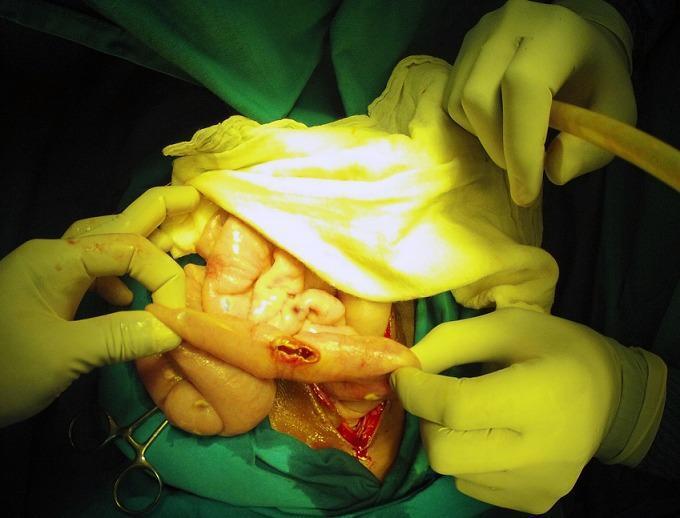
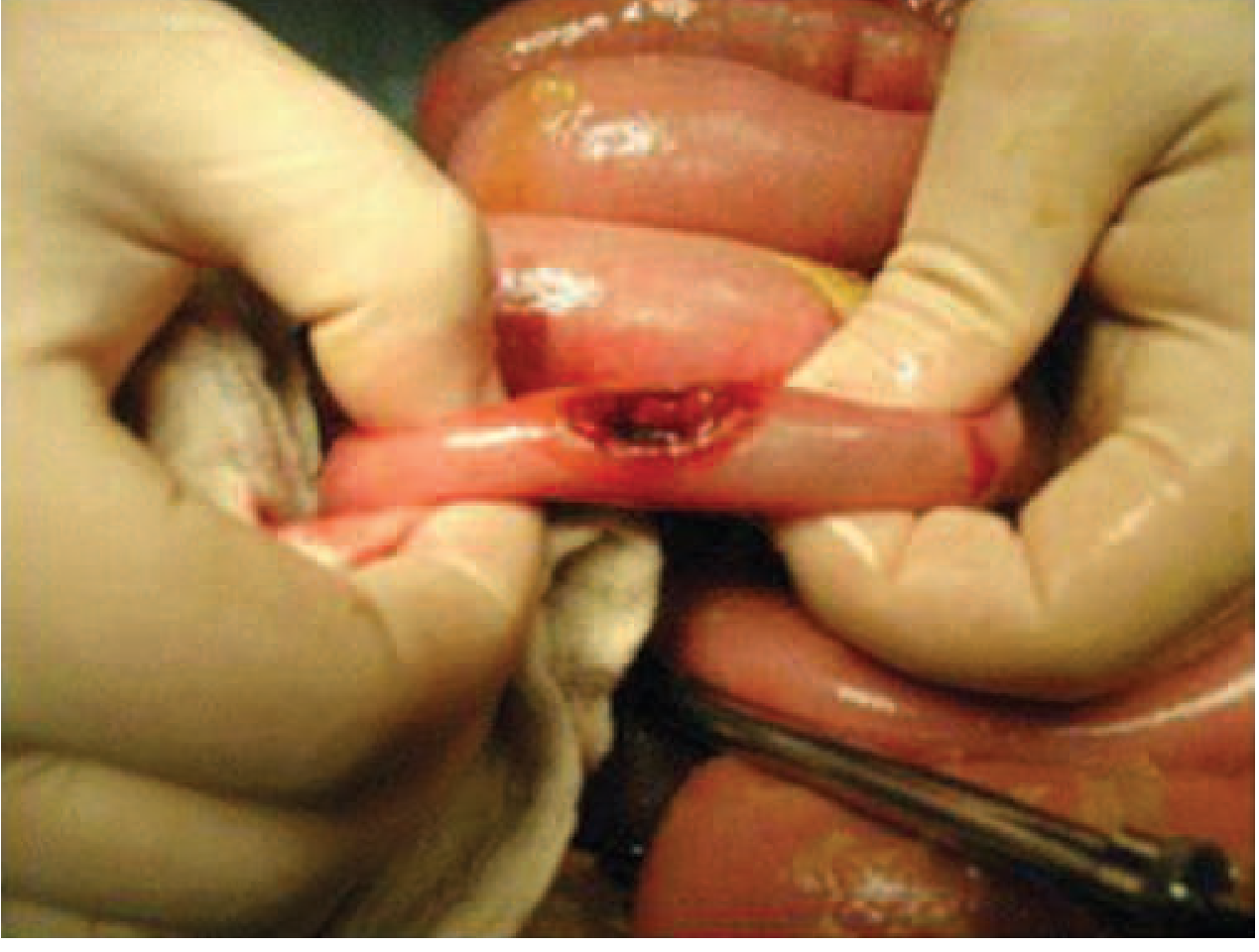
This clinical photograph shows an intraoperative view of an exploratory laparotomy, demonstrating a bowel perforation in the context of typhoid fever (Salmonella Typhi). The image focuses on a loop of small intestine held by a surgeon's gloved hands, revealing a discrete, longitudinal perforation with hyperemic and slightly everted edges. The surrounding serosa exhibits generalized inflammation and congestion. In the background, other segments of the intestine appear edematous and pale-pink, with visible white-yellowish peritoneal nodules and purulent exudate suggestive of concurrent miliary peritoneal tuberculosis and secondary peritonitis. Surgical instruments, suction tubing, and green sterile drapes define the surgical field. This image serves as an educational example of the severe gastrointestinal complications of typhoid fever, specifically intestinal perforation, and illustrates the challenging clinical scenario of co-infection with tuberculosis in an acute surgical setting.

This clinical photograph shows an intraoperative view of an exploratory laparotomy, demonstrating a bowel perforation in the context of typhoid fever (Salmonella Typhi). The image focuses on a loop of small intestine held by a surgeon's gloved hands, revealing a discrete, longitudinal perforation with hyperemic and slightly everted edges. The surrounding serosa exhibits generalized inflammation and congestion. In the background, other segments of the intestine appear edematous and pale-pink, with visible white-yellowish peritoneal nodules and purulent exudate suggestive of concurrent miliary peritoneal tuberculosis and secondary peritonitis. Surgical instruments, suction tubing, and green sterile drapes define the surgical field. This image serves as an educational example of the severe gastrointestinal complications of typhoid fever, specifically intestinal perforation, and illustrates the challenging clinical scenario of co-infection with tuberculosis in an acute surgical setting.

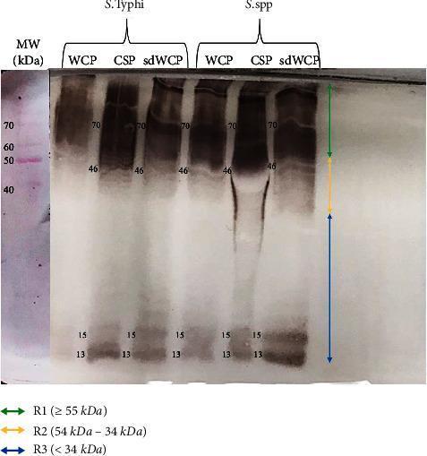
A diagnostic Western blot (WB) analysis image showing the immunoreactivity of antigens derived from Salmonella typhi and Salmonella spp. The image displays six experimental lanes representing three different protein extraction methods—Whole Cell Protein (WCP), Cell Surface Protein (CSP), and sodium deoxycholate-extracted WCP (sdWCP)—for each bacterial species. A molecular weight (MW) marker lane on the far left, stained with Ponceau S, shows reference bands from 40 kDa to 70 kDa. The WB profile is categorized into three vertical regions: R1 (≥ 55 kDa), R2 (54–34 kDa), and R3 (< 34 kDa). Prominent, high-intensity immunoreactive bands are observed in both species at approximately 70 kDa and 46 kDa, with a noticeable smear in the CSP lanes. Low molecular weight bands are consistently identified at 13 kDa and 15 kDa across all preparations. This visual provides comparative data on the antigenic profiles and antibody isotype reactivity against somatic and flagellar antigens, relevant for typhoid fever serodiagnosis research.

A diagnostic Western blot (WB) analysis image showing the immunoreactivity of antigens derived from Salmonella typhi and Salmonella spp. The image displays six experimental lanes representing three different protein extraction methods—Whole Cell Protein (WCP), Cell Surface Protein (CSP), and sodium deoxycholate-extracted WCP (sdWCP)—for each bacterial species. A molecular weight (MW) marker lane on the far left, stained with Ponceau S, shows reference bands from 40 kDa to 70 kDa. The WB profile is categorized into three vertical regions: R1 (≥ 55 kDa), R2 (54–34 kDa), and R3 (< 34 kDa). Prominent, high-intensity immunoreactive bands are observed in both species at approximately 70 kDa and 46 kDa, with a noticeable smear in the CSP lanes. Low molecular weight bands are consistently identified at 13 kDa and 15 kDa across all preparations. This visual provides comparative data on the antigenic profiles and antibody isotype reactivity against somatic and flagellar antigens, relevant for typhoid fever serodiagnosis research.

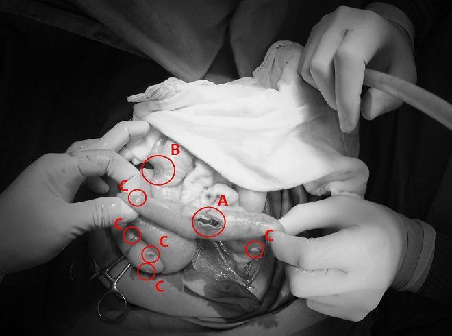
This monochrome clinical photograph captures an intraoperative view of the abdominal cavity during a surgical procedure for bowel perforation. The image illustrates two distinct infectious pathologies concurrently affecting the intestines and peritoneum. Label 'A' identifies a typhoid intestinal perforation, appearing as an irregular, longitudinal breach in the seromuscular layer of the small bowel, typical of advanced Salmonella typhi infection. Label 'B' shows a non-perforated typhoid lesion, characterized by localized inflammation and thickening without a full-thickness defect. Surrounding these intestinal segments, multiple small, discrete, whitish nodules are indicated by label 'C', representing miliary peritoneal tuberculosis. These nodules are scattered across the serosal surfaces and mesentery, demonstrating a grain-like distribution characteristic of disseminated mycobacterial infection. The image serves as a high-level educational example of acute surgical abdomen complications resulting from co-infection of typhoid fever and miliary tuberculosis, highlighting diagnostic landmarks such as transmural perforation and widespread peritoneal seeding.

This monochrome clinical photograph captures an intraoperative view of the abdominal cavity during a surgical procedure for bowel perforation. The image illustrates two distinct infectious pathologies concurrently affecting the intestines and peritoneum. Label 'A' identifies a typhoid intestinal perforation, appearing as an irregular, longitudinal breach in the seromuscular layer of the small bowel, typical of advanced Salmonella typhi infection. Label 'B' shows a non-perforated typhoid lesion, characterized by localized inflammation and thickening without a full-thickness defect. Surrounding these intestinal segments, multiple small, discrete, whitish nodules are indicated by label 'C', representing miliary peritoneal tuberculosis. These nodules are scattered across the serosal surfaces and mesentery, demonstrating a grain-like distribution characteristic of disseminated mycobacterial infection. The image serves as a high-level educational example of acute surgical abdomen complications resulting from co-infection of typhoid fever and miliary tuberculosis, highlighting diagnostic landmarks such as transmural perforation and widespread peritoneal seeding.

Reading File
Reading File
Reading File
Loading Image
I now have comprehensive, authoritative information from Harrison's Principles, Tintinalli's Emergency Medicine, and Park's Preventive Medicine. Let me compose the full, in-depth answer.

Typhoid Fever - Comprehensive Overview

Sources: Harrison's Principles of Internal Medicine 22E (2025), Tintinalli's Emergency Medicine, Park's Textbook of Preventive & Social Medicine

1. Definition

Typhoid fever (also called enteric fever) is a severe systemic febrile illness caused by Salmonella enterica serovar Typhi (S. Typhi) or, less commonly, S. Paratyphi A, B, or C. It is characterized by prolonged fever, abdominal symptoms, and involvement of lymphoid tissues. The name "typhoid" was coined for its clinical resemblance to typhus, but by 1869 the term "enteric fever" was proposed based on the anatomic site of infection - Harrison's, p. 1359.

2. Microbiology / Etiology

  • Organism: Salmonella enterica serovar Typhi - a Gram-negative, motile, non-spore-forming, facultative intracellular bacillus of the family Enterobacteriaceae
  • Antigens: Three main surface antigens - O (somatic/LPS), H (flagellar), and Vi (virulence capsular polysaccharide). Vi antigen helps the organism evade phagocytosis
  • Phage typing: At least 80 phage types identified - useful epidemiological tool for outbreak tracing - Park's, p. 277
  • The organism survives intracellularly in tissues. It is killed by drying, pasteurization, and common disinfectants
  • Unlike other Salmonella serotypes, S. Typhi and S. Paratyphi have no known host other than humans - they are obligate human pathogens

3. Epidemiology

Global Burden

  • WHO estimates 11-21 million cases and 110,000-280,000 deaths annually
  • Highest incidence: Indian subcontinent (India, Pakistan, Bangladesh, Nepal), Eastern Mediterranean, sub-Saharan Africa - rates exceed 1,000 per 100,000 children in some urban areas
  • In the US, ~5,700 cases per year; ~78% are travel-associated (predominantly South Asia)

Risk Factors

  • Fecally contaminated drinking water or ice
  • Street vendor food and raw vegetables fertilized with sewage
  • Flooding and inadequate sanitation
  • Ill household contacts
  • Evidence of prior H. pylori infection (associated with reduced gastric acidity, lowering the infectious dose barrier)
  • Age group 5-19 years most commonly affected - Park's, p. 277

Reservoir and Transmission

  • Reservoir: Humans only - cases and carriers (temporary and chronic)
  • Chronic carriers: 2-5% of cases; organisms persist in the gallbladder/biliary tract; excretion may last decades (the famous "Typhoid Mary" caused >1,300 cases)
  • Sources: Primary = feces and urine of cases/carriers; Secondary = contaminated water, food, fingers, flies
  • Transmission is fecal-oral, dose-dependent - Harrison's, p. 1359

Antimicrobial Resistance (a major modern concern)

  • MDR strains emerged in China/SE Asia in the 1980s - resistant to chloramphenicol, ampicillin, and TMP-SMX
  • Decreased susceptibility to ciprofloxacin (DSC) emerged in the 1990s on the Indian subcontinent
  • XDR (extensively drug-resistant) S. Typhi - resistant to all first- and second-line drugs except azithromycin and carbapenems - emerged in Pakistan (2016), now spreading globally - Harrison's, p. 1359

4. Pathogenesis

The infection follows a well-defined sequence:
  1. Ingestion - organism ingested via contaminated food/water (infective dose ~10^5-10^9 organisms, but lower in MDR strains)
  2. Gastric passage - surviving organisms reach the small intestine; gastric acidity is a major barrier
  3. Mucosal invasion - bacteria invade the intestinal mucosa, particularly via M cells overlying Peyer's patches in the ileum
  4. Intracellular survival - S. Typhi is taken up by macrophages and survives within them using type III secretion systems (T3SS) encoded on Salmonella Pathogenicity Islands (SPI-1 and SPI-2)
  5. Lymphatic spread - bacteria multiply in mesenteric lymph nodes, then enter the thoracic duct
  6. Primary bacteremia - organisms seed the bloodstream and disseminate to liver, spleen, bone marrow, and gallbladder (Week 1)
  7. Secondary bacteremia - re-seeding from the reticuloendothelial system causes sustained high-grade bacteremia and the characteristic fever (Week 2)
  8. Hyperplasia and necrosis of Peyer's patches - the hallmark pathological finding; ulceration in Week 3 can lead to hemorrhage or perforation
The Vi capsular antigen and endotoxin (LPS) drive the systemic inflammatory response.

5. Clinical Features - The Classic Week-by-Week Course

Incubation Period

  • Mean: 10-14 days (range 5-21 days) - varies with inoculum size and host immunity

Week 1 - Prodrome

FeatureDetail
FeverRises in a step-ladder pattern - daily incremental rise
HeadachePresent in ~80% of cases
Malaise, myalgia, anorexiaProminent
Cough~30%
ConstipationMore common than diarrhea initially (30% constipated)
Relative bradycardiaPulse-temperature dissociation (Faget sign)

Week 2 - Established Disease

  • Fever reaches a plateau: 38.8-40.5°C (101.8-104.9°F), sustained high-grade
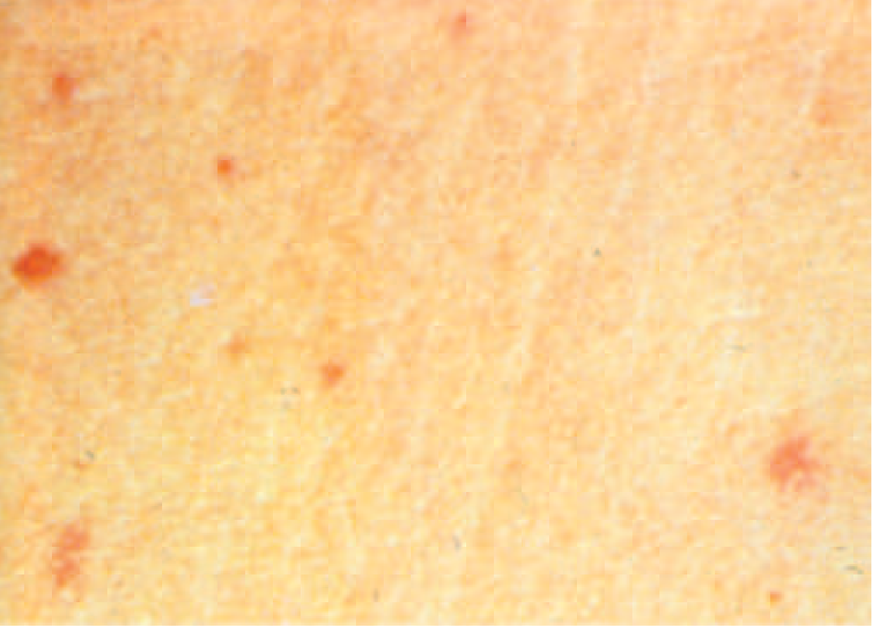
  • Rose spots (see image below) - faint, salmon-pink, blanching maculopapular lesions, 2-3 mm, located on trunk/chest; visible in ~30% of patients, especially fair-skinned individuals; appear during end of first week/early second week; each spot fades in 3-4 days
  • Splenomegaly develops
  • Abdominal distension and "pea soup" diarrhea may appear
  • Coated tongue (51-56%)
  • Patient appears toxic, exhausted, prostrated
  • Leukopenia and positive blood, urine, stool cultures

Week 3 - Complications

  • Intestinal hemorrhage (~6%) and perforation (~1%) - most common life-threatening events
  • Neuropsychiatric features: "muttering delirium," "coma vigil" (picking at bedclothes)
  • Risk of relapse up to 2 weeks after defervescence

Week 4 - Resolution or Worsening

  • Untreated patients who survive begin to defervesce
  • May have relapse in 2-3 weeks with the same strain
"Rose spots" - the classic rash of typhoid fever:
Rose spots - salmon-pink blanching maculopapular rash on the trunk in typhoid fever
Figure: Characteristic rose spots on the trunk in enteric fever (S. Typhi). Small, discrete, salmon-colored, blanching papules. (Harrison's, Fig. 171-2)

6. Complications

Complications occur in ~27% of hospitalized patients. They correlate with delayed treatment, host factors, and antibiotic choice - Harrison's, p. 1360.

Gastrointestinal (most common, weeks 3-4)

  • Intestinal hemorrhage (6%): sudden temperature drop, shock, dark/fresh blood in stool - from ulceration of Peyer's patches
  • Intestinal perforation (1%): most life-threatening; requires emergency surgery + broad-spectrum antibiotics for peritonitis
Typical ileal perforation in typhoid fever
Figure: Ileal perforation - a surgical emergency occurring typically in week 3 of untreated typhoid (Harrison's Fig. 171-3)

Neurological (2-40%)

  • Meningitis
  • Guillain-Barré syndrome
  • Neuritis
  • "Muttering delirium" / coma vigil
  • Psychosis, ataxia, seizures, deafness

Cardiovascular

  • Myocarditis, pericarditis, endocarditis
  • Mycotic aneurysm
  • DIC

Other

  • Hepatitis, hepatic/splenic abscesses
  • Pancreatitis
  • Hemolytic-uremic syndrome
  • Orchitis, glomerulonephritis
  • Severe pneumonia
  • Osteomyelitis, septic arthritis
  • Chronic carrier state (2-5%) - associated with increased risk of gallbladder carcinoma

7. Laboratory Diagnosis

Gold Standard: Culture

SpecimenSensitivityOptimal TimingNotes
Blood culture40-80%Week 1-2Mainstay of diagnosis; sensitivity increases with volume
Bone marrow culture>90%AnytimeMost sensitive; positive even after antibiotic treatment
Stool culture30-40%Week 2-3S. Typhi shed in bile into gut
Urine culture25%Week 2-3Secondary bacteriuria
Rose spot cultureLowWeek 2Rarely performed

Serology

  • Widal test (Felix-Widal): Measures agglutinating antibodies against O and H antigens
    • O antibodies appear day 6-8; H antibodies day 10-12
    • Sensitivity and specificity are moderate
    • Can be negative in up to 30% of culture-proven cases (blunted by prior antibiotics)
    • Cross-reactions with other Salmonella serotypes limit specificity
    • Interpretation requires paired sera (4-fold rise in titer)
  • Vi antibody: Present in ~80% of chronic carriers - useful for carrier detection
  • Rapid antigen tests: Used in resource-limited settings; variable performance

Other Lab Findings (non-specific)

  • Leukopenia and neutropenia (15-25% of cases)
  • Leukocytosis: more common in children, early illness, or when complicated by perforation
  • Moderately elevated LFTs (hepatitis)
  • Elevated ESR, CRP
  • Mild anemia, thrombocytopenia

Newer Methods

  • PCR (blood, stool): High sensitivity and specificity; useful in early disease and when culture is negative; not widely available in endemic areas
  • Antigen detection (urine/stool): TUBEX, Typhidot-M

8. Differential Diagnosis

In a returning traveler with prolonged fever, consider:
  • Malaria (must exclude first)
  • Viral hepatitis (A, B, E)
  • Dengue fever
  • Bacterial enteritis
  • Rickettsial infections (typhus, scrub typhus)
  • Leptospirosis
  • Amebic liver abscess
  • Brucellosis
  • Acute HIV infection

9. Treatment

Antibiotic Therapy (Harrison's Table 171-1)

The initial choice depends on local resistance patterns:
IndicationAgentDose (Route)Duration
EmpiricalCeftriaxone2 g/day (IV)10-14 days
Ciprofloxacin500 mg BD (PO) or 400 mg q12h (IV)5-7 days
Azithromycin1 g/day (PO)10 days
Fully susceptibleCiprofloxacin or CeftriaxoneAs aboveAs above
AlternativesChloramphenicol25 mg/kg TID (PO/IV)14-21 days
Amoxicillin1 g TID (PO) / 2 g q6h (IV)14 days
TMP-SMX160/800 mg BD (PO)7-14 days
MDR (resistant to ampicillin, chloramphenicol, TMP-SMX)Ceftriaxone or Azithromycin or Ciprofloxacin (if susceptible)As aboveAs above
XDR (+ fluoroquinolone resistant, ± cephalosporin resistant)Azithromycin (mild-moderate)1 g/day5-7 days
Meropenem (severe)1 g q8h (IV)10-14 days
Key treatment notes:
  • Fluoroquinolones should no longer be used empirically for patients from the Indian subcontinent due to high rates of DSC strains - Harrison's, p. 1361
  • A 2022 systematic review of 27 RCTs found no significant difference between ceftriaxone, fluoroquinolones, and azithromycin in treatment failure, relapse, or convalescent carriage
  • For typhoid meningitis/encephalitis or septic shock: add dexamethasone (3 mg/kg IV loading dose, then 1 mg/kg q6h x 8 doses) to antibiotics
  • Chronic carriers: Treat with ampicillin/amoxicillin + probenecid for 6 weeks; or ciprofloxacin for 4 weeks; cholecystectomy if gallstones are present
  • Relapses (up to 10%): Occur 2-3 weeks after defervescence with the same strain and susceptibility pattern; re-treat with the same antibiotic

Supportive Care

  • IV fluid resuscitation and electrolyte correction
  • Antipyretics (paracetamol preferred)
  • Blood transfusion if significant GI bleeding
  • Surgical intervention for intestinal perforation (emergency laparotomy + bowel repair/resection + peritoneal lavage)

Case Fatality

  • Untreated: 10-20% (up to 30% historically)
  • With appropriate treatment: <1%
  • Hospitalized: ~4.5%

10. Prevention and Control

Three Lines of Defence (Park's, p. 279)

1. Control of Reservoir

Cases:
  • Early diagnosis and notification
  • Isolation until 3 bacteriologically negative stool and urine samples on separate days
  • Disinfection of stool/urine with 5% cresol for ≥2 hours
  • Follow-up stool/urine cultures at 3-4 months and 12 months post-discharge
Carriers:
  • Identification via culture, serology (Vi antibody in ~80%), duodenal drainage
  • Treatment: Ampicillin or amoxicillin (4-6 g/day) + probenecid (2 g/day) for 6 weeks - achieves eradication in ~70%
  • Cholecystectomy + antibiotics for carriers with gallstones
  • Chronic carriers should not work as food handlers or in healthcare

2. Control of Sanitation (the weakest link)

  • Safe water supply and proper sewage disposal
  • Food hygiene: proper cooking, avoiding raw vegetables irrigated with sewage, refrigeration
  • Pasteurization of milk
  • Hand hygiene after toilet use and before food preparation
  • Fly control

3. Immunization

VaccineTypeScheduleAgeEfficacyBooster
Ty21a (Vivotif)Oral live attenuatedDays 1, 3, 5, 7 (4 doses)≥6 years~50% at 2.5-3 yearsEvery 5 years
Vi CPS (Typherix, Typhim Vi)Injectable Vi polysaccharideSingle IM dose≥2 years~55% at 3 yearsEvery 2 years
Typhoid Conjugate Vaccines (TCV)Vi polysaccharide conjugated to carrier proteinSingle IM 0.5 mL dose≥6 months79-95%Antibody persists up to 7 years
TCVs (the current gold standard):
  • Typbar-TCV (Bharat Biotech) - WHO prequalified 2018
  • TYPHIBEV (Biological E) - WHO prequalified 2020
  • Effective in children <2 years (unlike unconjugated vaccines)
  • Recommended by WHO for high-incidence countries
  • Already introduced in national immunization programs in Pakistan, Nepal, Liberia, Zimbabwe, Malawi, Samoa - Harrison's, p. 1361
Indications for vaccination:
  • Travelers to endemic regions (South Asia, Africa, Latin America, SE Asia)
  • Laboratory workers handling S. Typhi cultures
  • Household contacts of known carriers
  • Vaccination does not replace food/water precautions - high inocula can overcome vaccine protection

11. Chronic Carrier State

  • Defined as shedding S. Typhi in stool or urine for >1 year after infection
  • Prevalence: 2-5% of cases
  • More common in: women, infants, those with biliary abnormalities, concurrent Schistosoma haematobium infection
  • Organisms form biofilms on gallstones and invade gallbladder epithelial cells
  • Long-term risk: gallbladder carcinoma (significantly increased in endemic areas)
  • Up to 10% of untreated patients shed S. Typhi for up to 3 months (transient carriers)

12. Notifiable Disease Status

Enteric fever is a notifiable disease in most countries. Reporting enables:
  • Identification of source patients and common-source outbreaks
  • Treatment of chronic carriers to prevent secondary cases
  • Surveillance of antimicrobial resistance trends

Summary Table

FeatureDetails
Causative agentS. enterica serovar Typhi (primarily)
TransmissionFecal-oral (contaminated water/food)
Incubation5-21 days (mean 10-14 days)
Classic fever patternStep-ladder rise → sustained plateau
Pathognomonic signRose spots (30%) on trunk
Relative bradycardiaFaget sign - pulse-temperature dissociation
DiagnosisBlood culture (gold standard); bone marrow culture (most sensitive)
First-line treatmentCeftriaxone, azithromycin (for Indian subcontinent/MDR); fluoroquinolone if fully susceptible
XDR treatmentAzithromycin (mild) or meropenem (severe)
Mortality (untreated)10-20%
Mortality (treated)<1%
Key complicationIntestinal perforation (3rd week)
Vaccine of choiceTyphoid Conjugate Vaccine (TCV) - 79-95% efficacy
Chronic carriage2-5%; gallbladder reservoir; linked to gallbladder cancer

References:

More differentials

Finding Sources
Finding Sources
Reading File
Finding Sources
Finding Sources
Finding Sources
Reading File
Finding Sources
Reading File
Finding Sources
I now have a comprehensive, structured differential table from Goldman-Cecil Medicine. Let me compile the full detailed answer.

Differential Diagnosis of Typhoid Fever

The differential is best organized by clinical context and incubation period, since typhoid presents as a nonspecific febrile illness. The single most important initial step is excluding malaria.

Framework: By Incubation Period in the Returning Traveler

This table is adapted from Goldman-Cecil Medicine (International Edition), Table 265-6:
IncubationDiagnoses to Consider
Short (<10 days)Malaria, arboviruses (dengue, chikungunya, Zika), hemorrhagic fevers (Ebola, Crimean-Congo, Lassa), enteric fever, spotted fever Rickettsiae, scrub typhus, leptospirosis, bacterial enteritis, respiratory infections, relapsing fever, amebic dysentery, meningococcemia, meningitis, fascioliasis, melioidosis, flaviviruses (TBE, JE)
Medium (10-21 days)Malaria, enteric fever, acute HIV, hemorrhagic fever viruses, rickettsial disease (epidemic/endemic typhus), Coxiella burnetii (Q fever), giardiasis, CMV, toxoplasmosis, brucellosis, leptospirosis, amebic dysentery, endemic fungi (histoplasmosis, coccidioidomycosis), babesiosis, East African trypanosomiasis, T. cruzi (acute), measles, melioidosis
Long (≥21 days)Malaria, schistosomiasis (acute/Katayama fever), TB (M. tuberculosis), acute HIV, viral hepatitis (A/B/E), filariasis, Q fever, syphilis (T. pallidum), EBV (acute), amebic liver abscess, leishmaniasis (visceral/kala-azar), brucellosis, CMV, babesiosis, West African trypanosomiasis (chronic), chronic bartonellosis

By Destination (Geographic Differential)

From Goldman-Cecil Table 265-5:
RegionPrimary Differentials
South-Central Asia (most likely typhoid)Dengue, enteric fever, malaria, chikungunya, Q fever, scrub typhus, visceral leishmaniasis
Southeast AsiaDengue, malaria, chikungunya, Zika, enteric fever, leptospirosis, melioidosis, scrub typhus, talaromycosis
Sub-Saharan AfricaMalaria, Rickettsiae, Katayama fever, dengue, acute HIV, African trypanosomiasis, amebic liver abscess, brucellosis, enteric fever, meningococcemia
Central/South AmericaDengue, malaria, chikungunya, Zika, enteric fever, leptospirosis, histoplasmosis, brucellosis, T. cruzi

By Clinical Feature Overlap

1. Malaria (must exclude first)

  • Mimics: Sustained or periodic high fever, rigors, headache, splenomegaly, anemia, thrombocytopenia
  • Distinguishes: Cyclical fever pattern (tertian/quartan), severe hemolysis, rapid diagnostic test (RDT) or thick/thin blood film positive; no rose spots; no step-ladder fever; leukocytosis in severe disease
  • Malaria can coexist with typhoid ("typhomalarial fever") in endemic areas

2. Dengue Fever

  • Mimics: Abrupt high fever, severe headache, myalgia, thrombocytopenia, leukopenia, rash
  • Distinguishes: Dengue has a very abrupt onset, retro-orbital pain, "breakbone" pain, saddle-back fever pattern; dengue rash is petechial/maculopapular beginning on trunk and spreading; NS1 antigen or IgM/IgG positive; no Faget sign; shorter duration (~7 days vs weeks)

3. Scrub Typhus (Orientia tsutsugamushi)

  • Mimics: Sustained fever, headache, myalgia, rash, hepatosplenomegaly, lymphadenopathy
  • Distinguishes: Eschar at mite bite site (pathognomonic, but absent in 30-50%); maculopapular rash; marked lymphadenopathy; exposure to scrub/vegetation; Weil-Felix OXK positive; responds rapidly to doxycycline
  • Common in SE Asia - can be confused with typhoid

4. Other Rickettsial Diseases (Spotted fevers, Epidemic/Endemic Typhus)

  • Mimics: Sustained fever, rash, headache, myalgia
  • Distinguishes: Spotted fever: petechial/purpuric rash on wrists/ankles spreading centripetally; epidemic typhus: louse-borne, severe prostration, macular rash starting on trunk; Weil-Felix reaction positive; respond to doxycycline

5. Leptospirosis

  • Mimics: Fever, headache, myalgia, abdominal pain, jaundice, occasionally meningism
  • Distinguishes: Exposure history (water/soil contact, animal exposure); biphasic illness (leptospiremic + immune phase); conjunctival suffusion (prominent); jaundice + renal failure (Weil's disease); marked myalgia especially calves; leukocytosis; MAT serology positive

6. Brucellosis

  • Mimics: Prolonged undulant fever, sweating, myalgia, arthralgia, hepatosplenomegaly, lymphadenopathy
  • Distinguishes: Animal contact (cattle, goats, unpasteurized dairy); undulant (wave-like) fever; marked diaphoresis with characteristic odor; spondylodiscitis on imaging; Brucella serology (Rose Bengal, SAT, ELISA) positive; blood culture on Castañeda medium; no rose spots

7. Visceral Leishmaniasis (Kala-azar, Leishmania donovani)

  • Mimics: Prolonged fever (weeks to months), massive splenomegaly, hepatomegaly, weight loss, leukopenia, anemia
  • Distinguishes: Very gradual onset; massive splenomegaly disproportionate to fever; hyperpigmentation of skin (Hindi: kala-azar = "black fever"); rK39 RDT positive; bone marrow/splenic aspirate shows amastigotes; no rose spots; no intestinal involvement

8. Malignant diseases (Lymphoma, leukemia)

  • Mimics: Prolonged fever, night sweats, weight loss, splenomegaly, lymphadenopathy, anemia, leukopenia
  • Distinguishes: Constitutional B-symptoms; painless lymphadenopathy; lactate dehydrogenase markedly elevated; bone marrow or lymph node biopsy; blood culture negative; no travel history required

9. Viral Hepatitis (A, B, E)

  • Mimics: Fever, anorexia, nausea, abdominal pain (RUQ), hepatomegaly, jaundice
  • Distinguishes: Prodromal phase followed by jaundice; markedly elevated transaminases (ALT/AST >500 IU/L); hepatitis serology positive (HAV IgM, HBsAg, HEV IgM); no rose spots; no splenomegaly typically; culture negative

10. Infectious Mononucleosis (EBV)

  • Mimics: Fever, pharyngitis, lymphadenopathy, hepatosplenomegaly, leukopenia
  • Distinguishes: Exudative pharyngitis, cervical lymphadenopathy, atypical lymphocytes on smear, Monospot/Paul-Bunnell test positive, EBV VCA IgM elevated; young adults; no intestinal complications; ampicillin causes a characteristic florid maculopapular rash

11. Bacterial Septicemia (other causes)

  • Mimics: High fever, rigors, prostration, splenomegaly
  • Distinguishes: Leukocytosis (not leukopenia); identifiable source (UTI, pneumonia, wound); CRP/procalcitonin markedly elevated; no step-ladder fever; blood cultures grow other organisms (E. coli, Klebsiella, Staphylococcus)

12. Tuberculosis (Miliary/Disseminated)

  • Mimics: Prolonged fever, night sweats, weight loss, hepatosplenomegaly, leukopenia, elevated ESR
  • Distinguishes: Chest X-ray shows miliary pattern; TB history or exposure; IGRA/Mantoux positive; AFB smear/culture of bone marrow, liver biopsy, or sputum; granulomata on histology; CT chest more sensitive

13. Amebic Liver Abscess

  • Mimics: Fever, right upper quadrant pain, hepatomegaly
  • Distinguishes: Localized RUQ tenderness; point tenderness over liver; ultrasound/CT shows hypoechoic lesion in right lobe; Entamoeba histolytica serology >90% sensitive; no rose spots; marked leukocytosis (not leukopenia); responds to metronidazole

14. Acute HIV Seroconversion

  • Mimics: Fever, lymphadenopathy, myalgia, pharyngitis, maculopapular rash, diarrhea
  • Distinguishes: High-risk sexual exposure or other HIV risk; generalized lymphadenopathy; oral ulcers; 4th-generation HIV Ag/Ab assay positive; very high HIV viral load; no prolonged 3-4 week course typical of typhoid

15. Q Fever (Coxiella burnetii)

  • Mimics: Prolonged fever, hepatitis, pneumonia
  • Distinguishes: Animal/parturient exposure; granulomatous hepatitis; atypical pneumonia; C. burnetii serology (Phase II IgM/IgG in acute, Phase I in chronic); no splenomegaly/rose spots typically

16. Melioidosis (Burkholderia pseudomallei)

  • Mimics: Sustained fever, septicemia, pulmonary involvement
  • Distinguishes: SE Asia/northern Australia exposure; pulmonary cavitation; abscess formation; B. pseudomallei on culture (safety concern); responds to ceftazidime or meropenem

17. Paratyphoid Fever (S. Paratyphi A, B, C)

  • The closest mimic of typhoid - clinically indistinguishable
  • Generally milder; more prominent gastrointestinal symptoms; a prospective study in Kathmandu found no reliable clinical distinction
  • Distinguished only by culture serology

Key Distinguishing Lab Clues at a Glance

FindingFavors TyphoidArgues Against Typhoid
Leukopenia + neutropenia
Leukocytosis✓ (suggests perforation or other cause)
Relative bradycardia (Faget sign)
Positive blood culture (S. Typhi)✓ (definitive)
Widal O ≥1:160 (rising)✓ (supportive)
Rose spots on trunk✓ (pathognomonic when present)
Positive malaria RDT/smear
NS1/dengue IgM positive
Eschar present✓ (scrub typhus)
ALT/AST >500 IU/L✓ (viral hepatitis)
Atypical lymphocytosis✓ (EBV/CMV)
Massive splenomegaly✓ (kala-azar, malaria)

Sources: Goldman-Cecil Medicine International Edition, Tables 265-5 & 265-6; Harrison's Principles of Internal Medicine 22E (Chapter 171); Tintinalli's Emergency Medicine
This is a shared conversation. Sign in to Orris to start your own chat.