
PE23.10 Perform independently examination of the cardiovascular system – look for precordial bulge, pulsations in the precordium, JVP and its significance in children and infants, relevance of percussion in Pediatric examination, Auscultation and other system
pediatric cardiovascular examination precordial bulge JVP auscultation heart sounds children
https://geekymedics.com/paediatric-cardiovascular-examinatio…
PE23.10 - Cardiovascular System Examination in Children and Infants
Overview: Approach to the Pediatric CVS Exam
1. Inspection of the Precordium
Precordial Bulge
- Cause: Long-standing cardiomegaly causing the compliant chest wall (especially in infants) to expand outward. This is a pediatric-specific finding because the bony thorax in young children is soft and cartilaginous.
- Significance:
- Indicates chronic cardiomegaly - the heart has been enlarged for a prolonged period
- Seen in large left-to-right shunts (VSD, PDA, ASD with significant shunting)
- Seen in chronic rheumatic heart disease, cardiomyopathies
- A right-sided bulge suggests right ventricular (RV) enlargement
- A left-sided or generalized bulge suggests left ventricular (LV) enlargement or biventricular enlargement
- Note: Adult chests are too rigid to develop this - it is almost exclusively a pediatric finding
Pulsations in the Precordium
| Pulsation | Location | Significance |
|---|---|---|
| Hyperdynamic apical impulse | 4th/5th ICS, MCL | Volume overload (VSD, MR, PDA) |
| Left parasternal heave (visible) | Left sternal border | RV hypertrophy / enlargement |
| Epigastric pulsation | Below xiphisternum | RV hypertrophy, aortic aneurysm |
| Visible thrill | Any precordial area | Turbulent flow - usually indicates significant shunt/stenosis |
| Suprasternal pulsation | Suprasternal notch | Aortic dilatation, PDA, coarctation |
| Carotid pulsation | Neck | Hyperkinetic circulation, AR, complete heart block |
- Left-to-right shunts with large pulmonary blood flow
- High-output states (anemia, fever, thyrotoxicosis)
- Anxiety/excitement in thin-chested children
2. Jugular Venous Pressure (JVP) - Pediatric Specifics
Assessment Technique
- Age restriction: Formal JVP assessment is only reliable and routinely performed in children older than 8 years
- The internal jugular vein (IJV) is used (not the external jugular vein, which is more easily visible but has valves and is unreliable)
- Position: semi-recumbent at 45°, head turned slightly to the left
- The IJV runs between the medial clavicle and the earlobe, under the sternocleidomastoid
- Measurement: Vertical distance from the sternal angle to the top of the venous pulsation column - normal is ≤3 cm (equivalent to Right Atrial Pressure = vertical ht. of blood column + 5 cm of H₂O; normal JVP < 8 cm H₂O or < 6 mmHg)
- The IJV shows a double waveform pulsation (a and v waves), distinguishing it from carotid artery pulsation
Why JVP is Difficult in Infants and Young Children
- Short, fat neck - the IJV is anatomically inaccessible
- Inability to cooperate - crying, squirming, head-turning all raise intrathoracic and thus venous pressure artifactually
- High resting heart rate - individual waveforms (a, c, x, v, y) cannot be discerned at rates >100 bpm
- In infants, hepatomegaly is a surrogate: Right atrial hypertension causes hepatomegaly before it causes visible JVD. A liver edge >3 cm below the right costal margin is the equivalent indicator of right-sided venous congestion in infants
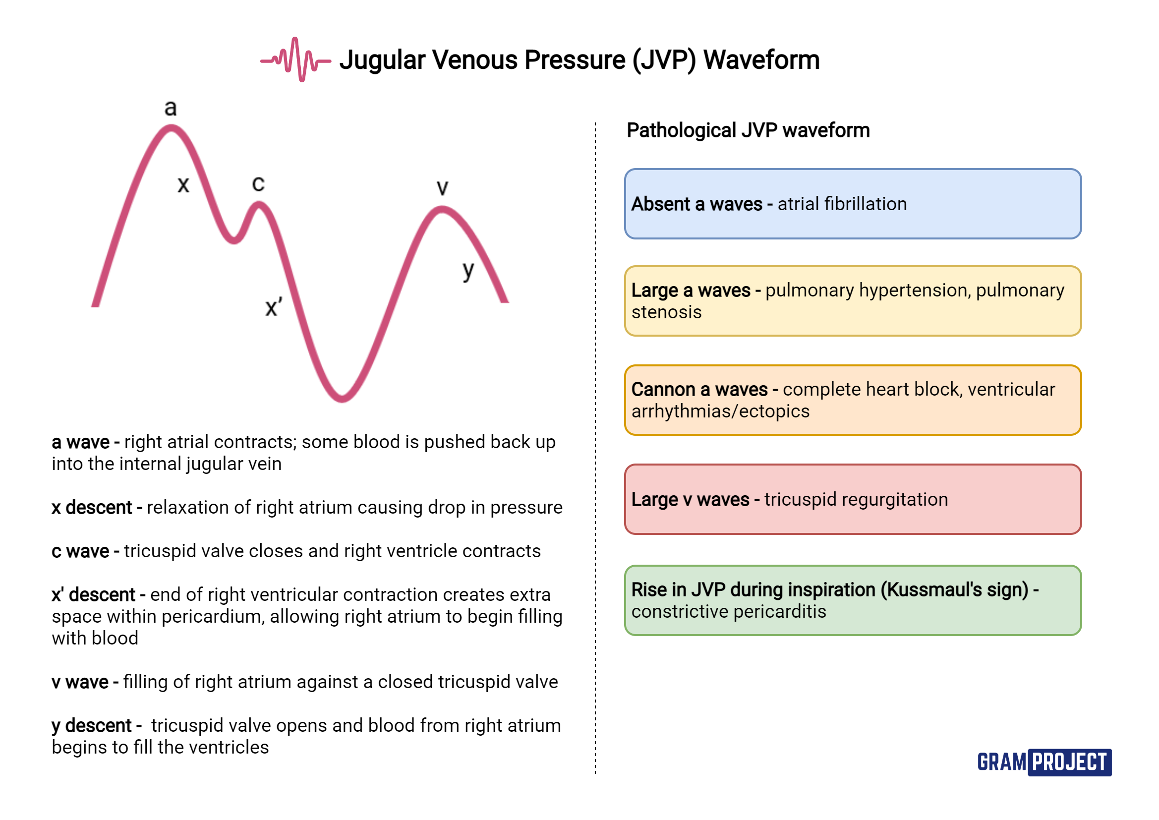
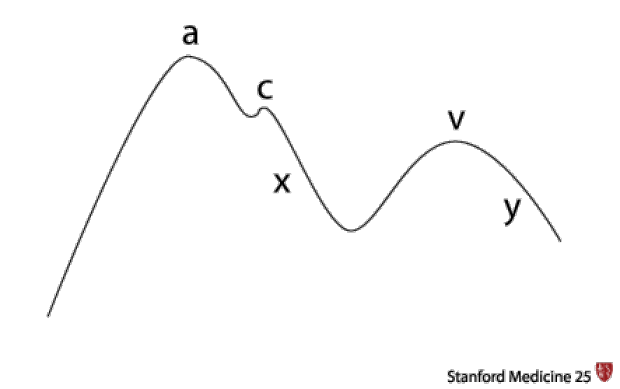
JVP Waveform Analysis (when assessable - older children)
| Wave/Deflection | Represents | Abnormality |
|---|---|---|
| a wave | Atrial contraction | Absent in AF; cannon a wave in CHB, PVST, TS |
| c wave | Tricuspid valve closure | Usually not visible |
| x descent | Atrial relaxation + RV systole | Exaggerated in cardiac tamponade |
| v wave | Venous return during ventricular systole | Giant v wave in TR |
| y descent | Tricuspid valve opening | Sharp y descent in constrictive pericarditis |
Clinical Significance of JVP in Children
| Finding | Clinical Meaning |
|---|---|
| Raised JVP | Right heart failure, constrictive pericarditis, cardiac tamponade, TR, TS |
| Low JVP | Hypovolemia, shock, dehydration |
| Kussmaul's sign (JVP rises on inspiration) | Constrictive pericarditis, cardiac tamponade, RV failure |
| Hepatojugular reflux | Right heart failure, TR |
| Friedrich's sign (rapid fall and rise) | TR, constrictive pericarditis |
3. Palpation of the Precordium
Apex Beat
- Normal location by age:
- Neonate/infant (<2 years): 4th ICS, lateral to mid-clavicular line
- 2-7 years: 4th-5th ICS, mid-clavicular line
-
7 years: 5th ICS, mid-clavicular line (adult position)
- Lateral displacement: LV dilatation (large VSD, MR, DCM)
- Sustained/heaving impulse: LV hypertrophy (AS, hypertension)
- Tapping apex: Palpable S1 - mitral stenosis
Left Parasternal Heave
- Palpate with the heel of the hand along the left sternal border
- Positive heave (lift): RV enlargement/hypertrophy - seen in pulmonary hypertension, large ASD, PS, TOF
Thrills
- Systolic thrill at LUSB: PS, AS, large VSD
- Diastolic thrill at apex: Mitral stenosis (rare in children)
- Continuous thrill: PDA
4. Percussion - Relevance in Pediatric Examination
Why Percussion Has Limited Utility in Children
- Chest X-ray is more accurate: The cardiothoracic ratio (CTR) on CXR is a far more reliable and reproducible method of assessing cardiac size. CTR >0.55 in children (>0.60 in infants) indicates cardiomegaly.
- Small chest dimensions: The compact thorax of infants/toddlers makes demarcating cardiac dullness from lung resonance unreliable.
- Overlying lung tissue variability: Lung hyperinflation (as in bronchiolitis, asthma) artificially narrows the area of cardiac dullness by percussion.
- ECG and echo are more informative: These modalities provide direct chamber dimension data.
When Percussion Is Still Useful
- Pleural effusion detection: Stony dull percussion note at the lung base can indicate pleural effusion (seen in heart failure, post-cardiac surgery)
- Pericardial effusion: Gross enlargement of the cardiac shadow can be appreciated as a wide area of cardiac dullness
- Dextrocardia: Percussion helps confirm cardiac position when echocardiography is unavailable - dullness on the right with resonance on the left
- Hepatomegaly: Percussion of the right upper quadrant defines the liver span - important surrogate for right heart congestion in infants where JVP is unreliable
5. Auscultation
Practical Tips
- Use a pediatric stethoscope with bell and diaphragm
- Bell (light pressure): detects low-frequency sounds - S3, S4, mid-diastolic murmurs (e.g., mitral stenosis rumble)
- Diaphragm (firm pressure): detects high-frequency sounds - S1, S2, systolic murmurs, pericardial rub
- In infants, perform auscultation first before any distressing maneuvers
- At high heart rates (>160 bpm in neonates), it can be difficult to distinguish systole from diastole - palpate the pulse simultaneously to orient yourself
Auscultation Areas in Children
- Mitral area (apex) - 4th/5th ICS MCL
- Tricuspid area - 4th ICS left sternal border (LSB)
- Pulmonary area - 2nd ICS left sternal border
- Aortic area - 2nd ICS right sternal border
- Erb's point - 3rd ICS LSB (useful for early diastolic murmurs of AR and PR)
- Axilla - radiation of MR
- Back - radiation of PS, coarctation, PDA
Heart Sounds
S1 (First Heart Sound)
- Closure of mitral (M1) and tricuspid (T1) valves
- M1 precedes T1; normally heard as a single sound
- Best heard at the apex and LLSB
- Loud S1: High output states, MS with pliable valve, short PR interval
- Soft S1: Poor LV function, MR, long PR interval
- Variable S1: Complete heart block, AF
S2 (Second Heart Sound)
- Closure of aortic (A2) and pulmonary (P2) valves
- Best heard at the base (2nd ICS)
- Physiological splitting of S2: A2 precedes P2; splitting increases on inspiration (increased RV filling → delayed P2) - normal finding in children and adolescents
- Wide fixed split S2: ASD (hallmark finding)
- Wide variable split: RBBB, PS, RV failure
- Narrow split / single S2: Pulmonary hypertension (P2 loud and early), severe AS, Eisenmenger syndrome
- Reversed (paradoxical) split: LBBB, severe AS
S3 (Third Heart Sound)
- Low-frequency sound at the apex, heard in early diastole (during rapid ventricular filling)
- Can be normal in children (physiological S3) - especially thin, athletic children
- Pathological S3: Dilated cardiomyopathy, large left-to-right shunt (e.g., large VSD), heart failure
- Best heard with the bell of the stethoscope
S4 (Fourth Heart Sound)
- Low-frequency presystolic sound (atrial gallop)
- Always pathological in children
- Indicates reduced ventricular compliance
- Seen in: severe hypertension, severe AS, HCM, cardiomyopathy
Gallop Rhythms
- S3 gallop (proto-diastolic): Suggests ventricular dysfunction or volume overload
- S4 gallop (presystolic/atrial): Suggests stiff, non-compliant ventricle
- Summation gallop: When S3 + S4 fuse (seen in tachycardia) - sounds like a horse's gallop
Murmurs: Key Features to Assess
| Attribute | Details |
|---|---|
| Site | Where loudest? (mitral, tricuspid, pulmonary, aortic area) |
| Radiation | Axilla (MR), carotid (AS), back (PS, PDA) |
| Intensity | Graded I-VI (Levine scale) |
| Character | Harsh, blowing, rumbling, machinery |
| Pitch | High, medium, low |
| Timing | Systolic, diastolic, continuous |
| Relation to posture/maneuvers | Valsalva, standing, squatting |
- Grade I: Very faint; only heard in quiet room
- Grade II: Faint but easily heard
- Grade III: Moderate; no thrill
- Grade IV: Loud; thrill present
- Grade V: Very loud; heard with stethoscope barely on chest
- Grade VI: Heard without stethoscope
Innocent vs. Pathological Murmurs
- Grade III/IV or higher with thrill
- Diastolic or continuous murmur (almost always pathological)
- Abnormal S2 (fixed split, loud P2, single S2)
- Pan-systolic or early systolic murmur
- Associated symptoms (cyanosis, poor weight gain, syncope, exercise intolerance)
- Radiation to axilla or back
- Harsh/blowing character
Common Murmurs in Children
| Murmur | Type | Location | Radiation | Character |
|---|---|---|---|---|
| VSD | Pansystolic | 3rd-4th ICS LSB | Whole precordium | Harsh |
| ASD | Systolic ejection | 2nd ICS LSB | - | Ejection; fixed split S2 |
| PDA | Continuous | 2nd ICS LSB | Back | "Machinery" murmur |
| PS | Systolic ejection | 2nd ICS LSB | Back | Ejection; wide split S2 |
| AS | Systolic ejection | 2nd ICS RSB | Carotid | Ejection; narrow S2 |
| MR | Pansystolic | Apex | Axilla | Blowing, high-pitched |
| AR | Early diastolic | 3rd ICS LSB | - | Blowing, high-pitched |
| MS | Mid-diastolic | Apex | - | Rumbling; loud S1, OS |
| Still's | Systolic | 3rd-4th ICS LSB | - | Musical/vibratory |
Pericardial Friction Rub
- Scratchy, to-and-fro sound heard in pericarditis
- Best heard at left sternal border with patient leaning forward
- Three components: atrial systole, ventricular systole, ventricular diastole
- Unlike pleural rub: persists when breath is held
6. Other Systems to Complete the CVS Examination
Peripheral Pulses
- Brachial (infants), radial (older children)
- Femoral pulses: Always palpate - radio-femoral delay indicates coarctation of the aorta (essential to not miss)
- Pulse character: Collapsing (PDA, AR), bounding (large shunts), weak/absent (coarctation, tamponade)
Blood Pressure
- Four-limb BP in suspected coarctation: >20 mmHg difference between upper and lower limbs is significant
- Age-appropriate cuff size is mandatory
Respiratory System
- Tachypnea is the earliest sign of cardiac decompensation in infants
- Pulmonary rales indicate left heart failure (less common in infants due to adaptive lymphatic drainage)
- Pleural effusion (stony dull, absent breath sounds) suggests decompensated heart failure
Abdomen
- Hepatomegaly (>3 cm below RCM in infants; >2 cm in children): surrogate for raised JVP and right heart congestion
- Splenomegaly: infective endocarditis, congestive splenomegaly
- Pulsatile liver: severe TR, markedly elevated right heart pressures
- Ascites: severe right heart failure, constrictive pericarditis
Periphery
- Clubbing: Cyanotic CHD, infective endocarditis
- Cyanosis: Central (tongue/mucous membranes) or peripheral
- Edema: Dependent (sacral in infants who lie flat, ankle in older children); periorbital in infants
- Splinter hemorrhages: IE
- Osler nodes / Janeway lesions: IE
Summary Framework for the Exam
General → Vitals (HR, RR, BP 4 limbs, SpO₂) → Peripheral signs (clubbing, cyanosis, edema)
→ Pulses (radial, femoral, character)
→ JVP (>8 years only; use hepatomegaly as surrogate in infants)
→ Precordium Inspection (bulge, visible pulsations, scars)
→ Palpation (apex beat, parasternal heave, thrills, pulsations)
→ Percussion (limited role; mainly for pleural effusion, hepatomegaly, dextrocardia)
→ Auscultation (S1, S2, added sounds, murmurs, rub)
→ Lung bases (rales, effusion)
→ Abdomen (hepatomegaly, splenomegaly)
- Precordial bulge = chronic cardiomegaly in children; adult chests are too rigid for this
- JVP is not reliably assessable below age 8; use hepatomegaly as the equivalent marker of right heart congestion in infants
- Percussion has limited utility; CXR, ECG, and echo are far more informative
- S3 can be physiological in children; S4 is always pathological
- Fixed split S2 = ASD until proven otherwise
- Loud P2 = pulmonary hypertension
- Disappearance of a murmur on standing is the strongest predictor of an innocent murmur (98% PPV)
- Always palpate femoral pulses to exclude coarctation
With images
precordial bulge pediatric cardiac examination image site:wikimedia.org OR site:radiopaedia.org OR site:teachmepediatrics.com
JVP waveform a c x v y waves diagram pediatric jugular venous pressure
heart auscultation areas pediatric diagram S1 S2 heart sounds children stethoscope placement
precordial bulge cardiac enlargement child chest photo clinical finding cardiac examination
heart murmur grading Levine scale diagram innocent pathological murmur pediatric location
PE23.10 - Cardiovascular System Examination: Children & Infants (With Images)
Overview: The Pediatric Approach
1. Inspection of the Precordium
Precordial Bulge
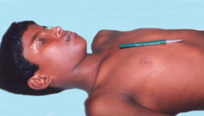
- Indicates long-standing cardiomegaly (the heart has been enlarged for a prolonged period)
- A left-sided bulge suggests LV enlargement or biventricular enlargement
- A right-sided (parasternal) bulge suggests chronic RV enlargement
- Causes: Large left-to-right shunts (VSD, PDA, large ASD), chronic rheumatic heart disease, dilated cardiomyopathy
Pulsations in the Precordium
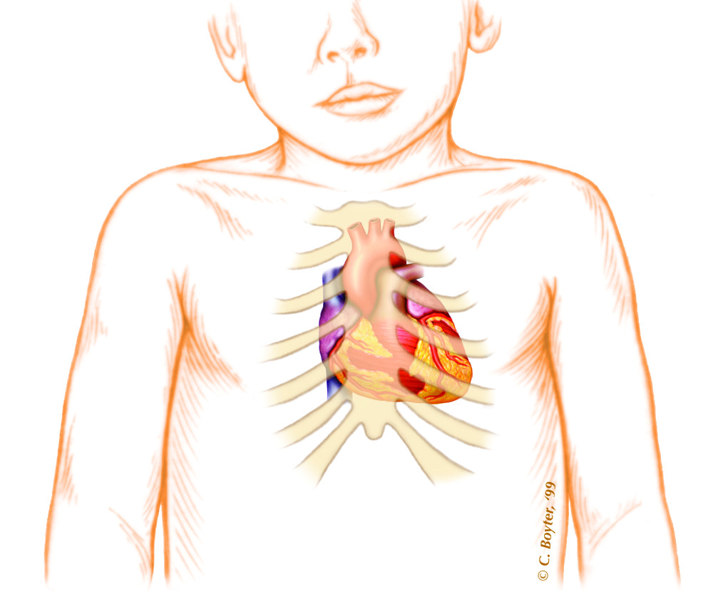
| Pulsation | Location | Significance |
|---|---|---|
| Hyperdynamic apical impulse | 4th-5th ICS, MCL | Volume overload (VSD, MR, PDA) |
| Left parasternal pulsation | Left sternal border | RV hypertrophy/enlargement |
| Epigastric pulsation | Below xiphisternum | RV hypertrophy, aortic dilatation |
| Visible thrill | Any precordial area | Significant turbulent flow |
| Suprasternal pulsation | Suprasternal notch | Aortic dilatation, PDA, coarctation |
2. Jugular Venous Pressure (JVP)
The JVP Waveform
JVP Assessment - Pediatric Specifics
- Short, fat necks make the IJV inaccessible
- Crying and movement falsely raise intrathoracic pressure
- High heart rates (>100 bpm) prevent discernment of individual waveforms
- Use hepatomegaly as the surrogate - liver >3 cm below the right costal margin in infants = right atrial hypertension
- Semi-recumbent position at 45°, head turned slightly left
- Identify the IJV running from the medial clavicle to the earlobe, under the sternocleidomastoid
- The IJV shows a double waveform (a + v) distinguishing it from the carotid (single pulse)
- Measure vertical distance from sternal angle to top of pulsation: normal ≤3 cm (= RAP <8 cmH₂O)
JVP Waveform Components
| Component | Represents | Abnormality |
|---|---|---|
| a wave | Right atrial contraction | Absent = AF; Cannon = CHB/VT; Giant = PS/TS |
| c wave | Tricuspid valve bulging into RA | Usually not visible clinically |
| x descent | Atrial relaxation + RV systole | Exaggerated = cardiac tamponade |
| v wave | Passive RA filling (tricuspid closed) | Giant v = tricuspid regurgitation |
| y descent | Tricuspid valve opens → RA empties | Prominent sharp y = constrictive pericarditis |
Clinical Significance of JVP Changes
| Finding | Clinical Meaning |
|---|---|
| Raised JVP | Right heart failure, constrictive pericarditis, tamponade, TR, TS |
| Low JVP | Hypovolemia, dehydration, shock |
| Kussmaul's sign (JVP rises on inspiration) | Constrictive pericarditis, cardiac tamponade, RV failure |
| Hepatojugular reflux positive | Right heart failure, TR |
| Friedrich's sign (rapid y descent) | TR, constrictive pericarditis |
3. Palpation of the Precordium
Apex Beat
- Normal location by age:
- Neonate/infant (<2 yrs): 4th ICS, lateral to mid-clavicular line
- 2-7 years: 4th-5th ICS, mid-clavicular line
-
7 years: 5th ICS MCL (adult position)
- Lateral displacement = LV dilatation
- Sustained/heaving impulse = LV hypertrophy
- Tapping apex = palpable S1 (mitral stenosis)
Parasternal Heave
- Heel of hand along left sternal border
- Positive lift/heave = RV enlargement (pulmonary hypertension, large ASD, PS, TOF)
Thrills (palpable murmurs)
- Systolic thrill at LUSB: PS, AS, large VSD
- Continuous thrill: PDA
- Diastolic thrill at apex: MS (rare in children)
4. Percussion - Relevance in Pediatric Examination
- Chest X-ray is more accurate - CTR >0.55 (>0.60 in infants) on CXR reliably indicates cardiomegaly
- Small chest - compact thorax makes demarcating cardiac dullness unreliable
- Lung hyperinflation (bronchiolitis, asthma) artificially narrows the area of cardiac dullness
- Echo and ECG are superior for chamber dimensions
- Detecting pleural effusion (stony dull at base) - seen in heart failure, post-cardiac surgery
- Confirming dextrocardia when imaging is unavailable
- Defining hepatomegaly (right upper quadrant percussion to define liver span - surrogate for right heart congestion in infants)
5. Auscultation
5 Areas for Auscultation

- Aortic - Right 2nd ICS
- Pulmonary - Left 2nd ICS
- Erb's point - Left 3rd ICS (AR, PR murmurs)
- Tricuspid - Left 4th ICS lower sternal border
- Mitral - Apex (5th ICS MCL)
- Use bell (light pressure) for low-frequency sounds: S3, S4, mid-diastolic rumble
- Use diaphragm (firm pressure) for high-frequency: S1, S2, systolic murmurs, pericardial rub
- In infants: auscultate first before any distressing maneuver; palpate pulse simultaneously to orient systole vs diastole at rapid heart rates
Heart Sounds
S1 - "LUB"
- Closure of mitral (M1) + tricuspid (T1) valves; marks start of systole
- Best heard at apex and LLSB
- Loud S1: MS (pliable valve), high output state, short PR
- Soft S1: Poor LV function, MR, long PR
- Variable S1: Complete heart block, AF
S2 - "DUB"
- Closure of aortic (A2) + pulmonary (P2) valves; marks end of systole
- Best heard at the base (2nd ICS)
- Physiological splitting (A2 before P2, widens on inspiration): normal in children and adolescents
| S2 Splitting Pattern | Clinical Meaning |
|---|---|
| Wide fixed split | ASD (hallmark) |
| Wide variable split | RBBB, PS, RV failure |
| Single S2 | Pulmonary hypertension (Eisenmenger), severe AS |
| Reversed (paradoxical) split | LBBB, severe AS |
| Loud P2 | Pulmonary hypertension |
S3 - Early diastolic (rapid ventricular filling)
- Low frequency; heard at apex with bell
- Can be physiological in children (thin, athletic)
- Pathological: Dilated cardiomyopathy, large VSD, heart failure
S4 - Presystolic (atrial gallop)
- Always pathological in children
- Reduced ventricular compliance
- Seen in: severe AS, HCM, cardiomyopathy, severe hypertension
Murmurs
Murmur Timing - Key Patterns

- Systolic ejection (crescendo-decrescendo): AS, PS
- Holosystolic/Pansystolic: VSD, MR, TR
- Mid-systolic click + late systolic: MVP
- Early diastolic decrescendo: AR, PR
- Mid-diastolic rumble: MS, relative tricuspid stenosis in ASD
- Continuous "machinery": PDA
Innocent vs. Pathological Murmurs - Locations & Types
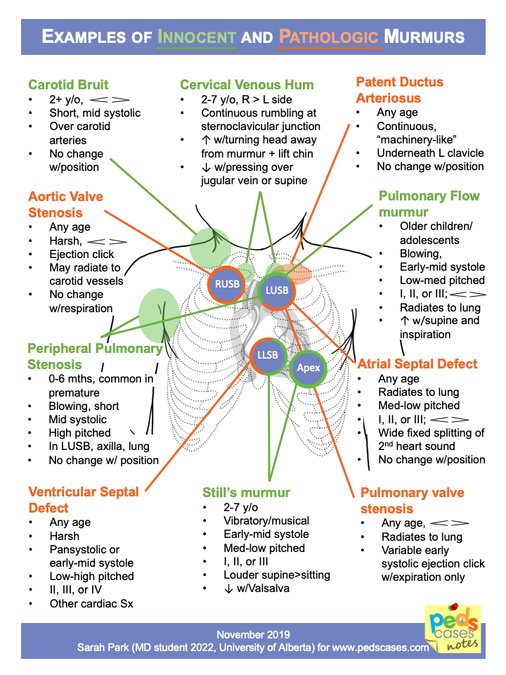
Levine Grading of Murmur Intensity
| Grade | Description | Thrill? |
|---|---|---|
| I | Very faint; only in quiet room | No |
| II | Faint but easily heard | No |
| III | Moderately loud | No |
| IV | Loud | Yes |
| V | Very loud; stethoscope barely on chest | Yes |
| VI | Heard without stethoscope | Yes |
Key Murmur Quick Reference
| Lesion | Type | Best Area | Radiation | S2 Change |
|---|---|---|---|---|
| VSD | Pansystolic | 3rd-4th ICS LSB | Whole precordium | Normal |
| ASD | Systolic ejection | 2nd ICS LSB | Lungs | Fixed wide split |
| PDA | Continuous | 2nd ICS LSB | Back, left clavicle | Normal |
| PS | Systolic ejection | 2nd ICS LSB | Back | Wide split |
| AS | Systolic ejection | 2nd ICS RSB | Carotid | Narrow/single |
| MR | Pansystolic | Apex | Axilla | Normal |
| AR | Early diastolic | 3rd ICS LSB | - | Single (loud A2) |
| Still's (innocent) | Ejection systolic | 3rd-4th ICS LSB | None | Normal |
Innocent Murmur Features (7 S's)
Pericardial Friction Rub
- Scratchy, to-and-fro, three-component sound (atrial systole + ventricular systole + diastole)
- Best heard: LLSB with patient leaning forward, breath held in expiration
- Distinguishing feature: persists when breath is held (unlike pleural rub)
- Seen in: pericarditis (viral, post-surgical, rheumatic, uraemic)
6. Completing the Examination - Other Systems
Peripheral Pulses
- Always palpate femoral pulses - radio-femoral delay = coarctation of aorta
- Collapsing/bounding pulse: PDA, AR, high-output states
- Weak/absent pulses: coarctation, tamponade, severe LV failure
Blood Pressure (4-limb)
-
20 mmHg upper-to-lower limb gradient = coarctation
Respiratory System
- Tachypnea = earliest sign of cardiac decompensation in infants
- Pulmonary rales = LVF (less common in infants due to good lymphatic drainage)
- Stony dull + absent breath sounds at lung base = pleural effusion
Abdomen
- Hepatomegaly (>3 cm below RCM in infants) = right heart congestion surrogate for JVP
- Pulsatile liver = severe TR / markedly raised right heart pressures
- Splenomegaly = infective endocarditis, congestive splenomegaly
Peripheral Signs
- Clubbing: Cyanotic CHD, infective endocarditis
- Central cyanosis (tongue, mucous membranes): Right-to-left shunts
- Edema: Sacral (infants lying flat), ankle (older children)
- Splinter hemorrhages / Osler nodes / Janeway lesions: Infective endocarditis
Summary Examination Framework
General inspection
    ↓
Vitals: HR, RR, SpO₂, BP (4 limbs)
    ↓
Peripheral signs: clubbing, cyanosis, edema, splinter hemorrhages
    ↓
Pulses: rate, rhythm, character, radio-femoral delay
    ↓
JVP: only >8 yrs; hepatomegaly as surrogate in infants
    ↓
Precordium INSPECTION: bulge → visible pulsations → scars
    ↓
PALPATION: apex beat → parasternal heave → thrills → epigastric pulsation
    ↓
PERCUSSION: limited role → pleural effusion, hepatomegaly, dextrocardia
    ↓
AUSCULTATION: S1 → S2 (splitting) → S3/S4 → murmurs → rub
    ↓
Lung bases: rales, dullness (effusion)
    ↓
Abdomen: hepatomegaly, splenomegaly, pulsatile liver
- Precordial bulge = chronic cardiomegaly; adult chests are too rigid for this
- JVP unreliable below age 8 - use hepatomegaly as the equivalent sign
- Percussion has limited utility; supplement with CXR, ECG, echo
- S3 can be physiological in children; S4 is always pathological
- Fixed split S2 = ASD until proven otherwise
- Loud P2 = pulmonary hypertension
- Murmur disappearing on standing = innocent with 98% positive predictive value
- Always palpate femoral pulses - missing radio-femoral delay = missing coarctation
Explain with images
Which part of the cardiovascular examination would you like explained with images? · What format works best for you?
JVP measurement technique position clinical photo 45 degrees internal jugular vein
clubbing fingers cyanosis pediatric cardiac examination clinical photo
S2 splitting diagram physiological fixed wide reversed ASD pulmonary hypertension heart sound
hepatomegaly infant right heart failure liver palpation technique pediatric clinical sign
parasternal heave apex beat palpation technique diagram cardiac exam RV hypertrophy
PE23.10 - Cardiovascular Examination in Children & Infants
Full Step-by-Step Illustrated Guide
STEP 1: General Inspection & Peripheral Signs
Clubbing + Cyanosis
- Place dorsal surfaces of two matching fingers together
- Normally a diamond-shaped window is visible at the nail bases
- In clubbing, this window is obliterated
- Causes in cardiac patients: cyanotic CHD, infective endocarditis
- Indicates SaO₂ <85% - significant right-to-left shunting
- Always check the tongue - peripheral cyanosis of cold hands can be normal
STEP 2: Inspection of the Precordium
Precordial Bulge
- Left-sided bulge → LV or biventricular enlargement (large VSD, PDA, DCM)
- Right-sided (parasternal) bulge → Chronic RV enlargement (large ASD, severe PS, TOF)
- Indicates long-standing cardiomegaly; cannot develop acutely
Visible Pulsations
| Pulsation Location | Significance |
|---|---|
| Hyperdynamic apex | Volume overload: VSD, MR, PDA |
| Left parasternal | RV hypertrophy (visible lift) |
| Epigastric | RV hypertrophy, aortic dilatation |
| Suprasternal | PDA, coarctation, aortic dilatation |
| Visible thrill | Significant shunt/stenosis |
STEP 3: Jugular Venous Pressure (JVP)
Anatomy and Measurement Technique
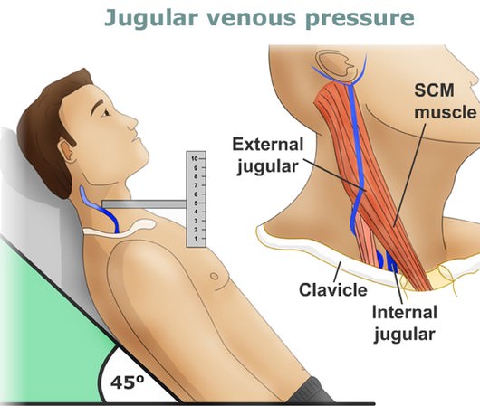
- Patient semi-recumbent at 45°, head turned slightly left
- Identify the IJV path: from medial clavicle → earlobe, running under the sternocleidomastoid
- The IJV shows a double waveform (a + v) - this distinguishes it from the carotid (single pulsation)
- Measure the vertical height from the sternal angle to the top of the pulsation column
- Normal = ≤3 cm (equivalent to RAP ≤8 cmH₂O)
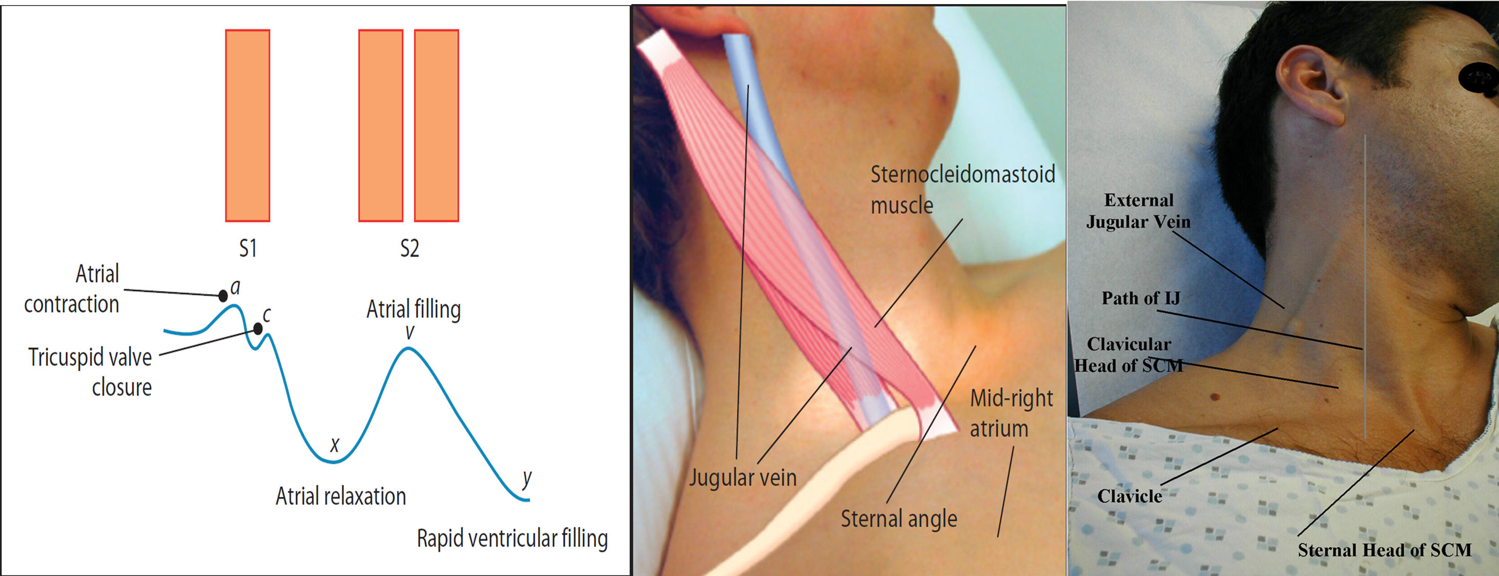
JVP Waveform Interpretation
| Wave/Descent | Mechanism | Pathology When Abnormal |
|---|---|---|
| a wave | RA contracts | Absent = AF; Cannon = CHB/VT; Giant = PS/TS |
| c wave | Tricuspid bulges into RA | Usually invisible clinically |
| x descent | Atrial relaxation | Exaggerated = cardiac tamponade |
| v wave | RA fills passively (tricuspid closed) | Giant v wave = TR |
| y descent | Tricuspid opens → RA empties | Sharp/prominent = constrictive pericarditis |
Why JVP Cannot Be Assessed in Infants and Young Children
| Problem | Consequence |
|---|---|
| Short, fat neck | IJV anatomically inaccessible |
| Crying/movement | Raises intrathoracic pressure artificially |
| Heart rate >100 bpm | Individual waveforms cannot be discerned |
| Cannot cooperate | Cannot maintain 45° position or relax neck |
STEP 4: Palpation
Apex Beat Character
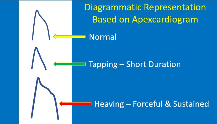
| Apex Beat Type | How it Feels | Meaning |
|---|---|---|
| Normal | Brief, localized tap | Normal LV |
| Tapping | Short, flicking, not displaced | Palpable S1 = Mitral stenosis |
| Heaving (sustained) | Forceful, lifts your finger, sustained | LV hypertrophy (AS, hypertension) |
| Hyperdynamic (thrusting) | Forceful but not sustained, displaced | LV volume overload (VSD, MR) |
| Displaced laterally | Apex >MCL | LV dilatation |
- Infant <2 years: 4th ICS, lateral to MCL
- 2-7 years: 4th-5th ICS, MCL
-
7 years: 5th ICS MCL (adult position)
Parasternal Heave
- Place the heel of the hand along the left sternal border
- A positive heave lifts your entire hand with each systole
- Indicates RV enlargement/hypertrophy
- Causes: pulmonary hypertension, large ASD, PS, TOF, mitral stenosis
Thrills (Palpable Murmurs = Grade IV+)
| Location of Thrill | Likely Cause |
|---|---|
| 2nd ICS LSB (pulmonary area) | PS, large VSD |
| 2nd ICS RSB (aortic area) | AS |
| 3rd-4th ICS LSB | VSD |
| Suprasternal notch | AS, PDA |
| Continuous machinery thrill | PDA |
STEP 5: Percussion
| Use of Percussion | What to Do | What to Find |
|---|---|---|
| Pleural effusion | Percuss lower chest | Stony dull = effusion (right > left in heart failure) |
| Hepatomegaly | Percuss RUQ downward | Defines liver upper border (used with palpation for liver span) |
| Dextrocardia | Percuss both sides | Dullness on right, resonance on left |
| Pericardial effusion | Precordial dullness | Unusually wide area of dullness |
STEP 6: Auscultation
5 Areas to Listen
- Aortic - Right 2nd ICS
- Pulmonary - Left 2nd ICS
- Erb's point - Left 3rd ICS (AR, PR early diastolic murmurs)
- Tricuspid - Left 4th ICS lower sternal border
- Mitral - Apex (5th ICS MCL)
- Bell (light touch): S3, S4, mitral stenosis rumble - low frequency sounds
- Diaphragm (firm pressure): S1, S2, all systolic murmurs, pericardial rub - high frequency
- In infants: auscultate before any painful/distressing step; palpate pulse simultaneously to identify systole at high heart rates
S2 Splitting Patterns - Clinical Significance
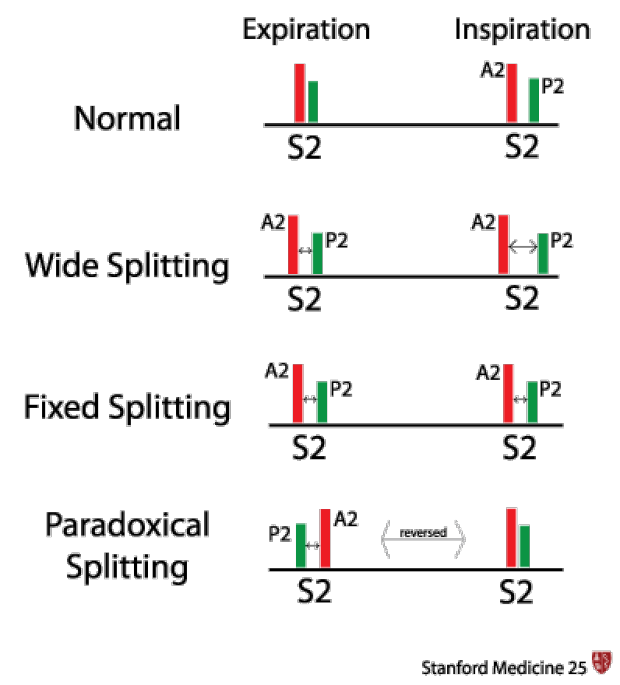

- Physiological split (widens on inspiration, disappears on expiration) = NORMAL in children and adolescents
- Fixed split (same in inspiration and expiration) = ASD until proven otherwise
- Loud P2 heard beyond the pulmonary area = pulmonary hypertension
- Single S2 = severe pulmonary hypertension (Eisenmenger), severe AS
Murmur Types - Timing Diagrams
Innocent vs. Pathological Murmurs - Location Map
- Location on the precordium
- Age of presentation
- Character and pitch
- Radiation
- Change with position
Innocent Murmur Features (7 S's)
| Feature | Detail |
|---|---|
| Soft | Grade I or II |
| Short | Not pan-systolic |
| Systolic only | Never diastolic or continuous |
| Symptom-free | No cyanosis, no FTT, no syncope |
| Supine louder | Decreases on sitting/standing |
| Single S2 | No fixed split, no loud P2 |
| No Spread | No radiation, no thrill |
Levine Grading Scale
| Grade | Description | Thrill? |
|---|---|---|
| I | Very faint, easily missed | No |
| II | Soft, easily heard | No |
| III | Moderately loud | No |
| IV | Loud + thrill | Yes |
| V | Very loud, stethoscope barely on chest | Yes |
| VI | Heard without stethoscope | Yes |
STEP 7: Completing the Examination - Abdomen
Hepatomegaly: The JVP Surrogate in Infants

- Start palpation from the right iliac fossa and move upward
- Lower border: palpation → the liver edge is felt below the right costal margin
- Upper border: percussion downward from the right lung → note where resonance becomes dull
- Liver span (normal by age): 4.5-5 cm in neonates, up to 8 cm in 12-year-olds
- Abnormal: Liver edge >3 cm below right costal margin in infants = hepatomegaly = right heart congestion
| Finding | Mechanism | Cause |
|---|---|---|
| Tender hepatomegaly | Venous congestion → capsule stretch | Right heart failure, constrictive pericarditis |
| Pulsatile liver | Systolic expansion with each beat | Severe TR, very high right heart pressures |
| Hard, non-tender liver | Cardiac cirrhosis | Chronic right heart failure |
| Splenomegaly | Venous congestion, IE | IE, congestive splenomegaly |
Summary: Examination Framework at a Glance
STEP 1: General + Peripheral Signs
        ↓ Clubbing, central cyanosis, edema, splinter hemorrhages

STEP 2: Vitals
        ↓ HR, RR, SpO₂, BP (4 limbs - femoral = coarctation screen)

STEP 3: Inspect Precordium
        ↓ Precordial bulge → visible pulsations → scars → chest shape

STEP 4: JVP (>8 yrs only; hepatomegaly as surrogate in infants)
        ↓ Height, waveform, Kussmaul's sign

STEP 5: Palpate Precordium
        ↓ Apex beat (site, character) → parasternal heave → thrills

STEP 6: Percuss
        ↓ Pleural effusion, hepatomegaly, dextrocardia

STEP 7: Auscultate
        ↓ S1 → S2 (splitting pattern) → S3/S4 → murmurs → rub
        ↓ Lung bases (rales, dullness)

STEP 8: Abdomen
        ↓ Hepatomegaly, splenomegaly, pulsatile liver
- Precordial bulge = chronic cardiomegaly - unique to children (compliant chest wall)
- JVP unreliable <8 years - use hepatomegaly as your right-heart pressure gauge
- Fixed split S2 = ASD until proven otherwise - never normal
- Murmur disappears on standing = innocent (98% PPV) - the single best bedside test
- Always palpate femoral pulses - radio-femoral delay = coarctation of aorta