
Why cardiac muscle cannot be tetanized
Cardiac muscle cannot be tetanized due to its very long refractory period, which lasts almost as long as the contraction itself. This means the muscle cannot be re-stimulated until it has nearly fully relaxed, preventing sustained (tetanic) contraction.
Searching sources...
Reading File
Finding Sources
Finding Sources
Reading File
Finding Sources
Reading File
Reading File
Reading File
Loading Image
Now I have everything needed for a thorough, well-sourced answer. Let me fetch the cardiac action potential figure from Costanzo as well.
Loading Image
Here is a complete, textbook-grounded explanation:
Why Cardiac Muscle Cannot Be Tetanized
The Short Answer
Tetanus in any muscle requires that a second stimulus arrive before the muscle has fully relaxed, so contractions summate. In cardiac muscle, the absolute refractory period (ARP) lasts almost as long as the contraction itself, making it physically impossible for a second stimulus to trigger another contraction before the first one is over. By the time the muscle can respond again, it has already relaxed.
1. The Cardiac Action Potential Has an Exceptionally Long Plateau (Phase 2)
Unlike skeletal muscle (action potential lasts ~1-2 ms), the cardiac ventricular action potential lasts ~250-300 ms, because of a sustained Phase 2 plateau driven by prolonged inward Ca²⁺ current through L-type Ca²⁺ channels (slow inward current). Outward K⁺ channels simultaneously close, keeping the membrane depolarized.
The five phases are:
| Phase | Name | Key Ion Current |
|---|---|---|
| 0 | Rapid depolarization (upstroke) | Rapid Na⁺ influx |
| 1 | Early repolarization | K⁺ efflux (transient) |
| 2 | Plateau | Inward Ca²⁺, reduced K⁺ efflux |
| 3 | Rapid repolarization | Ca²⁺ channels close; K⁺ efflux (IK) increases |
| 4 | Resting potential (-85 mV) | IK1 current (K⁺) |
The cardiac action potential action potentials in the ventricle, atrium, and SA node:
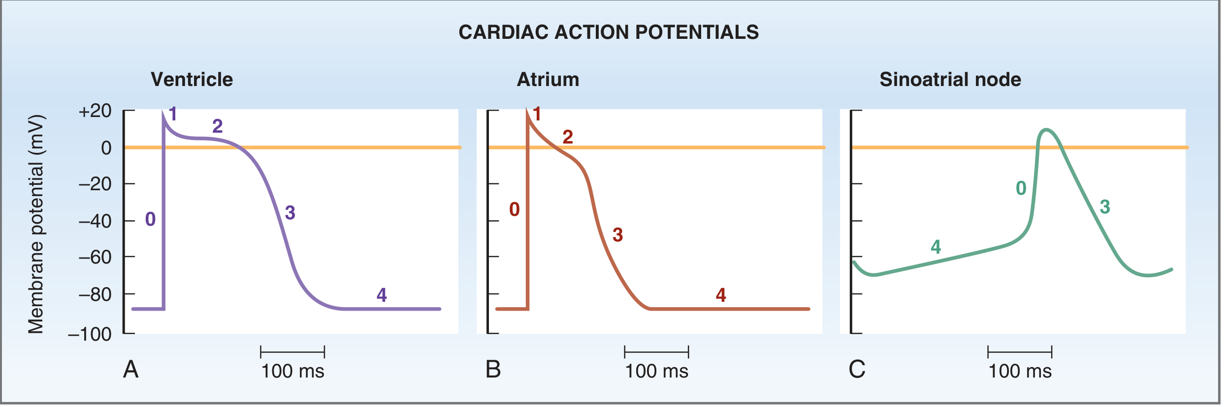
(Costanzo Physiology, Fig. 4.12)
2. The Long Plateau Directly Causes a Long Refractory Period
During Phases 0 through early Phase 3 (until the membrane potential reaches approximately -50 mV during repolarization), cardiac muscle is in absolute refractory period - it cannot be excited by any stimulus, no matter how strong. This is because the fast Na⁺ channels that mediate Phase 0 remain inactivated throughout the plateau.
- Absolute refractory period (ARP) of the ventricle: 0.25-0.30 seconds - essentially equal to the duration of the plateau action potential
- Relative refractory period (RRP): an additional ~0.05 seconds (late Phase 3 into Phase 4) during which only a very strong stimulus can re-excite the cell, but it produces a weaker-than-normal contraction
- The ARP in atria is shorter (~0.15 seconds), which is why atrial flutter/fibrillation is possible
"During phases 0 to 2 and about half of phase 3 (until the membrane potential reaches approximately -50 mV during repolarization), cardiac muscle cannot be excited again; that is, it is in its absolute refractory period. It remains relatively refractory until phase 4. Therefore, tetanus of the type seen in skeletal muscle cannot occur."
- Ganong's Review of Medical Physiology, 26th Ed.
3. Contraction Duration vs. Refractory Period - The Key Comparison
The contractile response of cardiac muscle begins at depolarization and lasts about 1.5 times as long as the action potential itself. The refractory period extends through virtually this entire mechanical contraction. Compare:
| Property | Skeletal Muscle | Cardiac Muscle |
|---|---|---|
| Action potential duration | ~1-2 ms | ~250-300 ms |
| Refractory period | ~2 ms | ~250-300 ms |
| Twitch duration | ~100 ms | ~300 ms |
| Tetanus possible? | Yes | No |
In skeletal muscle, the refractory period ends long before the twitch is over, so stimuli can pile up and summate. In cardiac muscle, the refractory period and twitch duration are nearly identical - there is no "window" in which a new stimulus can arrive while the fiber is still contracted.
4. Why This Is Physiologically Essential
As stated by Guyton & Hall: premature contractions in cardiac muscle do not cause wave summation the way they do in skeletal muscle (see figure below). Any stimulus delivered during the ARP is simply ignored. Even a premature beat during the RRP produces only a weakened, incomplete contraction - not summation.
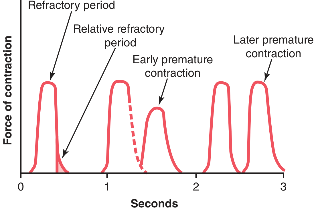
(Guyton & Hall, Fig. 9.6 - Note: premature contractions do not cause wave summation)
This is not a flaw - it is a protective design. As Ganong's notes: "Tetanization of cardiac muscle for any length of time would have lethal consequences" - a sustained (tetanic) contraction would prevent ventricular relaxation (diastole), halting cardiac filling and output entirely.
Summary
The reason cardiac muscle cannot be tetanized comes down to one mechanism:
The cardiac action potential plateau (Phase 2) prolongs the absolute refractory period to ~250-300 ms, which is approximately equal to the duration of the mechanical contraction. No new action potential can be triggered while the previous one is still active - so stimuli cannot summate, and tetanus is impossible.
This is a fundamental difference from skeletal muscle, where the 1-2 ms refractory period is far shorter than the ~100 ms twitch, allowing summation and tetanus to occur with repeated stimulation.
Sources: Guyton & Hall Textbook of Medical Physiology; Costanzo Physiology, 7th Ed.; Ganong's Review of Medical Physiology, 26th Ed.
Why in stokes adam syndrome after fainting condition improves
In Stokes-Adams syndrome, fainting occurs due to sudden complete heart block causing ventricular asystole. The condition improves after fainting because when the patient falls horizontal, cerebral perfusion is restored even with a slow ventricular escape rhythm. Additionally, lower pacemakers in the ventricle eventually "take over" and fire spontaneously.
Searching sources...
Reading File
Finding Sources
Finding Sources
Reading File
Reading File
Finding Sources
Finding Sources
Finding Sources
Finding Sources
Finding Sources
Finding Sources
Reading File
Finding Sources
I now have all the information needed for a complete, well-sourced answer. Here it is:
Stokes-Adams Syndrome: Why the Condition Improves After Fainting
What Is Stokes-Adams Syndrome?
Stokes-Adams syndrome refers to sudden episodes of loss of consciousness (syncope) caused by complete (third-degree) AV block, in which conduction between the atria and ventricles is completely interrupted. The ventricles either stop beating entirely (asystole) or beat at an extremely slow idioventricular rate - averaging only ~35 beats/min (sometimes as low as 15 beats/min), which is far too slow to maintain adequate cerebral perfusion. This results in dizziness and fainting (cerebral ischemia).
"The ventricular rate is lower; it averages 35 beats/min, but in individual cases it can be as low as 15 beats/min. In such individuals, there may also be periods of asystole lasting a minute or more. The resultant cerebral ischemia causes dizziness and fainting (Stokes-Adams syndrome)."
- Ganong's Review of Medical Physiology, 26th Ed.
Why Does the Condition Improve After Fainting?
There are three interlocking mechanisms that explain the spontaneous recovery:
1. The Horizontal (Supine) Posture Restores Cerebral Perfusion
This is the most immediate and important mechanism. When the patient is standing, the heart must pump blood upward against gravity to perfuse the brain. Even a low-output ventricular escape rhythm cannot overcome this.
When the patient falls, they become horizontal. Now:
- Gravity no longer opposes cerebral blood flow
- Even a very slow ventricular rate (e.g., 20-30 beats/min) can generate enough cardiac output to perfuse the brain adequately in the supine position
- Venous return to the heart also improves (less pooling in the legs), boosting stroke volume
This is why the recovery of consciousness in Stokes-Adams attacks is typically rapid once the patient is on the ground.
"Regaining of consciousness generally is rapid."
- Bradley and Daroff's Neurology in Clinical Practice
2. A Ventricular Escape Pacemaker Eventually "Takes Over"
When the SA node's impulses are completely blocked from reaching the ventricles, the ventricles do not remain in permanent asystole. Lower subsidiary pacemakers within the ventricular conduction system possess inherent automaticity - they can spontaneously depolarize and drive a slow but regular ventricular rhythm (idioventricular rhythm).
The key hierarchy of cardiac pacemakers:
| Pacemaker Site | Intrinsic Rate |
|---|---|
| SA node (normal) | 60-100 bpm |
| AV node / junctional (AV nodal block) | ~45 bpm |
| Bundle of His / Purkinje / ventricular muscle (infranodal block) | 15-40 bpm |
"When conduction from the atria to the ventricles is completely interrupted, complete (third-degree) heart block results, and the ventricles beat at a low rate (idioventricular rhythm) independently of the atria. In patients with AV nodal block, the remaining nodal tissue becomes the pacemaker and the rate of the idioventricular rhythm is approximately 45 beats/min."
- Ganong's Review of Medical Physiology, 26th Ed.
This escape pacemaker may take 5-10 seconds to "warm up" after the SA node impulses are cut off - this delay is precisely the window during which the patient loses consciousness. Once the escape rhythm establishes itself, cardiac output begins again and the patient recovers.
3. Sympathetic Reflexes Are Activated During the Attack
The sudden fall in blood pressure and cardiac output during asystole triggers a baroreceptor-mediated sympathetic surge. This:
- Increases the rate and reliability of ventricular escape foci (catecholamines enhance automaticity)
- Causes vasoconstriction to redirect blood toward the brain
- Helps stabilize and speed up the idioventricular rhythm
The Full Sequence of a Stokes-Adams Attack
SA node impulse blocked → Ventricular asystole (5-15 sec)
        ↓
Cerebral hypoperfusion → Syncope (patient falls horizontal)
        ↓
Horizontal posture → Cerebral blood flow restored even at low CO
        ↓
Ventricular escape pacemaker fires → Idioventricular rhythm established
        ↓
Sympathetic surge → Speeds escape rhythm, vasoconstriction
        ↓
Patient regains consciousness (often within 10-60 seconds)
Why Attacks Recur
The underlying complete heart block persists. Each subsequent attack follows the same pattern: the escape rhythm may fail transiently again, causing repeated syncopal episodes. This is why permanent cardiac pacemaker implantation is the definitive treatment - it provides a reliable backup rate that eliminates the asystolic pauses entirely.
Sources: Ganong's Review of Medical Physiology, 26th Ed.; Bradley and Daroff's Neurology in Clinical Practice; Adams and Victor's Principles of Neurology, 12th Ed.
Sign and symptoms of wpw syndrome
WPW (Wolff-Parkinson-White) syndrome features palpitations, syncope, and sometimes sudden cardiac death due to an accessory conduction pathway (Bundle of Kent) bypassing the AV node. The classic ECG shows a short PR interval, delta wave, and wide QRS complex.
Searching sources...
Finding Sources
Finding Sources
Reading File
Reading File
Reading File
Reading File
Reading File
Reading File
Now let me get the ECG images from Harrison's and Tintinalli for visual support.
Loading Image
Finding Sources
Reading File
Reading File
Loading Image
Excellent - I now have comprehensive textbook data and ECG images. Here is the full answer:
Signs and Symptoms of Wolff-Parkinson-White (WPW) Syndrome
What Is WPW?
WPW syndrome is defined as the combination of ventricular preexcitation on ECG (during sinus rhythm) plus episodes of tachyarrhythmia. It is caused by a congenital accessory pathway (AP) - commonly called the Bundle of Kent - that directly connects atrial to ventricular muscle, bypassing the AV node. It occurs in approximately 1 in 1,500-2,000 people. Most have structurally normal hearts, but WPW is associated with Ebstein's anomaly and hypertrophic cardiomyopathy.
Clinical Symptoms
Symptoms range from none to life-threatening, depending on whether and what type of arrhythmia develops:
| Symptom | Notes |
|---|---|
| Asymptomatic | Many patients are diagnosed incidentally on routine ECG |
| Palpitations | Most common symptom; episodic, rapid, regular or irregular heartbeat |
| Dyspnea | During tachyarrhythmia episodes |
| Chest discomfort | From rapid ventricular rates |
| Near-syncope / dizziness | Reduced cardiac output during fast arrhythmias |
| Syncope | Occurs in ~25% - syncope in WPW is serious and associated with life-threatening arrhythmia via the accessory pathway |
| Sudden cardiac death | Rare but possible - due to AF conducting rapidly over the AP, degenerating to ventricular fibrillation |
"WPW syndrome may be asymptomatic or present with palpitations, dyspnea, near syncope, syncope, or sudden death. In some patients, the diagnosis may only be made after typical ECG findings are discovered on an ECG performed for some other reason."
- Symptom to Diagnosis: An Evidence-Based Guide, 4th Ed.
The Classic ECG Triad (During Sinus Rhythm)
These three findings are the hallmark signs of WPW and are visible during sinus rhythm - they usually disappear during tachycardia:
| ECG Feature | Finding | Mechanism |
|---|---|---|
| Short PR interval | < 120 ms (< 0.12 sec) | AP bypasses AV node, so the normal physiological delay at the node is skipped - impulse reaches ventricle faster |
| Delta wave | Slurred, slow initial upstroke of QRS | AP inserts directly into ventricular muscle (not His-Purkinje), so early ventricular depolarization spreads slowly cell-to-cell |
| Wide/widened QRS | Slightly prolonged (> 0.12 sec) | Fusion of slow AP-driven depolarization + normal His-Purkinje activation creates a broad, fused QRS |
Additionally, discordant ST-T changes (ST and T waves directed opposite to the delta wave and QRS) are secondary repolarization abnormalities from altered depolarization. The delta wave can also create pseudo-Q waves that mimic myocardial infarction on ECG.
ECG showing the classic WPW features - short PR, delta wave, and widened QRS in leads I, II, and V1:
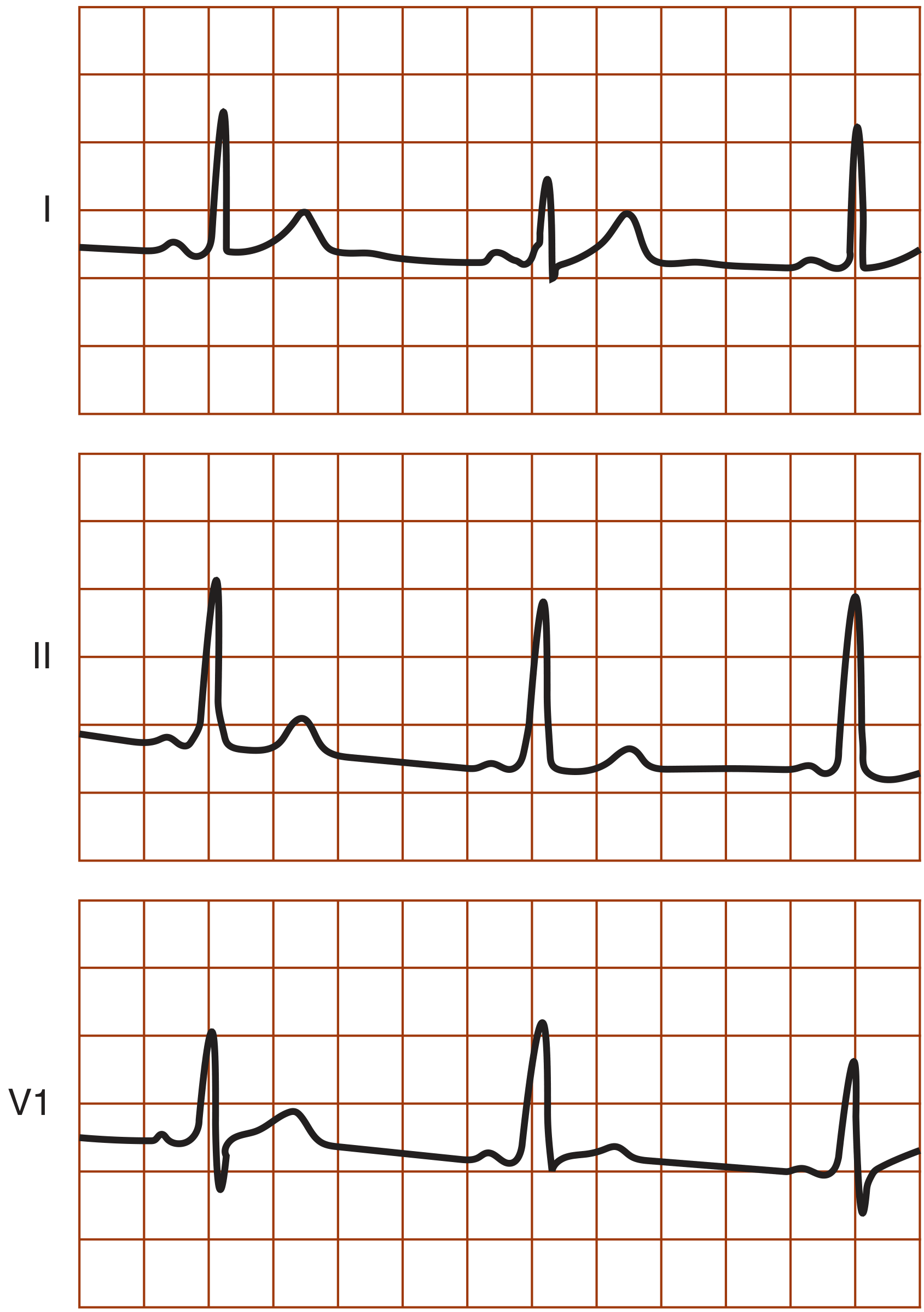
(Symptom to Diagnosis, Fig. 31-11)
Associated Arrhythmias (Signs During Tachycardia)
The ECG triad disappears during tachycardia. The three arrhythmias seen in WPW:
1. Orthodromic AV Reciprocating Tachycardia (AVRT) - 65% of cases
- Impulse travels DOWN the AV node normally, then back UP the AP in retrograde fashion
- QRS is narrow (no delta wave) because ventricles are activated via normal His-Purkinje
- Rate: 160-220 bpm, rapid and regular
- Difficult to distinguish from regular PSVT/AVNRT
2. Antidromic AVRT - 5-10% of cases
- Impulse travels DOWN the AP, returns UP via AV node
- QRS is wide and bizarre (delta wave present, pre-excitation)
- Rate: 160-220 bpm, rapid and regular
- Can be mistaken for ventricular tachycardia
3. Atrial Fibrillation with rapid AP conduction - ~25% of cases
- The most dangerous arrhythmia in WPW
- AF impulses bypass AV node and conduct directly over the AP, which has no filtering capacity
- Ventricular rates typically >200 bpm, sometimes 250-300 bpm
- ECG: wide, bizarre, irregular QRS with varying morphology and delta waves
- Risk of degeneration to ventricular fibrillation and sudden death
Conduction pathways in WPW - sinus rhythm, orthodromic, and antidromic AVRT:
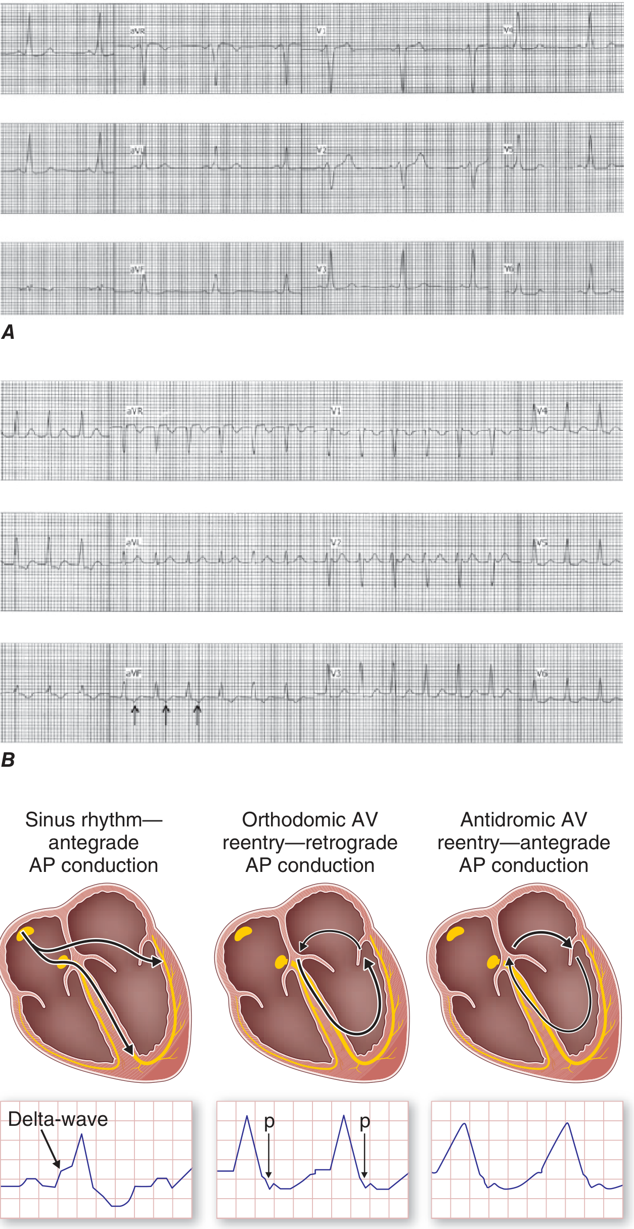
(Harrison's Principles of Internal Medicine 22E, Fig. 256-3)
Key Danger Sign: Syncope
Syncope in WPW is a red flag. Unlike vasovagal syncope, syncope in WPW is associated in 25% of cases with rapid, life-threatening conduction over the accessory pathway - it warrants urgent electrophysiological study (EPS) and likely radiofrequency catheter ablation.
Important Drug Warning
Avoid AV nodal blockers (digoxin, verapamil, diltiazem, adenosine) in WPW with AF. These drugs block the AV node but have no effect on - or actually accelerate - conduction through the AP, resulting in dangerously fast ventricular rates and possible VF. Procainamide or ibutilide (which block AP conduction) are the preferred pharmacological agents.
Summary Table
| Feature | WPW |
|---|---|
| Underlying defect | Accessory pathway (Bundle of Kent) bypassing AV node |
| Symptoms | Palpitations, dyspnea, dizziness, syncope, sudden death |
| ECG in sinus rhythm | Short PR (<120ms) + delta wave + wide QRS |
| Most common arrhythmia | Orthodromic AVRT (narrow QRS, 160-220 bpm) |
| Most dangerous arrhythmia | AF via AP → VF → sudden death |
| Definitive treatment | Radiofrequency catheter ablation (>95% success) |
Sources: Harrison's Principles of Internal Medicine, 22nd Ed.; Tintinalli's Emergency Medicine; Symptom to Diagnosis, 4th Ed.; Braunwald's Heart Disease; Ganong's Review of Medical Physiology, 26th Ed.
Oral contraceptive mechanism
Oral contraceptives (OCs) primarily work by suppressing ovulation through inhibition of the hypothalamic-pituitary-ovarian axis. Estrogen suppresses FSH (preventing follicle development) and progestin suppresses LH (preventing the LH surge and ovulation). Secondary mechanisms include thickening cervical mucus and altering the endometrium.
Searching sources...
Finding Sources
Finding Sources
Reading File
Reading File
Reading File
Finding Sources
Finding Sources
Reading File
Reading File
I have rich, authoritative content from four major pharmacology and physiology textbooks. Here is a comprehensive answer:
Mechanism of Action of Oral Contraceptives (OCs)
Oral contraceptives work through multiple simultaneous mechanisms - not just one. The combination pill (estrogen + progestin) is more effective than either component alone because they act synergistically at different levels of the reproductive axis.
Types of Oral Contraceptives
| Type | Components | Ovulation Inhibited? |
|---|---|---|
| Combined OCP (most common) | Estrogen (ethinyl estradiol) + Progestin | Yes - in virtually all cycles |
| Progestin-only pill ("minipill") | Progestin only | Only 60-80% of cycles |
| Monophasic | Fixed dose throughout cycle | Yes |
| Multiphasic | Varying doses (lower total steroid load) | Yes |
Primary Mechanism: Suppression of the Hypothalamic-Pituitary-Ovarian (HPO) Axis
This is the main contraceptive mechanism of combined OCPs.
Hypothalamus
    ↓  ← Estrogen + Progestin suppress GnRH pulse frequency
  GnRH ↓
    ↓
Anterior Pituitary
    ↓  ← Low GnRH → ↓ FSH and ↓ LH secretion
 FSH ↓  LH ↓
    ↓
Ovary
    ↓  ← No follicular development; no LH surge
No ovulation
Step by step:
-
Hypothalamic level - Progestin reduces the frequency of GnRH (gonadotropin-releasing hormone) pulses. The normal frequency of LH pulses is essential for ovulation; disrupting it is a major mechanism of action.
-
Pituitary level - Estrogen suppresses FSH secretion via negative feedback. Without adequate FSH, no follicle is recruited and no dominant follicle develops. Progestin suppresses LH secretion, eliminating the mid-cycle LH surge that normally triggers ovulation.
-
Ovarian level - In the absence of FSH stimulation, follicular development is minimal. No corpus luteum forms. The ovaries actually become smaller with chronic use.
"The contraceptive effectiveness of OCPs accrues from several actions. Like natural ovarian steroids, contraceptive steroids feed back both directly at the level of the hypothalamus (decreasing secretion of GnRH) and at the level of the gonadotrophs in the anterior pituitary. The net effect is suppressed secretion of the gonadotropins FSH and LH. The low FSH levels are insufficient to stimulate normal folliculogenesis; the low LH levels obviate the LH surge and therefore inhibit ovulation."
- Medical Physiology (Boron & Boulpaep)
Secondary Mechanisms (Backup Contraceptive Actions)
Even on cycles where ovulation may not be fully suppressed (especially with progestin-only pills), these secondary mechanisms provide additional protection:
1. Thickening of Cervical Mucus (Progestin)
- Progestin makes cervical mucus thick, viscid, and scanty - resembling post-ovulatory (hostile) mucus
- This physically blocks sperm penetration into the uterine cavity
- This is the primary mechanism of the progestin-only minipill
2. Endometrial Changes (Progestin)
- Progestin causes glandular atrophy and stromal changes in the endometrium
- The endometrium becomes inhospitable to implantation
- Specifically: reduced glandular glycogen production = diminished energy supply for blastocyst survival
- The "19-nor" progestins in particular produce marked glandular atrophy
3. Impaired Tubal Motility (Progestin)
- Progestins impair the peristaltic motility of the uterus and fallopian tubes
- This slows transport of both ova and sperm toward the normal fertilization site in the distal fallopian tube
4. Suppression of Ovarian Cysts (Estrogen + Progestin)
- Chronic use eliminates the cyclic follicular development that can form functional ovarian cysts
- This is a non-contraceptive benefit
Summary Diagram of All Mechanisms
COMBINED ORAL CONTRACEPTIVE PILL
         │
         ├─── HYPOTHALAMUS ──→ ↓ GnRH pulse frequency
         │
         ├─── PITUITARY ──────→ ↓ FSH → No follicle growth
         │                       ↓ LH  → No LH surge → No ovulation
         │
         ├─── CERVIX ─────────→ Thick, hostile mucus → Blocks sperm
         │
         ├─── ENDOMETRIUM ────→ Atrophy, ↓ glycogen → Impairs implantation
         │
         └─── FALLOPIAN TUBES →  ↓ Motility → Impairs sperm/egg transport
Progestin-Only Pill vs. Combined Pill
| Feature | Combined OCP | Progestin-Only (Minipill) |
|---|---|---|
| Ovulation suppression | ~100% | 60-80% of cycles |
| Cervical mucus effect | Yes | Yes (primary mechanism) |
| Endometrial changes | Yes | Yes |
| Tubal motility | Reduced | Reduced |
| Failure rate | ~0.3% (perfect use) | Higher |
| Who it suits | Most women | Nursing mothers, those with estrogen contraindications |
Additional (Non-Contraceptive) Benefits
OCs also:
- Reduce endometrial cancer risk by ~50%
- Reduce ovarian cancer risk by ~40%
- Suppress ovarian cysts
- Reduce dysmenorrhea and menorrhagia
- Improve hyperandrogenism (acne, hirsutism)
- Regulate menstrual cycles
Important Contraindications
Due to the estrogen component raising DVT risk (3-15 per 10,000 women-years):
- Smokers over 35 years of age
- History of DVT or thrombophilia
- Immediate postpartum period
- Migraine with aura
- History of estrogen-sensitive cancers
"The combinations of estrogens and progestins exert their contraceptive effect largely through selective inhibition of pituitary function that results in inhibition of ovulation. The combination agents also produce a change in the cervical mucus, in the uterine endometrium, and in motility and secretion in the uterine tubes, all of which decrease the likelihood of conception and implantation."
- Katzung's Basic and Clinical Pharmacology, 16th Ed.
Sources: Goodman & Gilman's Pharmacological Basis of Therapeutics; Katzung's Basic and Clinical Pharmacology, 16th Ed.; Medical Physiology (Boron & Boulpaep); Harrison's Principles of Internal Medicine, 22nd Ed.
This is a shared conversation. Sign in to Orris to start your own chat.