
from Guyton and Hall textbook of medical physiology, 4th South Asia edition write a 5 mark short note on Role of JGA in Regulation of Blood Pressure
Reading File
Finding Sources
Finding Sources
Finding Sources
Finding Sources
Finding Sources
Finding Sources
Finding Sources
Reading File
Finding Sources
Reading File
Reading File
Reading File
Loading Image
Reading File
Reading File
I now have all the key content from Guyton and Hall. Let me compile the comprehensive 5-mark short note.
Role of the Juxtaglomerular Apparatus (JGA) in Regulation of Blood Pressure
(Guyton and Hall Textbook of Medical Physiology)
Structure of the JGA
The juxtaglomerular apparatus (JGA) consists of two specialized cell types working in concert:
- Juxtaglomerular (JG) cells - modified smooth muscle cells in the walls of the afferent (and efferent) arterioles, immediately proximal to the glomeruli. They are the primary storage and secretion sites for renin.
- Macula densa cells - a specialized group of epithelial cells in the initial portion of the distal tubule, positioned in close contact with the afferent and efferent arterioles. They contain a Golgi apparatus directed toward the arterioles, suggesting paracrine secretory function.
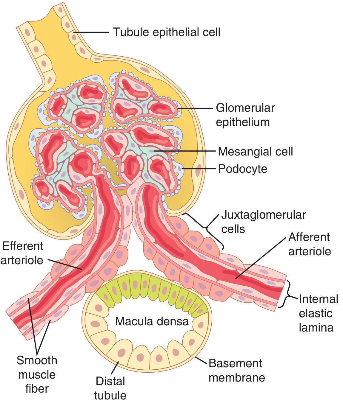
Mechanisms of Renin Release by JG Cells
When arterial pressure falls or salt intake is reduced, JG cells release renin by three major mechanisms:
-
Intrinsic baroreceptor mechanism: Pressure-sensitive baroreceptors within the JG cells respond directly to decreased stretch of the afferent arteriolar wall, triggering renin release.
-
Macula densa (tubuloglomerular feedback) mechanism: Decreased NaCl delivery to the macula densa cells (due to reduced GFR or reduced salt intake) sends a signal to the JG cells to increase renin secretion. Conversely, increased NaCl delivery suppresses renin release.
-
Sympathetic nervous system: Increased sympathetic activity stimulates renin release via beta-adrenergic receptors on JG cells. Sympathetic activation also enhances the sensitivity of both the baroreceptor and macula densa mechanisms.
The Renin-Angiotensin-Aldosterone System (RAAS) - Cascade
Once released, renin acts enzymatically:
Renin acts on angiotensinogen (an alpha-2 globulin synthesized in the liver) → cleaves it to form angiotensin I (10-amino acid peptide) → angiotensin-converting enzyme (ACE) in pulmonary endothelium (and kidneys, gut, vessels) converts it to angiotensin II (8-amino acid peptide, highly active).
Angiotensin II raises blood pressure through two principal mechanisms:
A. Acute: Vasoconstriction
- Angiotensin II is an extremely powerful vasoconstrictor, acting on arterioles (raising total peripheral resistance) and to a lesser degree on veins (increasing venous return and cardiac output).
- This response occurs within seconds to minutes and can restore arterial pressure at least halfway back to normal even after severe hemorrhage (as demonstrated experimentally - Fig. 19.10 in Guyton & Hall). However, it requires about 20 minutes to become fully active, making it slower than nervous reflexes but still powerful.
B. Chronic: Renal Salt and Water Retention
- Direct renal effects: Angiotensin II constricts efferent arterioles, reducing peritubular capillary pressure and increasing tubular fluid reabsorption. It also acts directly on tubular cells to increase sodium and water reabsorption.
- Aldosterone stimulation: Angiotensin II is one of the most powerful stimulators of aldosterone secretion from the adrenal cortex. Aldosterone markedly increases Na+ reabsorption in the collecting tubules, expanding extracellular fluid (ECF) volume over hours to days, raising blood pressure chronically.
- Combined, these effects can reduce urine output to less than one-fifth of normal.
Tubuloglomerular Feedback and Autoregulation of GFR
The JGA also participates in tubuloglomerular feedback (TGF) to maintain GFR:
- Increased NaCl at macula densa → ATP release → converted to adenosine → afferent arteriole vasoconstriction → reduced glomerular hydrostatic pressure → GFR returns to normal. Simultaneously, renin release is suppressed.
- Decreased NaCl at macula densa (e.g., with low arterial pressure) → afferent arteriole dilates (raises hydrostatic pressure) + renin released from JG cells → Angiotensin II constricts efferent arteriole → glomerular hydrostatic pressure restored → GFR normalized.
This dual-component mechanism ensures stable NaCl delivery to the distal tubule and prevents large fluctuations in renal excretion.
Long-Term Blood Pressure Buffering: Salt Intake Compensation
The JGA-RAAS axis plays a key role in maintaining arterial pressure despite wide variations in salt intake:
- High salt intake → suppresses renin → less angiotensin II and aldosterone → kidneys excrete more Na+ → BP stays normal.
- Low salt intake → stimulates renin → more angiotensin II and aldosterone → kidneys retain Na+ → BP stays normal.
- Blocking the RAAS (e.g., with ACE inhibitors) makes blood pressure highly sensitive to changes in salt intake, confirming the system's buffering importance.
- Quantitative analysis shows that complete absence of angiotensin II lowers equilibrium BP to ~75 mm Hg, while excess angiotensin II raises it to ~115 mm Hg.
Summary
| Mechanism | Timeframe | Effect on BP |
|---|---|---|
| JG baroreceptor → renin release | Seconds-minutes | ↑ via vasoconstriction |
| Macula densa → renin suppression/release | Minutes | Autoregulates GFR; modulates BP |
| Angiotensin II vasoconstriction | Minutes (~20 min full effect) | Acute ↑ BP |
| Angiotensin II → aldosterone → Na+ retention | Hours-days | Chronic ↑ BP |
| Efferent arteriole constriction (direct Ang II) | Minutes-hours | ↑ tubular reabsorption |
The JGA is therefore a central integrator of short-term and long-term blood pressure regulation, linking renal perfusion pressure, tubular fluid composition, and the endocrine RAAS cascade into a coordinated response that maintains circulatory homeostasis.
Source: Guyton and Hall Textbook of Medical Physiology, Chapters 19 and 27 (ISBN 9780443111013)
This is a shared conversation. Sign in to Orris to start your own chat.