
Venous system of brain
venous sinuses of the brain diagram superior sagittal sinus transverse sinus
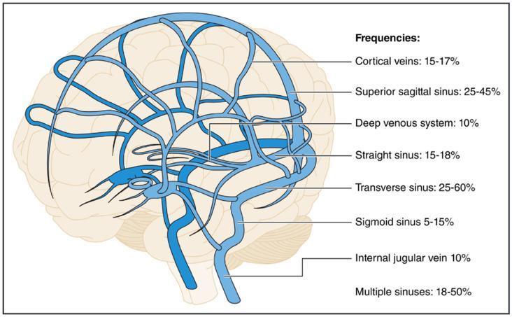
This medical anatomical diagram illustrates the cerebral venous system superimposed on a lateral view of the human brain. The illustration highlights the complex network of superficial and deep drainage pathways. Key anatomical structures labeled include the superior sagittal sinus along the dorsal midline, the cortical veins, the deep venous system (including the internal cerebral veins), the straight sinus, the transverse and sigmoid sinuses, and the internal jugular vein. The diagram serves as an educational infographic for Cerebral Venous Thrombosis (CVT), providing statistical frequencies of clot occurrence at specific anatomical sites: Transverse sinus (25-60%), Superior sagittal sinus (25-45%), Multiple sinuses (18-50%), Straight sinus (15-18%), Cortical veins (15-17%), Deep venous system (10%), Internal jugular vein (10%), and Sigmoid sinus (5-15%). The visual emphasizes the confluence of sinuses and the transition from dural venous sinuses to the extracranial venous system, providing clinical relevance for neurology and radiology residents studying stroke and vascular pathology.
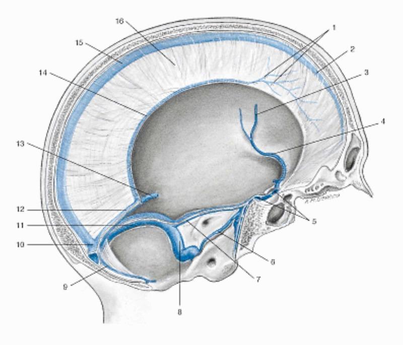
An anatomical diagram illustrating the venous sinuses of the dura mater in a sagittal section of the human skull. The illustration highlights the interconnected venous drainage system of the brain. Key structures include the superior sagittal sinus (2, 15) running along the superior border of the falx cerebri (16), and the inferior sagittal sinus (14) at its free margin. The straight sinus (12) is seen connecting the great cerebral vein of Galen (13) and the inferior sagittal sinus to the confluence of sinuses (10). Posteriorly, the occipital sinus (9) and transverse sinus (11) also drain into the confluence. Anterior venous structures include the cavernous sinus (5), sphenoparietal sinus (4), and middle cerebral vein (3). The drainage pathway continues through the superior (7) and inferior (6) petrosal sinuses toward the sigmoid sinus (8), which eventually leads to the internal jugular vein. This diagram serves as a primary educational resource for neuroanatomy, demonstrating the relationship between dural folds and cranial venous outflow.
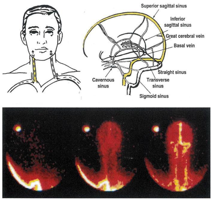
This composite educational graphic details the cerebral venous sinus system through schematic illustrations and diagnostic imaging. The upper panel includes a sagittal anatomical diagram labeling the superior and inferior sagittal sinuses, great cerebral vein, basal vein, straight sinus, cavernous sinus, transverse sinus, and sigmoid sinus. An accompanying coronal sketch shows the bilateral internal jugular veins in the neck. The lower panel displays a Technetium-99 radioisotope scan (scintigraphy) of the dural sinuses from a posterior view, illustrating a lateralized drainage pattern. In this clinical example, the superior sagittal sinus—which drains the cerebral cortex—predominantly flows into the right transverse sinus and right internal jugular vein. Simultaneously, the straight sinus—draining deep subcortical structures—flows into the left internal jugular vein. This visualization emphasizes anatomical variations in venous lateralization, which is critical for clinical procedures such as selective jugular venous sampling used to quantify regional brain monoamine turnover.
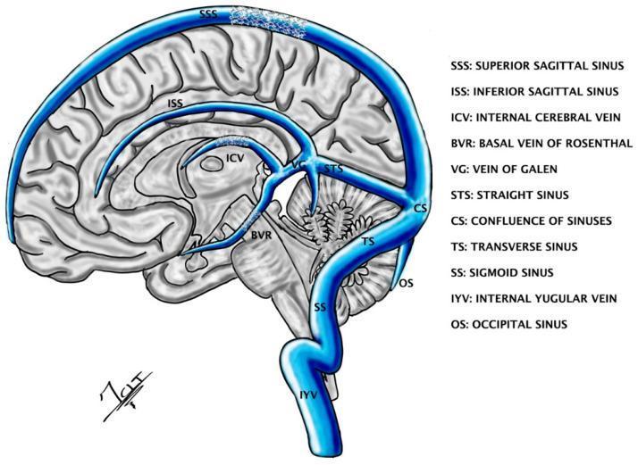
A medical anatomical diagram showing a sagittal cross-section of the human brain with the cerebral venous sinus system overlaid in blue. Key structures labeled include the superior sagittal sinus (SSS) running along the superior margin, the inferior sagittal sinus (ISS) within the falx cerebri, and the straight sinus (STS) connecting to the confluence of sinuses (CS). Deep venous structures shown include the internal cerebral vein (ICV), basal vein of Rosenthal (BVR), and the vein of Galen (VG). The posterior drainage pathway is illustrated via the transverse sinus (TS), sigmoid sinus (SS), and occipital sinus (OS), ultimately draining into the internal jugular vein (IYV). The diagram highlights the theoretical placement of 'STENTrodes' within the SSS and deep veins (BVR/ICV) for endovascular neuromodulation of cortical and subcortical targets like the thalamus and brainstem. This illustration serves as an educational tool for understanding the transvenous approach to deep brain stimulation and the spatial relationship between dural venous sinuses and deep neuroanatomy.
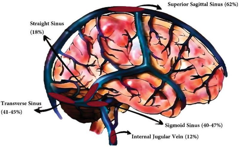
This medical illustration depicts the lateral view of the human brain with an anatomical overlay of the dural venous sinuses, highlighting the distribution and incidence of cerebral venous thrombosis (CVT). The venous structures are rendered in blue, superimposed on a reddish-toned representation of the cerebral hemispheres showing sulci and gyri. Key anatomical structures labeled include the superior sagittal sinus, straight sinus, transverse sinus, sigmoid sinus, and internal jugular vein. Each label is accompanied by a percentage indicating the reported incidence of thrombosis at that site based on clinical data. The superior sagittal sinus is identified as the most common site of involvement (62%), followed by the transverse (41-45%) and sigmoid (40-47%) sinuses. The diagram serves as an educational tool for neurology and radiology, illustrating the superficial and deep venous systems of the brain and their relative susceptibility to thrombotic events. It effectively demonstrates the confluence of sinuses and the drainage pathway into the internal jugular vein within a clinical context.
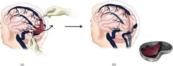
Two-part medical illustration depicting a neurosurgical resection of a tumor involving the dural venous sinuses. (a) This anatomical diagram shows a sagittal-lateral view of the brain with a large, reddish, vascularized mass (identified in context as a hemangiopericytoma) invading the confluence of sinuses (CoS), the posterior third of the superior sagittal sinus (SSS), and the proximal transverse sinus. Surgeon hands are illustrated using microsurgical instruments to perform silk knot ligatures at the tumor margins and the invaded sinuses. (b) The follow-up diagram shows the post-resection state. The tumor has been removed en-bloc and is displayed in a surgical kidney dish. The primary brain illustration depicts the empty surgical bed at the occipital region, showing the ligated ends of the SSS and transverse sinuses. The diagram highlights the surgical management of complex dural sinus tumors and the radical resection technique required for aggressive vascular lesions.
Venous System of the Brain
1. Cerebral Veins
- Superior cerebral veins (8-12) drain the superolateral and medial surface of the hemisphere upward into the superior sagittal sinus.
- Superficial middle cerebral vein runs along the lateral sulcus and drains into the cavernous or sphenoparietal sinus.
- Superior and inferior anastomotic veins (veins of Trolard and Labbé) connect the superficial middle cerebral vein to the superior sagittal sinus and transverse sinus respectively, providing important collateral pathways.
- Inferior cerebral veins drain the inferior surface of the hemisphere into the transverse and cavernous sinuses.
- Paired internal cerebral veins (formed by the union of the thalamostriate and choroidal veins) run in the roof of the third ventricle.
- The basal vein (of Rosenthal) drains the orbital surface of the frontal lobe, insula, and midbrain.
- The two internal cerebral veins join the basal veins to form the unpaired great cerebral vein (of Galen), which curves around the splenium of the corpus callosum and joins the inferior sagittal sinus to form the straight sinus.
2. Dural Venous Sinuses
| Sinus | Location | Receives |
|---|---|---|
| Superior sagittal sinus | Superior border of falx cerebri | Superior cerebral veins, diploic/emissary veins, CSF (via arachnoid granulations) |
| Inferior sagittal sinus | Inferior free margin of falx cerebri | A few cerebral veins, falx cerebri veins |
| Straight sinus | Junction of falx cerebri and tentorium cerebelli | Inferior sagittal sinus + great cerebral vein, posterior cerebral veins, superior cerebellar veins |
| Occipital sinus | Falx cerebelli | Connects confluence of sinuses with vertebral venous plexus |
| Confluence of sinuses | Internal occipital protuberance | Superior sagittal, straight, and occipital sinuses |
| Transverse sinus (paired) | Attachment of tentorium cerebelli | Confluence of sinuses (right usually continues SSS, left usually continues straight sinus), superior petrosal sinus, cerebral/cerebellar/diploic/emissary veins |
| Sigmoid sinus (paired) | S-shaped groove to jugular foramen | Continuation of transverse sinus -> internal jugular vein |
| Cavernous sinus (paired) | Either side of sella turcica | Superior/inferior ophthalmic veins, sphenoparietal sinus, superficial middle cerebral vein; drains via superior petrosal sinus (to sigmoid) and inferior petrosal sinus (to IJV) |
| Superior/inferior petrosal sinuses | Petrous temporal bone borders | Connect cavernous sinus to sigmoid sinus / internal jugular vein |
- The superior sagittal sinus usually deviates right and the straight sinus usually deviates left at the confluence of sinuses (torcular Herophili) - clinically relevant for lateralized venous drainage.
- The cavernous sinus is unique in that the internal carotid artery and abducens nerve pass through it, while CN III, IV, V1, and V2 run in its lateral wall - making it a site where infections (e.g. from the face via the ophthalmic veins) or thrombosis produce characteristic cranial nerve palsies.
- Right and left cavernous sinuses communicate via intercavernous sinuses.
3. Emissary and Diploic Veins
- Diploic veins lie within the spongy bone of the skull vault and connect dural sinuses to superficial scalp veins (frontal, anterior/posterior temporal, occipital diploic veins).
- Emissary veins pass through skull foramina to directly connect intracranial sinuses with extracranial veins (e.g. parietal emissary vein: superior sagittal sinus <-> superficial temporal vein; mastoid emissary vein: sigmoid sinus <-> occipital vein; condylar emissary vein: sigmoid sinus <-> vertebral venous plexus).
- Clinically important because they are valveless, so they can serve as a route for extracranial infection (e.g. scalp or facial infection) to spread into the dural sinuses (Gray's Anatomy for Students, p. 1024; Color Atlas of Human Anatomy Vol. 2, p. 134-135).
Diagram
Clinical Relevance
- Gray's Anatomy for Students, p. 1024-1025
- Color Atlas of Human Anatomy, Vol. 2: Internal Organs, p. 133-136
- Adams and Victor's Principles of Neurology, 12th Edition (Thrombosis of cerebral veins and venous sinuses)
Cerebral venous thrombosis treatment
"cerebral venous thrombosis" AND anticoagulation
cerebral venous thrombosis treatment guidelines 2024 anticoagulation direct oral anticoagulants
Cerebral Venous Thrombosis (CVT) - Treatment
1. Acute-Phase Anticoagulation (first-line, mainstay)
- Start anticoagulation immediately, even in patients who already have a venous hemorrhagic infarct - CVT is the one situation where anticoagulants are given despite intracranial hemorrhage, because the hemorrhage is due to venous congestion/outflow obstruction rather than an actively bleeding arterial source (Goldman-Cecil Medicine, "Acute Treatment").
- Options: low-molecular-weight heparin (LMWH) subcutaneously or intravenous unfractionated heparin (UFH).
- LMWH is generally preferred over UFH (fewer complications, easier dosing) - a "weak recommendation" per emergency medicine guidance (Rosen's Emergency Medicine).
- UFH may be preferred if there is a high bleeding risk, renal impairment, or if rapid reversal might be needed (e.g. planned intervention), since it is more easily reversed with protamine and more titratable.
- Historical trial data (Einhäupl et al. vs. de Bruijn and Stam) gave mixed but net-positive signal for heparin benefit; anticoagulation became the accepted standard despite the small trial sizes (Adams and Victor's Principles of Neurology, 12th ed.).
2. Transition to Long-Term Oral Anticoagulation
- After clinical stabilization on heparin, patients transition to oral anticoagulation for 3-12 months depending on whether the CVT was provoked (transient risk factor) or unprovoked/has persistent risk factors (e.g. thrombophilia).
- Vitamin K antagonists (warfarin), target INR 2.0-3.0, remain the guideline-endorsed default per the 2011 AHA/ASA and 2017 European Stroke Organization (ESO) statements.
- Direct oral anticoagulants (DOACs) - dabigatran, rivaroxaban, apixaban - are increasingly used and supported by growing evidence:
- The RE-SPECT CVT trial and subsequent DOAC-CVT prospective cohort study found DOACs had similar efficacy (recanalization, recurrence) and a more favorable bleeding profile compared with warfarin (Goldman-Cecil Medicine; Lancet Neurology 2025 DOAC-CVT study).
- Recent meta-analyses (2024-2025, including one pooling ~4,929 patients) support DOACs as non-inferior to VKAs with less bleeding, though these are still based on observational/cohort data rather than large acute-phase RCTs.
- Example dosing cited in textbook: rivaroxaban 20 mg daily for 3-6 months with imaging follow-up (MR or CT venography) to confirm sinus recanalization.
- Important caveat: no randomized trial data yet support DOAC use in the acute phase of CVT - the 2024 German S2k consensus guideline explicitly recommends clinical stabilization with heparin first before considering a switch, and current major guidelines still favor VKA over DOAC for that reason, with DOAC as a reasonable option mainly for the maintenance/long-term phase in stable patients without antiphospholipid syndrome.
3. Endovascular / Thrombolytic Therapy (reserved for severe/refractory cases)
- Local intra-sinus thrombolysis (tPA) or mechanical thrombectomy is reserved for patients who deteriorate despite adequate anticoagulation - e.g. progressive stupor/coma, extensive thrombosis, or markedly raised intracranial pressure (Adams and Victor's Principles of Neurology).
- Systematic reviews (2023-2024) of endovascular treatment show it can be effective in this severe subgroup, but is not first-line and carries added hemorrhage risk; it has not been shown superior to anticoagulation alone in the overall CVT population (per 2024 systematic reviews on endovascular thrombolysis/thrombectomy for CVT).
4. Supportive and Adjunctive Management
- Seizure control: antiepileptics for patients who have had a seizure (not given prophylactically without seizures).
- Raised intracranial pressure: managed with head elevation, osmotherapy (mannitol/hypertonic saline) if needed; acetazolamide or therapeutic lumbar puncture/optic nerve sheath fenestration/shunting may be used for CVT-associated intracranial hypertension with papilledema threatening vision.
- Decompressive hemicraniectomy for large venous infarcts with life-threatening mass effect/herniation.
- Treat the underlying cause: antibiotics if septic (e.g. mastoiditis, sinusitis causing cavernous or sigmoid sinus thrombosis - now rare but still seen, per Scott-Brown's Otorhinolaryngology); stop offending drugs (e.g. oral contraceptives); manage prothrombotic states (pregnancy/postpartum, thrombophilia, malignancy).
- Glucocorticoids are not recommended routinely.
Summary Algorithm
- Confirm diagnosis (MR/CT venography) -> start LMWH or UFH immediately, regardless of hemorrhagic venous infarction.
- Monitor for neurological worsening/raised ICP - escalate to thrombolysis/thrombectomy or decompressive surgery only if deteriorating despite anticoagulation.
- Once stable, transition to oral anticoagulation (warfarin traditionally; DOAC increasingly used) for 3-12 months based on provoking factors.
- Manage seizures, intracranial hypertension, and the underlying etiology in parallel.
- Adams and Victor's Principles of Neurology, 12th Edition - "Treatment of Cerebral Venous Thrombosis"
- Goldman-Cecil Medicine, International Edition - "Acute Treatment" (Cerebral Venous Thrombosis)
- Rosen's Emergency Medicine - "Cerebral Venous Thrombosis"
- Scott-Brown's Otorhinolaryngology Head & Neck Surgery, Vol. 2 - "Sigmoid Sinus Thrombosis"
- PubMed: PMID 41358859 (2025 meta-analysis, DOAC vs VKA), PMID 38772568 (2024 RCT meta-analysis), German S2k Guideline 2024 (Neurological Research and Practice)