
Function of platelets
platelet function hemostasis diagram
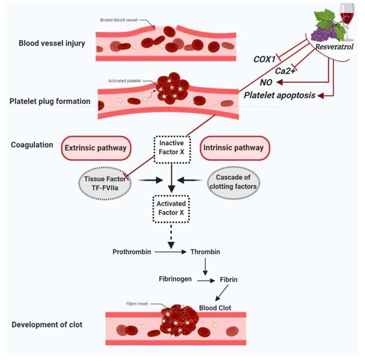
This pathophysiology diagram illustrates the process of hemostasis and the inhibitory mechanisms of resveratrol on platelet function. The central vertical flow depicts the sequence of events following a blood vessel injury: (1) Blood vessel injury, (2) Platelet plug formation involving activated platelets, (3) Coagulation, and (4) Development of a blood clot with a fibrin mesh. The coagulation cascade highlights the convergence of the extrinsic pathway (Tissue Factor: TF-FVIIa) and the intrinsic pathway (cascade of clotting factors) onto Inactive Factor X. Activated Factor X then facilitates the conversion of Prothrombin to Thrombin, and Fibrinogen to Fibrin. Interacting with these pathways, the diagram shows Resveratrol's antiplatelet effects through the inhibition of COX1, reduction of calcium (Ca2+) and nitric oxide (NO) concentrations, and the induction of platelet apoptosis. Additionally, Resveratrol is shown to inhibit the TF-FVIIa complex within the extrinsic pathway. The illustration uses a combination of anatomical cross-sections and biochemical flowcharts to detail secondary hemostasis and pharmacodynamic interactions relevant to cardiovascular health.
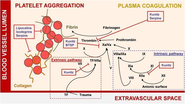
A pathophysiology diagram illustrating the mechanisms of hemostasis and the inhibitory effects of tick salivary proteins on platelet aggregation and plasma coagulation. The diagram is divided into the 'Blood Vessel Lumen' and the 'Extravascular Space.' On the left, 'Platelet Aggregation' is shown as a cluster of red spheres representing platelets cross-linked by fibrin strands; this process is inhibited by Lipocalins, Ixodegrins, and Serpins. On the right, the 'Plasma Coagulation' cascade is detailed, featuring both the Extrinsic pathway (initiated by Tissue Factor/TF due to trauma) and the Intrinsic pathway (activated via anionic surfaces). The diagram highlights key enzymatic conversions, such as Factor X to Xa/Va, Prothrombin to Thrombin, and Fibrinogen to Fibrin. Red annotation boxes identify specific tick protein families—including Kunitz-type inhibitors, BTSPs, and Serpins—that target and inhibit various coagulation factors like Thrombin, Factor Xa, and the XIIa/XIa complex. This visual resource effectively demonstrates the interference of exogenous biological agents with human blood clotting mechanisms.
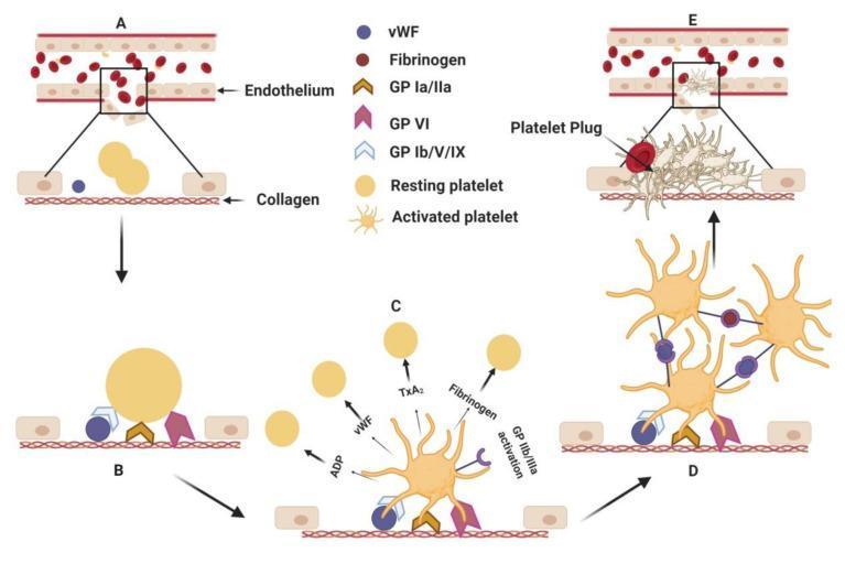
This hematology diagram illustrates the multistep pathophysiology of primary hemostasis following endothelial injury. The process is divided into five sequential stages (A–E). (A) Endothelial disruption exposes subendothelial collagen. (B) Platelet adhesion occurs as resting platelets (smooth, discoid) bind to collagen via surface glycoprotein receptors GP Ia/IIa, GP VI, and GP Ib/V/IX mediated by von Willebrand factor (vWF). (C) Platelet activation results in a dramatic morphological transformation, with platelets developing multiple cytoplasmic pseudopodia (starburst shape). This stage shows the release of granular contents including ADP, TxA2, vWF, and fibrinogen, which recruit additional platelets. (D) Platelet aggregation is shown, where activated GP IIb/IIIa receptors form cross-links between adjacent platelets using vWF and fibrinogen as molecular bridges. (E) The sequence concludes with the formation of a stable platelet plug at the injury site, effectively sealing the vascular breach. This educational visual explains the cellular signaling and mechanical interactions required for initial thrombus formation.
Functions of Platelets (Thrombocytes)
1. Primary Hemostasis - Forming the Platelet Plug
A. Adhesion
- vWF (von Willebrand factor) acts as a molecular "glue," binding simultaneously to subendothelial collagen and the platelet receptor GpIb/V/IX - especially critical under high shear stress
- Direct collagen receptors on the platelet surface - GpIa/IIa and GpVI
B. Activation
- Shape change: smooth discs transform into spiky "sea urchin" forms with greatly increased surface area
- Phospholipid translocation: negatively charged phosphatidylserine moves to the outer leaflet, providing nucleation sites for coagulation factor complexes (critical for secondary hemostasis)
- Granule secretion (release reaction):
- Dense granules: release ADP, serotonin, Ca²⁺
- Alpha (α) granules: release fibrinogen, vWF, PDGF, factor V, platelet factor 4
- Thrombin - acts via protease-activated receptor-1 (PAR-1)
- ADP - acts via G protein-coupled receptors P2Y1 and P2Y12; causes recruitment (positive feedback loop)
- TxA₂ (thromboxane A2) - synthesized from arachidonic acid via cyclooxygenase; potent vasoconstrictor and aggregation inducer
Note: Aspirin irreversibly inhibits cyclooxygenase, blocking TxA₂ synthesis. Clopidogrel/prasugrel block P2Y12. GP IIb/IIIa inhibitors block aggregation directly. - Schwartz's Principles of Surgery, 11e
C. Aggregation
- Platelet activation causes conformational change in GpIIb/IIIa (integrin αIIbβ3), increasing its affinity for fibrinogen
- Fibrinogen is a bivalent plasma protein that forms bridges between adjacent activated platelets, cross-linking them
- The initial wave of aggregation is reversible; thrombin then stabilizes the plug through further activation and irreversible platelet contraction (cytoskeleton-dependent)
- This forms the primary hemostatic plug
2. Secondary Hemostasis - Supporting the Coagulation Cascade
- Phosphatidylserine on the activated platelet surface provides an anionic scaffold for assembly of the tenase and prothrombinase complexes
- These amplify thrombin generation, which converts fibrinogen to fibrin, cementing the platelet plug into a definitive secondary hemostatic plug
- Fibrin cross-linking by factor XIIIa (also activated by thrombin) creates a tough, stable clot
3. Vasoconstriction
- Released serotonin and TxA₂ cause local vasoconstriction, reducing blood flow to the injury site and minimizing blood loss while the plug forms
4. Clot Retraction
- Platelet contraction, driven by the cytoskeleton (actin-myosin interaction), pulls fibrin strands together and consolidates the clot
- This mechanical contraction squeezes out serum and produces a compact, firm thrombus
5. Wound Healing and Vessel Repair
- Alpha granules release PDGF (Platelet-Derived Growth Factor) and TGF-β
- PDGF stimulates smooth muscle cell proliferation and fibroblast recruitment, initiating vessel wall repair after injury - Robbins & Kumar Basic Pathology, p.85
6. Immune and Inflammatory Functions
- Activated platelets express P-selectin on their surface, which mediates adherence of leukocytes (neutrophils, monocytes) to the platelet plug
- Platelets can release cytokines and antimicrobial peptides, contributing to innate immunity
Summary Diagram

Quick Reference Table
| Function | Key Molecules | Clinical Relevance |
|---|---|---|
| Adhesion | GpIb, vWF, GpIa/IIa, GpVI | Bernard-Soulier, von Willebrand disease |
| Activation | Thrombin (PAR-1), ADP (P2Y12), TxA₂ | Targets of aspirin, clopidogrel |
| Aggregation | GpIIb/IIIa, fibrinogen | Glanzmann thrombasthenia; GP IIb/IIIa inhibitors |
| Coagulation scaffold | Phosphatidylserine, Ca²⁺ | Amplifies thrombin generation |
| Vasoconstriction | Serotonin, TxA₂ | Reduces blood loss |
| Wound repair | PDGF, TGF-β | Smooth muscle/fibroblast proliferation |
| Immunity | P-selectin, cytokines | Leukocyte recruitment |


Make notes on this
Platelets (Thrombocytes) - Notes
Structure & Basic Facts
- Very small, disc-shaped cell fragments, 2-4 µm in diameter
- Budded off from the cytoplasm of megakaryocytes in red bone marrow
- No nucleus - cytoplasm packed with granules containing substances essential for clotting
- Contain contractile actin fibres - allow platelets within a developing clot to retract
- Normal count: 200-350 × 10⁹/L (200,000-350,000/mm³)
- Lifespan: 8-11 days; unused platelets destroyed by macrophages mainly in the spleen
- About 1/3 of platelets stored in the spleen as an emergency reserve, released when needed
- Thrombopoietin (from the liver) stimulates platelet production
Haemostasis - Overview
- When a blood vessel is damaged, haemostasis stops blood loss through overlapping processes
- Occurs in 3 stages: Vasoconstriction → Platelet plug formation → Coagulation
- More severe damage = faster coagulation (can begin within 15 seconds of injury)
Stage 1: Vasoconstriction
- Platelets contact damaged vessel wall → surface becomes sticky → adhere to the site
- Release serotonin (5-HT) and thromboxanes → constrict the vessel → reduce blood flow
- Other vasoconstrictors (e.g. endothelins) also released by the damaged vessel itself
Stage 2: Platelet Plug Formation
- Sticky platelets clump together and release ADP (adenosine diphosphate)
- ADP attracts more platelets → positive feedback loop → rapid platelet accumulation
- Platelet plug is usually complete within 6 minutes of injury
- Acts as a temporary seal - soft, easily disrupted, precursor to the more durable blood clot
Stage 3: Coagulation (Blood Clotting)
- Complex cascade involving clotting factors (mainly enzymes, produced in the liver, circulate as inactive forms)
- Clotting factor numbers reflect order of discovery, not order of action in the cascade
| Factor | Name |
|---|---|
| I | Fibrinogen |
| II | Prothrombin |
| VII | Proconvertin |
| VIII | Antihemophilic factor A |
| IX | Antihemophilic factor B |
| X | Stuart-Prower factor |
| XII | Hageman factor |
| XIII | Fibrin stabilising factor |
Vitamin K is essential for synthesis of factors II, VII, IX, and X
- Prothrombin activator is formed (final step of either pathway)
- Prothrombin activator converts prothrombin → thrombin
- Thrombin converts fibrinogen → insoluble fibrin threads
- Fibrin threads form a 3D meshwork that stabilises the platelet plug
- The maturing blood clot traps RBCs and plasma proteins including plasminogen (which will eventually dissolve the clot)
- Result: a much stronger, durable clot
| Feature | Extrinsic Pathway | Intrinsic Pathway |
|---|---|---|
| Trigger | Damaged tissue releases thromboplastin (tissue factor) | Blood contacts damaged vessel lining (endothelium) |
| Speed | Fast - within seconds | Slow - 3-6 minutes |
| Importance | More important of the two | Slower backup pathway |
Clot Retraction
- After clot forms, actin fibres in platelets contract
- Clot shrinks (retracts), squeezing out serum
- Pulls the edges of the damaged vessel together → closes the hole → reduces blood loss
Key Points Summary
| Feature | Detail |
|---|---|
| Origin | Megakaryocytes, red bone marrow |
| Size | 2-4 µm |
| Count | 200,000-350,000/mm³ |
| Lifespan | 8-11 days |
| Regulator | Thrombopoietin (liver) |
| Haemostasis steps | Vasoconstriction → Plug → Coagulation |
| Key chemicals released | Serotonin, thromboxanes, ADP |
| Coagulation end-product | Fibrin clot (via prothrombin → thrombin → fibrinogen → fibrin) |
| Vitamin K role | Synthesis of factors II, VII, IX, X |
Make a visual flowchart diagram of the haemostasis cascade
A detailed medical educational flowchart diagram of the haemostasis cascade, showing three parallel sections: Left column titled "Stage 1: Vasoconstriction" with steps: Blood vessel injury → Platelets contact damaged wall → Platelets become sticky and adhere → Release serotonin (5-HT) and thromboxanes → Vasoconstriction (reduced blood flow). Also shows endothelins released by damaged vessel. Middle column titled "Stage 2: Platelet Plug Formation" with steps: Sticky platelets clump together → Release ADP → ADP attracts more platelets (positive feedback loop arrow) → Platelet accumulation at injury site → Temporary platelet plug forms (complete within 6 minutes). Labeled as soft and temporary. Right column showing two parallel pathways converging - "Extrinsic Pathway" (triggered by tissue thromboplastin/tissue factor, fast - seconds) and "Intrinsic Pathway" (triggered by blood contact with damaged endothelium, slow - 3-6 minutes). Both converge downward to "Stage 3: Coagulation" section showing: Prothrombin Activator → Prothrombin → Thrombin → Fibrinogen → Loose Fibrin Threads → Stabilised Fibrin Clot (with Factor XIII crosslinking). Arrow showing Fibrin meshwork traps RBCs and platelets. Final box: Clot Retraction (actin fibres contract, serum expelled, vessel edges pulled together). Clean white background, color-coded sections (blue for vasoconstriction, orange for platelet plug, green for coagulation), bold clear arrows, professional medical textbook style, all text clearly readable.
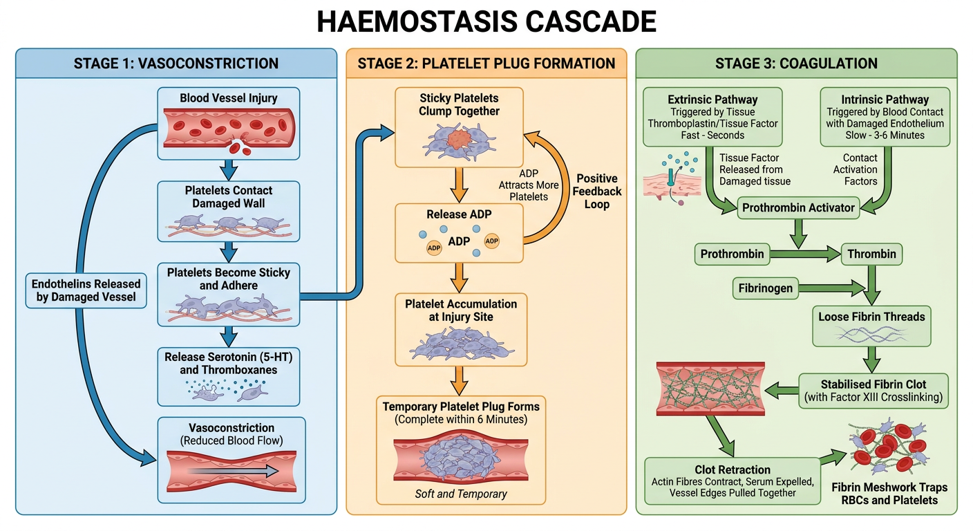
- Blue (Stage 1) - Vasoconstriction: platelet adhesion → serotonin/thromboxane release → vessel constriction
- Orange (Stage 2) - Platelet plug: ADP-driven positive feedback loop → temporary plug within 6 minutes
- Green (Stage 3) - Coagulation: the extrinsic (fast, seconds) and intrinsic (slow, 3-6 min) pathways converging on the final common pathway → prothrombin → thrombin → fibrinogen → stabilised fibrin clot → clot retraction