
Recent advances in management of oesophageal cancer
I'll start with a quick overview, then pull in textbook content and recent literature simultaneously.
Oesophageal cancer management has evolved significantly, with key advances in:
- Multimodal therapy: neoadjuvant chemoradiotherapy (CROSS protocol) remains standard for resectable disease
- Immunotherapy: checkpoint inhibitors (nivolumab, pembrolizumab) now approved in first-line and adjuvant settings
- Targeted therapy: trastuzumab for HER2+ adenocarcinoma
- Minimally invasive surgery: robotic and laparoscopic oesophagectomy gaining ground
- Endoscopic techniques: expanding role for early-stage disease (ESD, EMR)
Searching sources...
Reading File
Searching PubMed
oesophageal cancer management advances
Searching the Web
recent advances oesophageal cancer treatment 2024 2025 immunotherapy surgery
Now let me query the medical library for textbook content and get abstracts of the most relevant PubMed papers in parallel.
Finding Sources
Reading Article
PMID: 39712027
Reading Article
PMID: 38127239
Reading Web Page
https://aoe.amegroups.org/article/view/8536/html
Finding Sources
Searching PubMed
esophageal cancer nivolumab pembrolizumab checkpoint inhibitor
Reading Web Page
https://emedicine.medscape.com/article/277930-treatment
Finding Sources
Searching PubMed
esophageal cancer minimally invasive esophagectomy robotic outcomes
Reading File
Reading Article
PMID: 38960881
Reading Article
PMID: 40701573
Reading File
Searching PubMed
esophageal cancer adjuvant nivolumab CheckMate 577 overall survival
Reading File
Loading Image
I now have comprehensive data from textbooks, recent RCTs, meta-analyses, and the latest FDA approvals. Let me compile the full answer.
Recent Advances in Management of Oesophageal Cancer
Oesophageal cancer management has undergone a substantial shift toward personalised, multimodal strategies with immunotherapy now embedded across multiple disease stages. Below is a structured synthesis of current and emerging evidence.
1. Staging and Diagnosis
Enhanced staging precision is now achievable with:
- High-resolution endoscopic ultrasound (EUS) with fine-needle aspiration for T/N staging
- 18F-FDG PET-CT for detection of occult metastasis and response assessment
- AI-assisted endoscopy - machine learning algorithms are being validated for early detection and margin delineation in ESCC and Barrett's-related EAC (systematic review, 2025, PMID 40205603)
- Circulating tumour DNA (ctDNA) as a biomarker for treatment response monitoring and minimal residual disease detection - now actively studied in perioperative trials
Molecular profiling of both ESCC and EAC is now standard to guide targeted therapy selection (HER2, PD-L1 CPS, CLDN18.2).
2. Early-Stage Disease: Expanding Endoscopic Therapies
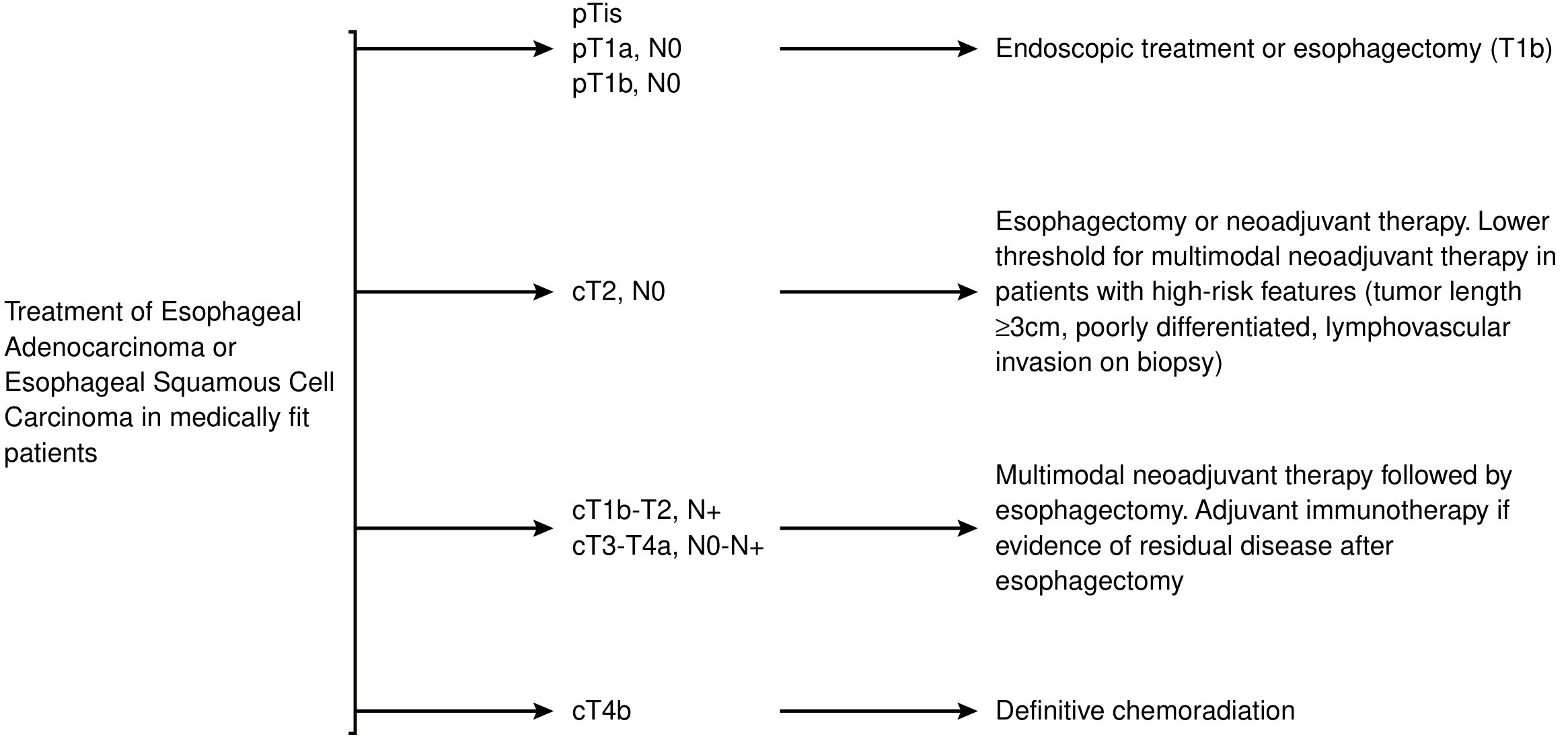
For pTis/T1a lesions (mucosal), endoscopic submucosal dissection (ESD) and endoscopic mucosal resection (EMR) provide curative outcomes with minimal invasiveness and are now preferred over surgery in specialist centres. The JUPITER trial data have further refined criteria for endoscopic treatment in early ESCC. T1b (submucosal) disease is managed with esophagectomy or endoscopic resection depending on depth, as illustrated in the treatment algorithm below.
3. Multimodal Therapy for Locally Advanced Disease
CROSS Protocol (still the cornerstone)
The landmark CROSS trial (neoadjuvant carboplatin/paclitaxel + 41.4 Gy radiation, then surgery) remains the standard of care for resectable esophageal/GEJ cancer. Long-term data show a survival benefit for both EAC and ESCC, with pCR rates of ~23% in EAC and ~49% in ESCC.
Perioperative Chemotherapy for EAC
- FLOT4 regimen (docetaxel, oxaliplatin, leucovorin, 5-FU, 4 pre- and 4 post-operative cycles) is preferred over ECF/ECX for EAC/GEJ adenocarcinoma, with median survival increasing from 35 to 50 months vs. MAGIC ECF
- ESOPEC trial directly compared CROSS vs. FLOT4 and found FLOT4 superior for EAC (5-year OS 50.7% vs. 36.3%)
- Total neoadjuvant therapy (moving all chemoradiation to the preoperative window) is under active investigation with FOLFOX + RT and FLOT4 + RT protocols
Current Surgical Therapy 14e, p. 102-106
Definitive Chemoradiotherapy
Remains the treatment for cT4b unresectable disease and as an alternative to surgery in ESCC with complete clinical response (organ preservation strategy, analogous to rectal cancer).
4. Immunotherapy - The Major Advance
Immune checkpoint inhibitors (ICIs) have transformed the treatment landscape across all stages.
Adjuvant Immunotherapy
- CheckMate 577: Nivolumab (1 year, adjuvant) after trimodality therapy (CROSS + surgery) in patients with residual pathological disease doubled disease-free survival (DFS 22.4 vs. 11.0 months; HR 0.69). This is now standard of care for residual disease post-trimodality therapy. The treatment algorithm (above) now explicitly includes "adjuvant immunotherapy if evidence of residual disease after esophagectomy."
Perioperative Immunochemotherapy (Neoadjuvant + Surgery ± Adjuvant)
A 2024 network meta-analysis of 14 studies (1,139 patients) found that neoadjuvant immunochemotherapy significantly outperforms traditional neoadjuvant therapy for pCR, MPR, ORR, and DCR:
- Camrelizumab + chemotherapy: highest pCR and MPR rates
- Pembrolizumab + chemotherapy: highest ORR and DCR
- No significant increase in grade ≥3 TRAEs vs. chemotherapy alone
- Most common immune-related AEs: rash (4-22%), thyroid dysfunction (6-17%), pneumonitis (4-6%)
PMID 39712027 - Systematic Review & Network Meta-Analysis, Front Immunol 2024
The ESCORT-NEO trial added further evidence for neoadjuvant chemo-IO in resectable ESCC.
First-Line Metastatic/Unresectable
Multiple PD-1/PD-L1 inhibitors are now FDA-approved with chemotherapy:
- Nivolumab + chemotherapy (CheckMate 648, 2022): approved for advanced/metastatic ESCC with PD-L1 ≥1% - first-line
- Nivolumab monotherapy (CheckMate 648): approved for tumour mutational burden (TMB)-high ESCC
- Pembrolizumab + chemotherapy (KEYNOTE-590): first-line ESCC and EAC/GEJ with PD-L1 CPS ≥10
- Tislelizumab (Tevimbra): FDA-approved March 2025 for first-line treatment of unresectable/metastatic ESCC with PD-L1 ≥1% (in combination with platinum-based chemotherapy); also approved 2024 as second-line monotherapy post-chemotherapy
- Durvalumab + FLOT: FDA-approved November 2025 for resectable gastric/GEJ adenocarcinoma, increasing pCR rate from 7% to ~19% (MATTERHORN trial) - first FDA-approved perioperative immunotherapy for this setting
A 2024 systematic review of the immunotherapy landscape confirmed that ICI integration improves tumour response and survival with acceptable perioperative safety and minimal impact on operative outcomes (PMID 38127239).
New Immunotherapy Formulations (2024-2025)
- Nivolumab + hyaluronidase-nvhy (Opdivo Qvantig): subcutaneous formulation, FDA-approved 2024 - eliminates IV infusion
- Pembrolizumab + berahyaluronidase alfa-pmph (Keytruda Qlex): subcutaneous pembrolizumab, FDA-approved 2025
5. Targeted Therapy
HER2-Positive EAC/GEJ
- Trastuzumab + chemotherapy (ToGA regimen) remains standard for HER2-overexpressing advanced GEJ/EAC
- Trastuzumab deruxtecan (T-DXd): antibody-drug conjugate showing marked activity in HER2+ gastroesophageal cancers in later-line settings; phase 3 data expected
- Zanidatamab (bispecific HER2-targeting antibody) in trials
CLDN18.2-Positive EAC/GEJ
- Zolbetuximab (anti-CLDN18.2 cytolytic antibody): FDA-approved 2024 for first-line HER2-negative, CLDN18.2-positive locally advanced unresectable or metastatic gastric/GEJ adenocarcinoma, in combination with fluoropyrimidine/platinum (SPOTLIGHT and GLOW trials). This is a landmark shift in treatment strategy requiring routine CLDN18.2 testing at diagnosis.
ASCO Guideline Update (2026)
The ASCO published an updated guideline on Immunotherapy and Targeted Therapy for Advanced Gastroesophageal Cancer (J Clin Oncol, April 2026), reflecting the rapidly evolving approvals landscape.
6. Surgical Advances
Minimally Invasive Esophagectomy (MIE) and Robotic-Assisted (RAMIE)
A 2025 network meta-analysis of 8 RCTs (1,776 patients) confirmed (PMID 40701573):
- MIE and RAMIE both reduce pulmonary complications by ~50% vs. open (RR 0.46-0.48) and vs. hybrid esophagectomy (RR 0.54-0.57)
- Reduced intraoperative blood loss and shorter hospital stays
- Equivalent anastomotic leak rates, in-hospital mortality, and oncological radicality vs. open approaches
- Equivalent outcomes between MIE and RAMIE
The REVATE RCT (2024, PMID 38960881) - a multicentre trial comparing robot-assisted (RAO) vs. video-assisted thoracoscopic (VAO) oesophagectomy - showed RAO achieved superior left recurrent laryngeal nerve (RLN) lymph node dissection (88% vs. 69% success, p<0.001) and lower permanent RLN palsy rates (5.8% vs. 20%, p=0.003), with more mediastinal lymph nodes harvested.
Prehabilitation
Structured prehabilitation programs (exercise, nutritional optimisation, smoking cessation) before surgery are now actively promoted to reduce operative risk, particularly in frail patients with dysphagia-related malnutrition.
AI in Surgery
Artificial intelligence applications in esophageal surgery - including intraoperative navigation, margin assessment, and anastomotic leak prediction - are under investigation (PMID 41099978).
7. Oligometastatic and Unresectable Disease
- Conversion surgery (after systemic therapy/immunotherapy leading to tumour downstaging) is increasingly pursued in selected patients with initially unresectable or T4b disease - a strategy borrowed from hepatic metastasis concepts
- Thoracic endovascular aortic repair (TEVAR) combined with resection is being evaluated for T4b tumours abutting the aorta (PMID 39058341)
- Stereotactic ablative body radiotherapy (SABR) for oligometastatic disease
8. Active Areas of Investigation
| Area | Status |
|---|---|
| FLOT + IO perioperative (ESOPEC-2 concepts) | Active trials |
| Total neoadjuvant therapy (induction chemo + CRT, all preop) | Active RCTs |
| PD-L1/CPS, TMB, ctDNA as response biomarkers | Validation ongoing |
| Organ preservation (watch-and-wait after CR to CRT) | Emerging evidence |
| HER2/neu + ICI combinations | Phase 2/3 |
| CLDN18.2 testing as routine biomarker | Now standard in GEJ/EAC |
| Vaccine-based immunotherapy | Early phase |
| Microbiome modulation to improve ICI response | Preclinical/early clinical |
Summary Table: Key Milestones 2022-2026
| Approval/Trial | Agent | Setting | Year |
|---|---|---|---|
| CheckMate 577 | Nivolumab (adjuvant) | Post-trimodality, residual disease | 2021 (practice-changing) |
| CheckMate 648 | Nivolumab + chemo | 1st-line advanced ESCC | 2022 |
| KEYNOTE-590 | Pembrolizumab + chemo | 1st-line ESCC/EAC | 2021 (ongoing use) |
| Zolbetuximab | Anti-CLDN18.2 | 1st-line CLDN18.2+ GEJ/EAC | 2024 |
| Tislelizumab | Anti-PD-1 | 2nd-line ESCC monotherapy | 2024 |
| Nivolumab SC | Subcutaneous nivolumab | Multiple | 2024 |
| Tislelizumab + chemo | Anti-PD-1 | 1st-line ESCC | March 2025 |
| Durvalumab + FLOT | Anti-PD-L1 + FLOT | Perioperative GEJ/gastric EAC | November 2025 |
| ASCO Guideline | -- | Advanced gastroesophageal cancer | April 2026 |
Key take-away: The biggest paradigm shift is the incorporation of ICI at every treatment stage - adjuvant nivolumab for residual post-CROSS disease, neoadjuvant immunochemotherapy for locally advanced ESCC (most data from Asian trials), and first-line ICI + chemotherapy for advanced/metastatic disease. CLDN18.2 testing has emerged as a mandatory new biomarker for GEJ/EAC. Surgical outcomes continue to improve with robotic approaches, particularly for RLN dissection. Molecular profiling (HER2, PD-L1 CPS, TMB, CLDN18.2) is now foundational to treatment planning.
Write a note on Gastro-intestinal Anastomosis i.e.,factors influencing, sites and various Techniques.
Searching sources...
Reading File
Finding Sources
Finding Sources
Finding Sources
Finding Sources
Finding Sources
Finding Sources
Excellent - I found the best sources. Let me now read the primary anastomosis sections from Pye's Surgical Handicraft, Fischer's Mastery of Surgery, Maingot's, and Schwartz's in parallel.
Reading File
Reading File
Reading File
Reading File
Reading File
Finding Sources
Reading File
Reading File
Reading File
Finding Sources
Now let me fetch the key images (anastomosis technique diagram from Schwartz's, and the GI wound healing collagen balance graph) and get additional content on specific GI anastomosis sites.
Loading Image
Loading Image
Finding Sources
I now have comprehensive textbook content from Sabiston, Schwartz's, Fischer's Mastery, and Maingot's. Let me compile the full, detailed note.
Gastro-intestinal Anastomosis
A gastrointestinal (GI) anastomosis is a surgically created communication between two segments of the alimentary canal - either restoring continuity after resection or creating a bypass. The success of an anastomosis depends on a precise interplay of patient factors, technical choices, and the biology of GI wound healing.
I. Healing of a GI Anastomosis
Understanding anastomotic healing is fundamental to appreciating what influences its success or failure.
Layers of the Bowel Wall
The submucosa is the strongest layer of the bowel wall and must be incorporated in every anastomosis. It provides the tensile strength and suture-holding capacity of the anastomosis. The mucosa, muscularis propria, and serosa all contribute but are structurally weaker.
Phases of Healing
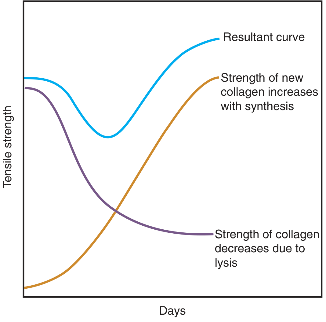
- Lag phase (days 0-4) - Inflammatory phase. Collagenase activity predominates; existing collagen is lysed faster than new collagen is synthesised. Anastomotic strength actually decreases in this window. This is the period of maximum vulnerability.
- Proliferative phase (days 4-14) - Fibroblast proliferation and new collagen synthesis accelerates. Anastomotic strength rises sharply.
- Remodelling phase (days 14 onwards) - Type III collagen matures to type I; final tensile strength approaches pre-resection levels.
The diagram below illustrates the delicate balance between collagen synthesis and collagenolysis that governs anastomotic strength:
II. Factors Influencing GI Anastomosis
Anastomotic healing and integrity depend on a complex interplay of factors. Schwartz's Principles of Surgery and Sabiston's Textbook classify these as follows.
A. Local Technical Factors (Surgeon-Controlled)
| Factor | Requirement for Success |
|---|---|
| Blood supply | Both bowel ends must be well vascularised. Ischaemia is the single most important technical cause of leak. Mesentery should not be stripped excessively from the bowel wall. |
| Tension | The anastomosis must be completely tension-free. Tension tears sutures, strangulates the anastomosis, and leads to dehiscence. Adequate mobilisation of both ends is mandatory. |
| Airtight and watertight seal | Full-thickness bites incorporating the submucosa must be taken. No gaps in the suture line. |
| Suture technique | Bites must be evenly spaced, not too tight (which strangulates tissue) and not too loose (which leaves gaps). The submucosa must be included. |
| Calibre match | Disparity in lumen size is addressed by choosing the appropriate configuration (see below). |
| Contamination | Operation in a contaminated field (faecal peritonitis, abscess) dramatically raises leak risk. |
| Haemostasis | A haematoma adjacent to the anastomosis impairs healing and acts as a nidus for infection. |
B. Patient/Systemic Factors (Definitive)
-
Nutritional status - Protein malnutrition directly impairs collagen synthesis, fibroblast function, and immune response. Hypoalbuminaemia (<3 g/dL) is one of the strongest predictors of anastomotic leak. Preoperative nutritional optimisation (including enteral/parenteral nutrition where needed) is essential.
-
Blood supply and perfusion - Systemic hypotension, shock, and coagulopathy reduce microvascular perfusion at the anastomotic edge. Over-zealous fluid resuscitation causes tissue oedema and third-space losses, also compromising small vessel flow.
-
Sepsis - Systemic sepsis and localised intra-abdominal infection raise collagenase levels, actively degrading newly formed collagen at the suture line.
-
Immunosuppression - Steroids, immunosuppressants (e.g. methotrexate, infliximab), and chemotherapy all impair wound healing.
-
Radiotherapy - Prior irradiation causes obliterative endarteritis and fibrosis in the bowel wall, profoundly impairing healing. Anastomoses in irradiated bowel carry substantially higher leak rates.
-
Smoking - Vasoconstriction and impaired tissue oxygenation.
-
Obesity - Poor tissue quality, difficult mobilisation, increased abdominal pressure.
-
Diabetes - Impairs leucocyte function and microvascular circulation.
-
Anaemia - Reduces oxygen delivery to the healing tissue.
-
Deficiencies - Vitamin C, iron, zinc, and copper are all required for normal collagen synthesis.
C. Local Bowel-Related Factors
- Crohn's disease - Transmural inflammation, dense adhesions, and impaired healing.
- Compromised distal lumen - Anastomosis performed proximal to an unrecognised partial obstruction will fail due to elevated intraluminal pressure.
- Bowel oedema - Reduces suture-holding capacity.
- Radiation damage - As above.
D. Location in the GI Tract
The risk of leak increases progressively further down the GI tract:
- Oesophageal anastomoses - Highest risk; oesophagus lacks a serosa and has a tenuous blood supply. Cervical oesophageal anastomoses leak more often than thoracic but the consequences are usually less severe.
- Gastroduodenal / gastrojejunal - Good blood supply; generally heal reliably.
- Small bowel - Excellent blood supply, heals well.
- Ileocolic - Good outcomes; side-to-side/functional end-to-end stapled anastomosis has the lowest leak rate.
- Colorectal - Significant risk, especially below the peritoneal reflection.
- Coloanal / low anterior - Highest risk in colorectal surgery. Routine defunctioning proximal stoma is often used as protection.
III. Sites of GI Anastomosis
Anastomoses are created at various levels of the GI tract depending on the surgical procedure:
Upper GI
| Anastomosis | Clinical Context |
|---|---|
| Oesophagogastric | Oesophagectomy; gastric conduit anastomosed to oesophageal remnant (cervical or intrathoracic) |
| Oesophagojejunal | Total gastrectomy; jejunum brought up as Roux limb |
| Gastroduodenostomy (Billroth I) | Distal gastrectomy for gastric cancer or peptic ulcer; restores gastroduodenal continuity |
| Gastrojejunostomy (Billroth II) | Distal gastrectomy where duodenum cannot be used; isoperistaltic jejunal loop attached to gastric remnant |
| Roux-en-Y gastrojejunostomy | Total or subtotal gastrectomy; avoids bile reflux into gastric remnant by creating a 40-60 cm Roux limb |
| Gastrojejunostomy (bypass) | Palliation of gastric outlet obstruction (e.g. unresectable pancreatic cancer) |
Biliary/Pancreatic
| Anastomosis | Context |
|---|---|
| Hepaticojejunostomy / choledochojejunostomy | Bile duct injuries, hilar cholangiocarcinoma, Whipple procedure |
| Pancreaticojejunostomy / pancreaticogastrostomy | Whipple procedure (pancreaticoduodenectomy) |
Small Bowel
| Anastomosis | Context |
|---|---|
| Enteroenterostomy | Small bowel resection for ischaemia, Crohn's, tumour, trauma |
| Ileocolic anastomosis | Right hemicolectomy; ileocaecal/ileocolic resection for Crohn's disease or cancer |
Large Bowel / Anorectal
| Anastomosis | Context |
|---|---|
| Colocolic | Left/right hemicolectomy, segmental colectomy |
| Colorectal | Anterior resection for rectal cancer; anastomosis above or below peritoneal reflection |
| Coloanal | Ultra-low anterior resection; anastomosis at dentate line (often with transanal technique or J-pouch) |
| Ileorectal | Subtotal colectomy for Crohn's, ulcerative colitis |
| Ileoanal pouch (IPAA) | Restorative proctocolectomy for UC or FAP; ileal J/W/S-pouch anastomosed to anal canal |
IV. Techniques of GI Anastomosis
A. Configuration (Geometry)
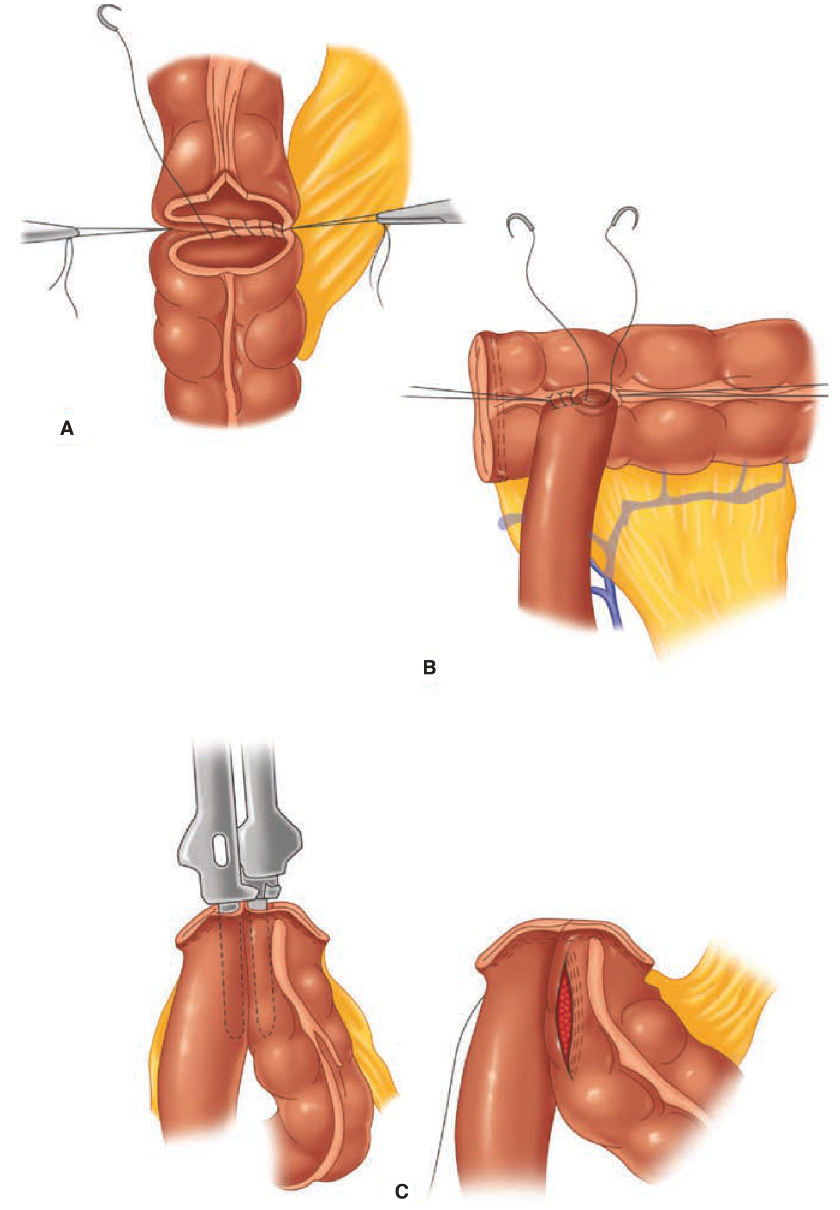
Illustrated below from Schwartz's Principles of Surgery (Fig. 29-13):
1. End-to-End (EE)
- Used when both bowel ends are of roughly equal calibre.
- Most physiological - restores normal anatomical continuity.
- Most common in: rectal resections (circular stapler EEA), small bowel anastomoses, colocolostomy.
- Risk: if there is size disparity, one end can be antimesenteric-ally cut obliquely ("fish-mouthed") to enlarge it.
2. End-to-Side (ES)
- One end of bowel is joined to the side of another segment.
- Useful when the proximal bowel is smaller than the distal bowel (e.g. chronic obstruction leads to proximal dilatation and distal collapse).
- Common examples: hepaticojejunostomy, Roux-en-Y jejunojejunostomy, oesophagojejunostomy after total gastrectomy.
3. Side-to-End (SE)
- The side of the proximal bowel is joined to the end of the distal bowel.
- Used when the proximal is of smaller calibre than the distal.
- Example: ileorectal anastomosis (small ileum to wider rectum).
- May have a slightly better blood supply than end-to-end.
4. Side-to-Side (SS) / Functional End-to-End
- Two antimesenteric walls of bowel are joined together using a linear cutting stapler.
- Creates a wide-calibre, well-vascularised anastomosis.
- Widely favoured in ileocolic and small bowel anastomoses.
- In Crohn's disease, a wider side-to-side anastomosis may delay the time to symptomatic re-stricturing.
- The open end after the stapled anastomosis is then closed with a further linear stapler or hand-sutured.
B. Suture Techniques (Hand-Sewn)
1. Single-Layer Anastomosis
The simplest and most commonly used technique today.
- Full-thickness interrupted or continuous sutures, taking a generous bite of submucosa on each side.
- Interrupted single-layer: most commonly 2-0 or 3-0 absorbable sutures (polyglycolic acid, polyglactin). Allows individual knot security.
- Continuous single-layer: faster to construct; slightly higher risk of purse-stringing the anastomosis if overly tightened.
- Used for colonic, ileocolic, small bowel, and oesophageal anastomoses.
2. Double-Layer (Two-Layer) Anastomosis
Classical technique, originally described by Lembert (1826):
- Inner layer: continuous all-layers (full-thickness) suture using absorbable material (e.g. 3-0 Vicryl) to achieve mucosal apposition and haemostasis.
- Outer layer: interrupted seromuscular (Lembert) sutures using non-absorbable or slowly absorbable suture (e.g. 3-0 silk or PDS), inverting the inner layer and bringing serosa into apposition (serosa-to-serosa contact aids healing in bowel, unlike the oesophagus which lacks serosa).
- Historically considered more secure; no longer shown to be superior to single-layer in controlled trials.
- More tissue inversion may narrow the anastomotic lumen.
3. Suture Material Considerations
| Type | Material | Advantage |
|---|---|---|
| Absorbable | Vicryl (polyglactin), PDS (polydioxanone), Monocryl | Reduced long-term foreign body reaction |
| Non-absorbable | Silk, Prolene | Traditional outer layer; silk causes more tissue reaction over time |
| Monofilament | PDS, Prolene, Nylon | Reduced capillary action - less infection risk |
| Braided | Vicryl, Silk | Easier to handle; more tissue friction |
Neither absorbable nor non-absorbable sutures have been proven superior for anastomotic outcomes. The choice is largely based on surgeon preference and location.
C. Stapled Techniques
Introduced in the 1970s, mechanical stapling devices have revolutionised GI anastomosis by reducing operating time and enabling anastomoses in anatomically inaccessible locations (e.g. deep pelvis). Clinically, stapled anastomoses have not been shown to have a major advantage over hand-sewn ones, except that stapled ileocolic anastomoses show slightly lower leak rates than hand-sewn in meta-analyses.
1. Linear Cutting Stapler (GIA - Gastrointestinal Anastomosis Stapler)
- Simultaneously cuts and places two rows of B-shaped staples on each side of the cut.
- Used for: bowel transection, side-to-side anastomosis (functional end-to-end), closure of enterotomies.
- Creates the commonest modern anastomosis: the functional end-to-end (side-to-side) ileocolic anastomosis.
- The residual common enterotomy opening is then closed with a further linear stapler or hand-sutured.
2. Linear Non-Cutting Stapler (TA - Thoraco-Abdominal Stapler)
- Places two or three rows of staples without cutting.
- Used for: closure of a bowel stump (e.g. Hartmann's procedure), closing the common enterotomy after a side-to-side anastomosis.
3. Circular Stapler (EEA - End-to-End Anastomosis Stapler)
- An instrument with an anvil and a circular cartridge that simultaneously cuts a ring of tissue and places a double row of circular staples, creating an end-to-end anastomosis.
- The anvil is placed in one bowel end (secured with pursestring suture); the instrument body is inserted per anus or through a stab incision in the other end.
- Key uses:
- Low anterior resection / colorectal anastomosis - where hand-suturing in the deep pelvis is technically impossible
- Oesophagogastric anastomosis (Ivor Lewis oesophagectomy) - circular or ORVIL linear stapler
- Total gastrectomy - oesophagojejunostomy
- Step-by-step: purse-string in each end → insert anvil → dock anvil to instrument body → close and fire → withdraw, inspect two complete "doughnuts" of resected tissue (confirms full-thickness bites taken on both sides) → test with air insufflation under saline.
Technique of circular stapled colorectal anastomosis (from Schwartz's, Fig. 29-14):
Patient in modified lithotomy position. After resection and purse-string placement, the circular stapler is inserted through the anal canal. Both purse-strings are tied to secure bowel to anvil and rod. The stapler is closed and fired, leaving a circular end-to-end anastomosis. The two "doughnuts" are inspected for completeness.
4. Comparison: Hand-Sewn vs. Stapled
| Feature | Hand-Sewn | Stapled |
|---|---|---|
| Flexibility | Any configuration, any location | Best for EE (circular) and SS (linear) |
| Cost | Negligible | Expensive devices |
| Time | Longer | Faster |
| Deep pelvis / poor access | Difficult | Superior (circular EEA) |
| Leak rate (ileocolic) | Slightly higher | Slightly lower (meta-analysis) |
| Stricture risk | Lower | Slightly higher (especially oesophageal) |
| Proven superiority | Not established overall | Not established overall |
D. Special Techniques
1. Mucosa-to-Mucosa (Biliary Anastomosis)
For biliary-enteric anastomoses (hepaticojejunostomy, choledochojejunostomy): precise mucosa-to-mucosa apposition using fine absorbable interrupted sutures (4-0 or 5-0 PDS) in a tension-free, well-vascularised manner.
- Maingot's Abdominal Operations: "A biliary-enteric anastomosis is performed using a mucosa-to-mucosa technique in a tension-free manner."
2. Transanal Coloanal Anastomosis
After ultra-low anterior resection, when the rectal stump is too low for a stapler:
- Manual mucosectomy of the anal canal
- Coloanal anastomosis created at the dentate line using interrupted absorbable sutures transanally
- Often protected by a proximal defunctioning loop ileostomy
3. Ileal Pouch (IPAA)
- After proctocolectomy for UC or FAP
- An ileal reservoir (J-, W-, or S-pouch) constructed from terminal ileum using linear staplers
- Pouch-anal anastomosis either hand-sewn (after mucosectomy) or stapled (double-stapled technique, preserving transitional zone)
4. Wide-Lumen (Kono-S) Anastomosis for Crohn's Disease
A newer technique: an anti-mesenteric side-to-side anastomosis with a supporting column of bowel wall on the mesenteric side, creating a wider lumen and potentially reducing re-stricturing rates at the ileocolic anastomosis.
V. Complications of Anastomosis
| Complication | Comment |
|---|---|
| Anastomotic leak | The most serious early complication; highest risk in colorectal, oesophageal, and pancreatic anastomoses. |
| Anastomotic stricture | Late complication; more common after circular stapled anastomoses, after ischaemia, or after leak (scarring). Treated by endoscopic dilatation. |
| Anastomotic bleeding | Early; usually minor; treated conservatively or endoscopically. |
| Marginal ulceration | At gastrojejunal anastomosis; due to acid exposure, NSAIDs, smoking, H. pylori, or surgical technique. |
| Anastomotic recurrence | Crohn's disease recurs most often at the ileocolic anastomosis; wider anastomoses may delay this. |
VI. Principles to Ensure a Safe Anastomosis (Summary)
The golden principles from Schwartz's Principles of Surgery (11e) and Sabiston's Textbook of Surgery:
- Adequate blood supply to both ends - healthy pink, pulsating vessels, no blue/dusky bowel.
- No tension - mobilise both limbs sufficiently before constructing the anastomosis.
- Submucosa included in every stitch or staple line.
- Airtight and watertight - test by air insufflation or rectal wash.
- Well-nourished, optimised patient - correct malnutrition, treat sepsis, stop immunosuppressants where possible.
- Avoid contaminated fields - in gross faecal peritonitis, consider a temporary stoma rather than a primary anastomosis.
- Calibre match - choose configuration appropriate to the lumen sizes.
- Protect if in doubt - a proximal defunctioning stoma (loop ileostomy) for high-risk low rectal or coloanal anastomoses.
"Accurate approximation of two well-vascularised, healthy limbs of bowel without tension in a normotensive, well-nourished patient almost always results in a good outcome."
- Schwartz's Principles of Surgery, 11e, p. 1301
References (Textbooks Consulted):
- Schwartz's Principles of Surgery, 11e - Anastomoses, Chapter 29, p. 1301-1303
- Sabiston Textbook of Surgery, The Biological Basis of Modern Surgical Practice - Anastomotic Leak, Chapter 26
- Fischer's Mastery of Surgery, 8e - Anastomosis, Chapter on Oesophagectomy and Gastric Reconstruction
- Maingot's Abdominal Operations - Anastomosis in Crohn's Disease, Chapter on Intestinal Surgery
- Current Surgical Therapy, 14e - Multimodal Therapy and Anastomotic Technique
Blind loop syndrome
Blind loop syndrome (also called stagnant loop or bacterial overgrowth syndrome) results from stasis of intestinal contents in a segment of bowel, leading to bacterial overgrowth and malabsorption. Key features include diarrhoea, steatorrhoea, vitamin B12 deficiency, and weight loss.
Searching sources...
Finding Sources
Finding Sources
Excellent - found dedicated sections. Let me now read all the primary sources in parallel.
Reading File
Finding Sources
Finding Sources
Reading File
Reading File
Reading File
Reading File
Finding Sources
Finding Sources
Finding Sources
Finding Sources
Finding Sources
Reading File
Reading File
Reading File
Reading File
Reading File
I now have comprehensive information from all primary sources. Let me compile a thorough, well-structured note.
Blind Loop Syndrome (Stagnant Loop Syndrome / Small Intestinal Bacterial Overgrowth)
Definition
Blind loop syndrome is a condition in which a segment of the small intestine is excluded from the normal flow of intestinal contents - creating a "blind" or stagnant loop - leading to bacterial overgrowth within it. The resulting bacterial proliferation causes a characteristic syndrome of malabsorption, manifesting as diarrhoea, steatorrhoea, megaloblastic anaemia, weight loss, deficiencies of fat-soluble vitamins, and neurological disturbances.
The modern term Small Intestinal Bacterial Overgrowth (SIBO) is broader and encompasses all conditions where excess bacteria colonise the upper small bowel, whether from a true anatomical blind loop or from other causes (dysmotility, hypochlorhydria, etc.).
Normal Intestinal Flora
Under normal conditions:
- The upper small intestine (duodenum and proximal jejunum) contains fewer than 10³ bacteria/mL, predominantly gram-positive aerobes and facultative anaerobes (lactobacilli, enterococci, streptococci)
- The distal ileum contains 10⁶-10⁸ organisms/mL - a mix of aerobes and anaerobes
- The colon contains >10¹¹ organisms/mL - predominantly anaerobes (Bacteroides, Clostridium)
Key protective mechanisms keeping the upper gut sterile:
- Gastric acid - bactericidal effect; achlorhydria markedly increases risk of SIBO
- Intestinal motility (peristalsis) - the "migrating motor complex" (MMC) sweeps bacteria distally
- Intestinal immunoglobulins (IgA) - mucosal antibacterial defence
- Ileocaecal valve - prevents retrograde colonic contamination
- Mucus layer and intact epithelium
- Bile salts - have direct bacteriostatic properties
Goldman-Cecil Medicine: "Motility and acid are the most important factors in keeping the number of bacteria in the upper small bowel low."
Aetiology and Causes
Blind loop syndrome results from any condition producing stasis or recirculation of intestinal contents:
A. Anatomical Blind Loops (Surgical)
| Cause | Mechanism |
|---|---|
| Billroth II gastrectomy / afferent loop | The afferent (duodenal) limb becomes a blind-ending loop with stasis of bile and pancreatic secretions |
| Roux-en-Y anastomosis | The excluded limb if poorly constructed or elongated |
| Side-to-side anastomosis bypasses | Bypassed loop excluded from flow |
| Enteroenteric bypass (historic jejunoileal bypass for obesity) | Entire excluded loop of intestine |
| Surgical blind pouches / pouches | Ileoanal or other pouches |
B. Anatomical Conditions (Non-Surgical)
| Cause | Mechanism |
|---|---|
| Jejunoileal diverticulosis | Multiple diverticula act as blind pockets; when bacterial overgrowth occurs, patients develop malabsorption, steatorrhoea, and megaloblastic anaemia (Maingot's) |
| Meckel's diverticulum | Single blind outpouching |
| Strictures (Crohn's disease, radiation, tuberculosis) | Stasis proximal to obstruction |
| Intestinal fistulas (ileocolic, enteroenteric) | Allows colonic bacteria to recirculate into small bowel |
| Duodenal/jejunal diverticula | Periampullary diverticula are particularly prone |
C. Motility Disorders
| Condition | Comment |
|---|---|
| Scleroderma (systemic sclerosis) | Small bowel dysmotility; may produce the "hide-bound" appearance (crowded folds) on barium studies; bacterial overgrowth is a major cause of malabsorption |
| Chronic intestinal pseudo-obstruction (CIPO) | Absent effective peristalsis |
| Diabetic autonomic neuropathy | Impaired MMC |
| Hypothyroidism | Reduced motility |
| Surgical vagotomy | Loss of normal motility |
D. Hypochlorhydria / Achlorhydria
- Prolonged PPI use
- Pernicious anaemia / atrophic gastritis
- Post-gastrectomy state
E. Immune Deficiency
- Common variable immunodeficiency (CVID)
- IgA deficiency
- HIV/AIDS
F. Miscellaneous
- Elderly (decreased gastric acid, impaired motility)
- Cirrhosis / portal hypertension (impaired gut immunity)
- Radiation enteritis
Pathophysiology
When bacterial overgrowth is established in the small bowel, coliforms and anaerobes (Bacteroides, Clostridium, anaerobic lactobacilli, enterococci) proliferate. These bacteria are normally only found in the colon. The consequences are:
1. Fat Malabsorption (Steatorrhoea)
Anaerobic bacteria release cholyamidases (bile salt hydrolases) that deconjugate conjugated bile salts into unconjugated bile salts. The effects:
- Unconjugated bile salts have a higher pKa and are passively absorbed in the proximal small bowel before they can participate in micelle formation
- The intraluminal concentration of bile salts falls below the critical micellar concentration (2-3 mmol/L)
- Micelle formation fails → impaired absorption of fat and fat-soluble vitamins (A, D, E, K)
- Unabsorbed fatty acids stimulate colonic secretion → watery diarrhoea
- Resulting in: steatorrhoea, osteomalacia (vitamin D), night blindness (vitamin A), coagulopathy (vitamin K), vitamin E deficiency (spinocerebellar degeneration)
2. Vitamin B12 Deficiency (Megaloblastic Anaemia)
- Bacteria, especially anaerobes in the distal small bowel, competitively consume vitamin B12 preferentially before it can be absorbed by the terminal ileum
- Some bacteria also produce proteases that degrade ileal B12-intrinsic factor complexes
- Leads to vitamin B12 deficiency → megaloblastic anaemia and subacute combined degeneration of the spinal cord (posterior and lateral column degeneration)
- Key distinguishing feature: in blind loop syndrome, B12 deficiency is NOT corrected by adding intrinsic factor (unlike pernicious anaemia), but IS corrected by antibiotics
3. Folate - Paradox
- Anaerobic bacteria synthesise folate; therefore serum folate levels are normal or elevated in SIBO
- This is a useful diagnostic distinction from tropical sprue (and pernicious anaemia), where both B12 and folate are low
4. Carbohydrate Malabsorption
- Bacteria release proteases that degrade brush-border disaccharidases (e.g. lactase, sucrase) → impaired carbohydrate digestion → osmotic diarrhoea and gas/bloating
- Fermentation of unabsorbed carbohydrates produces hydrogen and CO₂ → flatulence and abdominal distension
5. Protein Malabsorption
- Relatively uncommon and less severe; bacterial proteases can degrade luminal proteins
- Enterocyte damage from toxic bacterial metabolites contributes
6. Mucosal Damage
- Toxic bacterial metabolites and unconjugated bile salts directly damage the enterocyte brush border → partial villous atrophy → worsens all malabsorption
Clinical Features
Patients may present with a variable combination of:
Gastrointestinal
- Diarrhoea (watery or fatty/greasy stools)
- Steatorrhoea - pale, bulky, offensive, difficult to flush stools
- Abdominal pain and cramps
- Bloating, flatulence, borborygmi
- Anorexia and weight loss
- Nausea and vomiting
Haematological
- Megaloblastic anaemia - macrocytic anaemia with hypersegmented neutrophils
- Due to vitamin B12 deficiency
- Note: folate is usually normal or elevated (anaerobic bacteria synthesise it)
Neurological
- Subacute combined degeneration of the spinal cord (posterior columns + lateral corticospinal tracts)
- Peripheral neuropathy (paraesthesiae, numbness)
- Dorsal column loss (proprioception, vibration)
- Upper motor neuron signs if severe
- Spinocerebellar ataxia (vitamin E deficiency)
Nutritional/Metabolic
- Osteomalacia (vitamin D deficiency) - bone pain, proximal myopathy, pathological fractures
- Coagulopathy (vitamin K deficiency) - easy bruising, prolonged prothrombin time
- Glossitis and angular stomatitis (generalised nutritional deficiency)
- Peripheral oedema (hypoproteinaemia)
Dermatological (Bowel-Associated Dermatitis-Arthritis Syndrome)
In patients with bacterial overgrowth in blind loops, bacterial antigens enter the circulation and form immune complexes that deposit in skin and synovium, causing:
- Pustular or vesicopustular skin lesions
- Arthralgia / arthritis (non-destructive, seronegative)
- Dermatology 5e: "In bowel-associated dermatitis-arthritis syndrome, there is usually bacterial overgrowth in a blind loop of bowel."
Investigations
Laboratory Tests
| Test | Finding |
|---|---|
| FBC | Macrocytic (megaloblastic) anaemia |
| Blood film | Macro-ovalocytes, hypersegmented neutrophils |
| Serum vitamin B12 | Low |
| Serum folate | Normal or elevated (bacteria synthesise folate) |
| Serum albumin | Low (protein malnutrition) |
| Fat-soluble vitamins (A, D, E, K) | Low; raised PT/INR (vitamin K deficiency) |
| Faecal fat | Elevated (steatorrhoea confirmed) |
Specific Diagnostic Tests
1. Gold Standard - Small Intestinal Aspirate and Culture
- Duodenal / jejunal aspirate obtained via endoscope or nasojejunal tube
- Bacterial count >10³ CFU/mL in jejunal aspirate is diagnostic of SIBO
- Allows identification of organisms and antibiotic sensitivity
- Invasive; not widely used in routine practice
2. Hydrogen Breath Tests (Most Common Non-Invasive Test)
- Glucose hydrogen breath test: Glucose is absorbed in the proximal small bowel; if bacteria are present, they ferment glucose and produce an early rise in breath hydrogen
- Lactulose hydrogen breath test: Lactulose is not absorbed; a rise in breath hydrogen within 30 minutes of ingestion suggests early fermentation by small bowel bacteria
- Diagnostic criterion: early rise of ≥20 ppm above baseline within 30 minutes
- Tietz Textbook of Laboratory Medicine: "The early increase is diagnostic when it can be distinguished clearly from the later colonic phase"
- Limitations: poor sensitivity; variations in gastric emptying and transit times affect accuracy; false positives in rapid transit
3. ¹⁴C-Glycocholate Breath Test (Historical)
- Radiolabelled bile salt is administered; if bacteria deconjugate it, the ¹⁴C label is released and detected in breath
- Superseded by non-radioactive alternatives
4. ¹³C-Glycocholate / ¹³C-Xylose Breath Tests
- Non-radioactive versions; better safety profile
5. Schilling Test (Historical - No Longer Used)
As described in Sabiston's:
- Radiolabelled (⁵⁷Co) vitamin B12 absorption measured in urine
- In blind loop: urinary excretion is low (0-6% vs. normal 7-25%), resembling pernicious anaemia
- Key distinguishing feature: not corrected by intrinsic factor (unlike pernicious anaemia); IS corrected after a course of broad-spectrum antibiotics - this correction was pathognomonic of blind loop syndrome
- Now replaced by breath tests and direct culture
6. Barium / MRI Small Bowel Studies
- Identify structural abnormalities: diverticula, blind loops, strictures, fistulae, "hide-bound" appearance in scleroderma
- Useful for delineating the anatomy before surgical correction
7. CT Abdomen
- Identifies: diverticula, loops, strictures, fistulae, associated disease (Crohn's, scleroderma)
Treatment
Treatment aims to: (1) correct the underlying cause, (2) eradicate bacterial overgrowth, (3) correct nutritional deficiencies.
1. Correction of Underlying Cause (Definitive Treatment)
- Surgical correction of the anatomical abnormality (e.g. take down a blind loop, resect a stricture, repair a fistula) produces a permanent cure and is the treatment of choice when feasible
- Indicated when patients require multiple antibiotic courses or continuous antibiotic therapy
- Sabiston: "Surgical correction of the condition causing stagnation and blind loop syndrome produces a permanent cure and is indicated for patients who require multiple rounds of antibiotics or are receiving continuous therapy"
- Note: in diverticulosis or non-correctable dysmotility, surgery may not be possible; chronic medical management is required
2. Antibiotic Therapy
The cornerstone of medical management. Antibiotics must cover both aerobes and enteric anaerobes.
| Antibiotic | Dose | Notes |
|---|---|---|
| Rifaximin | 550 mg TDS for 14 days | Non-absorbable; minimal systemic side effects; first-line preferred; less effective in true blind intestinal loops |
| Tetracycline | 250-500 mg QDS for 10-14 days | Historical mainstay |
| Doxycycline | 100 mg BD for 10 days | Convenient dosing |
| Metronidazole | 250-400 mg TDS for 10 days | Excellent anaerobic cover |
| Ciprofloxacin | 500 mg BD for 10 days | Good broad-spectrum cover |
| Amoxicillin-clavulanate | 875 mg BD for 10 days | Broad spectrum; well tolerated |
| Trimethoprim-sulfa | 160/800 mg BD for 10 days |
- A single 7-10 day course is sufficient for most; the patient may remain symptom-free for months thereafter
- If symptoms recur, cyclic/rotating antibiotics (1-3 weeks out of every 4-6 weeks) with different antibiotic classes is used to minimise resistance
- Goldman-Cecil: "Rifaximin is effective, but less so in individuals with an excluded (blind) intestinal loop" - because the drug is non-absorbable and may not reach the excluded loop in adequate concentration
3. Nutritional Supplementation
| Deficiency | Supplement |
|---|---|
| Vitamin B12 | Parenteral B12 (cyanocobalamin/hydroxocobalamin) monthly IM injection; cannot rely on oral B12 as absorption is impaired |
| Fat-soluble vitamins | Vitamin D, A, K, E supplementation; monitor levels |
| Calcium | 500 mg BD-TDS orally |
| Protein malnutrition | Enteral/parenteral nutrition; medium-chain triglycerides (MCTs) do not require micelle formation and can be absorbed despite bile salt deficiency |
| Iron, zinc, copper | As required |
4. Prokinetic Agents
- Used in dysmotility-associated SIBO to improve intestinal transit and clearance
- Prucalopride (5-HT₄ agonist), metoclopramide, erythromycin (motilin receptor agonist)
- Octreotide (50 μg SC daily) - specifically useful in scleroderma-associated SIBO; stimulates the MMC and reduces bacterial overgrowth
- Generally have modest benefit; prokinetics alone are insufficient to eradicate established overgrowth
5. Stop/Reduce Acid-Suppressing Drugs
- PPIs and H₂ blockers should be withdrawn or reduced where possible, as achlorhydria is a contributing factor
Complications
If untreated, the syndrome leads to progressive:
- Severe malnutrition and cachexia
- Bone disease (osteomalacia, osteoporosis, pathological fractures)
- Irreversible neurological damage (subacute combined degeneration)
- Anaemia and immunosuppression
- In jejunoileal bypass patients - also oxalate nephrolithiasis and bowel-associated dermatitis-arthritis syndrome
Summary
| Feature | Detail |
|---|---|
| Cause | Stasis in a bowel segment → bacterial overgrowth with colonic flora |
| Key organisms | Bacteroides, Clostridium, anaerobic lactobacilli, coliforms, enterococci |
| Primary defect | Bile salt deconjugation → fat malabsorption + vitamin B12 consumption |
| Classic triad | Steatorrhoea + megaloblastic anaemia (low B12, normal folate) + weight loss |
| Gold standard test | Jejunal aspirate culture (>10³ CFU/mL) |
| Common diagnostic test | Hydrogen breath test |
| Key distinguishing test | B12 not corrected by intrinsic factor, but corrected by antibiotics |
| First-line antibiotic | Rifaximin (or metronidazole/ciprofloxacin) for 10-14 days |
| Definitive treatment | Surgical correction of the underlying anatomical abnormality |
| Must supplement | Parenteral vitamin B12, fat-soluble vitamins, calcium, MCTs |
Sources:
- Sabiston Textbook of Surgery, The Biological Basis of Modern Surgical Practice - Blind Loop Syndrome, Chapter 91, p. 1985
- Goldman-Cecil Medicine - Intestinal Bacterial Overgrowth, Chapter 126
- Bailey and Love's Short Practice of Surgery, 28e - Bacterial Overgrowth, Chapter 74
- Yamada's Textbook of Gastroenterology, 7e - Bacterial Overgrowth Syndrome, Chapters 11 & 25
- Maingot's Abdominal Operations - Jejunoileal Diverticulosis and Bacterial Overgrowth
- Goodman & Gilman's Pharmacological Basis of Therapeutics - SIBO, Table 54-7
- Tietz Textbook of Laboratory Medicine, 7e - Abnormalities of the Small Intestine Associated With Bacterial Overgrowth, Box 52.4
Tumor markers
Tumor markers are biological substances - proteins, hormones, enzymes, or antigens - produced by tumour cells or by the host in response to a tumour, detectable in blood, urine, or tissue. They are used for screening, diagnosis, monitoring treatment response, and detecting recurrence.
Searching sources...
Finding Sources
Finding Sources
Excellent - multiple dedicated sections found. Let me read the best sources in parallel.
Reading File
Reading File
Reading File
Reading File
Reading File
Reading File
Reading File
Reading File
Reading File
Reading File
Finding Sources
Reading File
Finding Sources
Reading File
Finding Sources
Finding Sources
Finding Sources
Finding Sources
Finding Sources
Reading File
I now have comprehensive data from all major textbooks. Let me compile the complete, well-organised note.
Tumor Markers
Definition
A tumor marker is any biological substance - a protein, enzyme, hormone, antigen, nucleic acid, or cell - that can be measured in the blood, urine, or tissue and whose presence or concentration reflects the existence, behaviour, or burden of a neoplasm. They may be produced directly by the tumour cell or by the host in response to the tumour.
"Tumor markers are indicators of cellular, biochemical, molecular, or genetic alterations by which neoplasia can be recognised. They are surrogate measures of the biology of the cancer, providing insight into the clinical behaviour of the tumor."
- Sabiston Textbook of Surgery
Ideal Characteristics of a Tumor Marker
The ideal tumor marker should fulfil three criteria (Sabiston):
- Produced exclusively by the specific tumour (100% specificity - never achieved in practice)
- Easy specimen collection (blood or urine)
- Assay is reproducible, rapid, and inexpensive
No current marker fully satisfies all three criteria for any cancer. In practice, tumor markers are limited by:
- Low sensitivity - especially in early-stage disease
- Low specificity - elevated in benign conditions
- Overlapping expression across multiple tumour types
"Biochemical assays for tumor-associated enzymes, hormones, and other tumor markers in the blood lack the sensitivity and specificity necessary to diagnose cancer; however, in concert with other tests, they may contribute to detection, and in many instances are useful in following response to therapy and detecting recurrence."
- Robbins & Kumar Pathologic Basis of Disease
Classification of Tumor Markers
Tumor markers fall into four broad categories (Sabiston):
1. Proteins
2. Whole cells (circulating tumour cells, CTCs)
3. RNA-based markers (mRNA, miRNA, lncRNA)
4. DNA-based markers (ctDNA, SNPs, gene fusions, methylation)
A. Protein Markers (Classical)
Proteins are the original and most clinically established tumor markers. They are broadly sub-classified as:
| Category | Examples |
|---|---|
| Oncofetal antigens | CEA, AFP |
| Hormones | hCG, calcitonin, ACTH, catecholamines |
| Enzymes | PSA (serine protease), LDH, ALP, NSE |
| Mucins / Glycoproteins | CA-125, CA 19-9, CA 15-3, CA 27-29, CYFRA 21-1 |
| Lineage-specific proteins | Immunoglobulins (M-protein), PSA, thyroglobulin |
| Structural proteins | Beta-2 microglobulin |
Individual Tumor Markers - Detailed
1. Carcinoembryonic Antigen (CEA)
- Nature: Oncofetal glycoprotein (MW 200 kDa); member of the immunoglobulin gene superfamily; normally present on the luminal surface of foetal intestinal epithelial cells; component of the glycocalyx.
- Normal value: <2.5 ng/mL (non-smokers); <5 ng/mL (smokers)
- Tumours: Colorectal (primary use), pancreatic, gastric, lung, breast carcinomas
| Application | Detail |
|---|---|
| Screening | NOT useful - sensitivity only 5-40% in localised disease |
| Prognosis | Elevated pre-op CEA is an independent predictor of poorer survival; correlates with stage |
| Monitoring (primary use) | CEA >5 ng/mL after colorectal cancer treatment = risk of recurrence; 10 ng/mL cutoff: sensitivity 68%, specificity 97% |
| Chemotherapy response | Falling CEA during chemotherapy for metastatic CRC correlates with longer survival |
Benign causes of elevation: IBD, pancreatitis, liver cirrhosis, COPD, smoking, hepatitis.
2. Alpha-Fetoprotein (AFP)
- Nature: Oncofetal single-chain polypeptide (MW 70 kDa); synthesised by hepatocytes and endoderm-derived GI tissues during foetal life; falls to <10 ng/mL after birth.
- Normal value: <10 ng/mL (non-pregnant adult)
- Tumours: Hepatocellular carcinoma (HCC), nonseminomatous germ cell tumours (yolk sac tumour 90-95%, embryonal carcinoma 10%, teratoma 20%), intrahepatic cholangiocarcinoma (sometimes)
| Application | Detail |
|---|---|
| Screening | Used alongside ultrasound in high-risk cirrhotic patients for HCC surveillance; combined AFP + US sensitivity 97% vs. 78% US alone |
| Diagnosis | AFP >100 ng/mL in cirrhotic patient with liver mass is highly suggestive of HCC; alone sensitivity only 31-54% |
| Prognosis | AFP >400 ng/mL associated with larger tumours and poorer prognosis; AFP doubling time correlates with worse outcome |
| Monitoring | Should fall to <10 ng/mL after complete resection; persistent elevation or rise = residual/recurrent disease; monitors transplant candidates |
Benign causes of elevation: Pregnancy, liver disease (hepatitis, cirrhosis), ataxia telangiectasia.
3. Prostate-Specific Antigen (PSA)
- Nature: A serine kallikrein protease; produced by prostatic epithelium and periurethral glands; liquefies seminal fluid.
- Normal value: Varies by age; generally <4.0 ng/mL (though age-specific norms are used)
- Tumour: Prostate adenocarcinoma
| Application | Detail |
|---|---|
| Screening | Controversial; USPSTF recommends shared decision-making (55-69 years). Prostate cancer may exist with normal PSA, and PSA can be elevated with benign disease |
| Diagnosis | Supports diagnosis but NOT definitive; tissue biopsy remains the gold standard |
| Monitoring (primary use) | Post-treatment PSA is the most valuable application - PSA nadir after radical prostatectomy should be undetectable; rising PSA = biochemical recurrence |
| Free:Total PSA ratio | Low free PSA (<25%) increases probability of malignancy over benign disease |
Benign causes of elevation: Benign prostatic hyperplasia (BPH), prostatitis, urinary tract instrumentation.
4. Cancer Antigen 125 (CA-125)
- Nature: A mucin-type glycoprotein expressed on the surface of epithelial ovarian cancer cells and normal peritoneal, pleural, and pericardial mesothelium.
- Normal value: <35 U/mL
- Tumours: Epithelial ovarian cancer (primary), fallopian tube, endometrial, cervical, pancreatic, colonic
| Application | Detail |
|---|---|
| Screening | NOT recommended (USPSTF Grade D). Low specificity in premenopausal women. UK CTOCS trial: no significant mortality reduction with multimodal screening using CA-125 |
| Diagnosis | Elevated in 50% of early-stage and 80% of advanced ovarian cancer. Sensitivity 83-90%, specificity 87-97% for ovarian mass in post-menopausal women |
| Prognosis | Correlates with stage: elevated in 50% Stage I, 70% Stage II, 90% Stage III, 98% Stage IV |
| Monitoring (primary use) | Most valuable application. Partial/complete response to therapy: CA-125 falls in >95% of patients. Rising CA-125 precedes clinical recurrence by a median 3 months; recurrent disease found ~90% of the time when CA-125 triggers second-look surgery |
Benign causes of elevation: Endometriosis, adenomyosis, uterine fibroids, PID, cirrhosis, ascites, pleural effusion, peritonitis.
5. Carbohydrate Antigen 19-9 (CA 19-9)
- Nature: A mucin-type glycoprotein expressed on pancreatic cancer cells; epitope is normally present within the biliary tree; requires the Lewis (Lea/Leb) blood group antigen for synthesis.
- Normal value: <37 U/mL
- Tumours: Pancreatic ductal adenocarcinoma (PDAC), biliary tract cancers, stomach, colon
| Application | Detail |
|---|---|
| Screening | NOT useful - positive predictive value <1% in general population |
| Diagnosis | In symptomatic patients: sensitivity 79-80%, specificity 82-90%. Cutoff of 100 U/mL: specificity 98%. Cannot be used in Lewis antigen-negative individuals (~10% of population) |
| Prognosis | Elevated pre-op CA 19-9 correlates with unresectability and reduced survival |
| Monitoring (primary use) | Guides chemotherapy decisions; falling CA 19-9 indicates response; rising levels indicate progression |
Benign causes of elevation: Obstructive jaundice (even benign biliary disease markedly elevates CA 19-9 - false positive), chronic pancreatitis, liver disease.
6. Human Chorionic Gonadotropin (hCG / β-hCG)
- Nature: A glycoprotein hormone (heterodimer: α and β subunits); normally produced by trophoblastic cells in pregnancy; β-subunit is measured specifically in oncology.
- Tumours: Gestational trophoblastic tumours (choriocarcinoma, hydatidiform mole), nonseminomatous germ cell tumours (choriocarcinoma >90%, some mixed GCTs), occasionally large cell lung carcinoma
| Application | Detail |
|---|---|
| Diagnosis | Choriocarcinoma: extremely elevated levels; essential diagnostic marker |
| Monitoring | The most sensitive and specific marker for gestational trophoblastic disease; can detect a single viable trophoblastic cell; used to monitor response to chemotherapy and detect relapse |
| Testicular GCT | Combined AFP + hCG: standard pre-orchidectomy and during post-treatment surveillance |
Note: Pure seminoma may produce modest hCG but NOT AFP; AFP elevation in a "seminoma" indicates a nonseminomatous component.
7. Lactate Dehydrogenase (LDH)
- Not tumour-specific but useful as a non-specific marker of tumour burden and tissue destruction.
- Elevated in: lymphomas, leukaemia, testicular germ cell tumours, melanoma, disseminated malignancy.
- In testicular GCT: LDH is the third serum marker (alongside AFP and hCG) used for staging and prognosis.
- Elevated LDH indicates aggressive disease, large tumour bulk, and rapid cell turnover.
8. CA 15-3 / CA 27-29
- Nature: Mucin glycoproteins (products of the MUC-1 gene); CA 27-29 is a more sensitive assay for the same antigen.
- Tumour: Breast cancer
- Used for monitoring response to therapy and detecting recurrence in metastatic breast cancer.
- NOT recommended for screening or primary diagnosis.
- Elevated in: cirrhosis, hepatitis, benign breast/ovarian disease, endometriosis.
9. Calcitonin
- Tumour: Medullary thyroid carcinoma (MTC) - produced by parafollicular C cells
- Elevated calcitonin is both diagnostic and a monitoring marker; stimulated by calcium/pentagastrin in equivocal cases.
- All patients with MTC and their first-degree relatives (MEN 2A, 2B) require calcitonin surveillance.
10. Thyroglobulin (Tg)
- Tumour: Differentiated thyroid carcinoma (papillary and follicular) post-thyroidectomy
- After total thyroidectomy + radioiodine ablation, serum Tg should be undetectable. Any detectable Tg indicates residual/recurrent differentiated thyroid cancer.
- Anti-thyroglobulin antibodies (anti-Tg Ab) can interfere with the assay.
11. Catecholamines and Metabolites
- Tumours: Phaeochromocytoma, paraganglioma, neuroblastoma
- Markers: Urinary vanillylmandelic acid (VMA), urinary metanephrines, plasma metanephrines (most sensitive)
- Plasma metanephrines: sensitivity ~99% for phaeochromocytoma
- 24-hour urinary catecholamines, metanephrines, VMA for neuroblastoma (also: urine homovanillic acid [HVA])
12. Immunoglobulins (M-protein / Paraprotein)
- Tumours: Multiple myeloma, Waldenström macroglobulinaemia, MGUS, plasmacytoma
- Serum protein electrophoresis (SPEP) demonstrates a monoclonal "M-spike"
- Quantification of the specific immunoglobulin (IgG, IgA, IgM) + serum free light chains (kappa/lambda ratio)
- Serum free light chains (FLC) are the most sensitive markers in light-chain only myeloma
- Used for diagnosis, staging, monitoring response, and detecting relapse
13. β-2 Microglobulin
- Non-specific marker of tumour burden
- Elevated in multiple myeloma, lymphoma, leukaemia
- Strong independent prognostic factor in multiple myeloma (forms part of staging systems)
14. Neuron-Specific Enolase (NSE)
- Tumours: Small cell lung cancer (SCLC), neuroblastoma, neuroendocrine tumours (NET), medullary thyroid carcinoma, carcinoid
- Used for monitoring response to treatment in SCLC
15. Chromogranin A (CgA)
- Tumours: Neuroendocrine tumours (functioning and non-functioning), carcinoid tumours, pheochromocytoma, paraganglioma
- Widely used for diagnosis and monitoring of NETs; correlates with tumour burden
- Can be elevated by PPIs (inhibit gastric acid → gastrin-driven CgA rise from ECL cells)
16. 5-HIAA (5-Hydroxyindoleacetic Acid)
- Tumour: Carcinoid tumour (functioning midgut)
- 24-hour urinary 5-HIAA: elevated in carcinoid syndrome
- Confirms carcinoid syndrome and monitors response to octreotide/surgery
Summary Table of Major Tumor Markers
| Marker | Primary Tumour(s) | Normal Value | Key Use | Benign Causes of Elevation |
|---|---|---|---|---|
| CEA | Colorectal, pancreas, gastric, lung, breast | <2.5 ng/mL | Post-op monitoring, recurrence | IBD, cirrhosis, COPD, smoking |
| AFP | HCC, yolk sac tumour | <10 ng/mL | HCC surveillance + monitoring; testicular GCT | Pregnancy, hepatitis, cirrhosis |
| PSA | Prostate | <4 ng/mL | Monitoring post-treatment; supports diagnosis | BPH, prostatitis |
| CA-125 | Ovarian (epithelial) | <35 U/mL | Monitoring response + recurrence | Endometriosis, PID, cirrhosis, ascites |
| CA 19-9 | Pancreatic, biliary | <37 U/mL | Monitoring PDAC; aids diagnosis in symptomatic pts | Obstructive jaundice, pancreatitis |
| β-hCG | Choriocarcinoma, testicular GCT (non-seminoma) | <5 mIU/mL | Diagnosis + monitoring of trophoblastic disease; GCT | Pregnancy |
| CA 15-3 / 27-29 | Breast cancer | <25 U/mL | Monitoring metastatic breast cancer | Cirrhosis, benign breast disease |
| LDH | Lymphoma, testicular GCT, melanoma | Normal range varies | Tumour burden; prognosis | Haemolysis, liver disease, MI |
| Calcitonin | Medullary thyroid carcinoma | <10 pg/mL | Diagnosis + monitoring of MTC | Hypercalcaemia, renal failure |
| Thyroglobulin | Differentiated thyroid carcinoma | Undetectable post-thyroidectomy | Post-treatment surveillance | Thyroiditis (mild elevation) |
| Catecholamines/VMA | Phaeochromocytoma, neuroblastoma | See lab reference | Diagnosis + monitoring | Physical stress, certain foods |
| Chromogranin A | NETs, carcinoid, pheochromocytoma | <100 ng/mL | Diagnosis + monitoring of NETs | PPI use, renal/hepatic disease |
| 5-HIAA (urine) | Midgut carcinoid | <6 mg/24h | Carcinoid syndrome diagnosis + monitoring | Certain foods (banana, avocado) |
| M-protein / FLC | Multiple myeloma, Waldenström | Absent | Diagnosis, staging, monitoring of myeloma | MGUS |
| NSE | SCLC, neuroblastoma | <12 ng/mL | Monitoring SCLC | Haemolysis (false positive) |
Clinical Uses of Tumor Markers
1. Screening
Limited value for most markers due to low positive predictive value in general populations. Exceptions where they contribute to surveillance:
- AFP + ultrasound for HCC in cirrhotic patients
- β-hCG for gestational trophoblastic disease follow-up
- Calcitonin for MTC in MEN 2 families
2. Diagnosis
No marker alone is sufficient for diagnosis. They are used as adjuncts to clinical, imaging, and histopathological diagnosis. A markedly elevated level in the correct clinical context can be highly suggestive (e.g. AFP >1000 ng/mL in a liver mass in a cirrhotic, hCG in gestational trophoblastic disease).
3. Staging and Tumour Burden
- Higher levels generally reflect greater tumour bulk
- AFP and hCG are part of the IGCCCG staging system for testicular germ cell tumours (good, intermediate, poor prognosis risk groups)
- β-2 microglobulin is a component of ISS staging for myeloma
4. Prognosis
- Pre-operative CEA: independent predictor of survival in CRC
- AFP level and doubling time: correlate with HCC prognosis
- CA-125 stage correlation in ovarian cancer
- LDH elevation: poor prognostic sign in lymphoma and testicular GCT
5. Monitoring Response to Therapy (Primary Clinical Use)
- After effective treatment, marker levels should fall. The rate of fall and the post-treatment nadir are clinically important.
- Persistent elevation or failure to reach expected nadir = residual disease
- Tietz: "Serial results are nearly always more useful than single isolated results because the main application of tumor markers is in monitoring."
6. Detection of Recurrence
- A rise in marker level (e.g. CEA post-colectomy, PSA post-prostatectomy, CA-125 post-chemotherapy) often precedes clinical or imaging evidence of recurrence by weeks to months
- Allows earlier intervention
Emerging / New Generation Markers
A. Tissue Molecular Markers (Predictive Biomarkers)
These guide selection of targeted therapies rather than detect tumour presence:
| Marker | Tumour | Therapeutic Implication |
|---|---|---|
| HER2 (ERBB2 amplification/overexpression) | Breast, gastric, GEJ | Trastuzumab, pertuzumab, T-DXd |
| EGFR mutation (exon 19 del, L858R) | NSCLC | Erlotinib, osimertinib |
| EGFR T790M mutation | NSCLC (acquired resistance) | Osimertinib |
| ALK rearrangement | NSCLC | Crizotinib, alectinib |
| BRAF V600E | Melanoma, colorectal, thyroid | Vemurafenib, dabrafenib |
| KRAS mutation | Colorectal, NSCLC | Predicts resistance to anti-EGFR therapy; now directly targetable (KRAS G12C: sotorasib) |
| ER/PR (oestrogen/progesterone receptors) | Breast | Tamoxifen, aromatase inhibitors |
| PD-L1 (CPS/TPS) | Multiple tumours | Pembrolizumab, nivolumab eligibility |
| MSI-H / dMMR | Colorectal, endometrial, others | Pembrolizumab (pan-cancer) |
| MYCN amplification | Neuroblastoma | Poor prognosis; high-risk treatment |
B. Circulating Tumor DNA (ctDNA) / Liquid Biopsy
A rapidly evolving field (Harrison's Principles of Internal Medicine 22e, Sabiston):
- Tumour-derived cell-free DNA shed into the bloodstream from apoptotic/necrotic cancer cells
- Detects tumour-specific mutations, gene fusions, copy number changes, methylation patterns
- Applications:
- Early detection: CancerSEEK assay (61-amplicon panel + 39 oncoproteins): sensitivity 69-98% for 5 cancer types, specificity >99%
- Residual disease detection: Post-surgical ctDNA predicts relapse in CRC
- Treatment guidance: EGFR T790M detected in plasma → osimertinib eligibility
- Treatment monitoring: ctDNA falls within 1-2 weeks in responsive tumours
- Acquired resistance detection: Non-invasive tumour evolution tracking
- FDA-approved applications: Cologuard (stool DNA + FIT for CRC), cobas EGFR Mutation Test (plasma EGFR)
C. Circulating Tumor Cells (CTCs)
- Intact malignant cells shed from the primary or metastatic tumour into blood
- In metastatic cancer, estimated at 1 CTC per billion blood cells
- CellSearch system (FDA-approved): EpCAM-based isolation
- Applications: prognosis in metastatic breast/colorectal/prostate cancer
D. RNA-Based Markers
- MicroRNA (miRNA): Stable in body fluids; differential expression in cancer
- NETest: 51-gene mRNA-based blood test for neuroendocrine tumours
E. Proteomic Profiling
- Mass spectrometry of serum generates complex protein "fingerprints"
- Potential for much higher diagnostic sensitivity/specificity than single markers
- Still largely investigational
Limitations of Tumor Markers
- Low sensitivity in early disease - the stage where treatment has the greatest impact
- Non-specificity - elevated in benign conditions (false positives cause anxiety and unnecessary investigation)
- Heterogeneity - not all tumours of the same type produce the marker (e.g. some CRCs do not produce CEA)
- No single tumour-specific marker exists for most cancers
- Assay variation between laboratories - serial measurements should be done on the same platform
- Rifaximin note: similarly, in oncology, "opportunistic screening with panels of tumor markers is not helpful" (Tietz Laboratory Medicine)
- Prozone / hook effect - extremely high antigen concentrations may give falsely low immunoassay results (especially β-hCG)
"Opportunistic screening with panels of tumor markers is not helpful. Serial results are nearly always more useful than single isolated results."
- Tietz Textbook of Laboratory Medicine
Conclusion
Tumor markers occupy an essential but supporting role in oncology practice. Their greatest value is in monitoring - detecting recurrence, guiding treatment decisions, and assessing response. They are not screening tools for the general population (with rare exceptions). The emerging landscape of liquid biopsy, ctDNA, and molecular/tissue biomarkers is transforming the field, moving from non-specific protein markers toward tumour-specific molecular fingerprints that guide precision oncology.
Sources:
- Robbins & Kumar Pathologic Basis of Disease (10e) - Tumor Markers, Chapter 7
- Robbins & Kumar Basic Pathology - Tumor Markers, Chapter 7
- Sabiston Textbook of Surgery - Tumor Markers, Chapter 60
- Tietz Textbook of Laboratory Medicine (7e) - Tumor Markers
- Harrison's Principles of Internal Medicine (22e) - Circulating Nucleic Acids as Liquid Biopsies, Chapter 503
- Henry's Clinical Diagnosis and Management by Laboratory Methods - Tumor Markers in Body Fluids
Sterotactic core biopsy
Stereotactic core biopsy is an image-guided needle biopsy technique that uses stereotactic (3D coordinate-based) localisation - most commonly with mammography - to precisely target non-palpable breast lesions (especially microcalcifications) that cannot be visualised by ultrasound.
Searching sources...
Finding Sources
Finding Sources
Reading File
Finding Sources
Reading File
Reading File
Finding Sources
Reading File
Reading File
Reading File
Finding Sources
Loading Image
Loading Image
Finding Sources
I now have all the information I need to compile a comprehensive note. Let me write it.
Stereotactic Core Biopsy
Definition
Stereotactic core biopsy (SCB) is the percutaneous sampling of non-palpable or indistinct breast lesions through spatial (3D coordinate-based) localisation of the target lesion within the breast using mammographic (radiographic) imaging. The word "stereotactic" is derived from Greek and Latin roots meaning "touching in space."
It is the procedure of choice for breast lesions - particularly microcalcifications, architectural distortions, and asymmetric densities - that are visible mammographically but not sonographically and therefore cannot be targeted by ultrasound guidance.
"Stereotactic breast biopsy is the sampling of nonpalpable or indistinct breast lesions through spatial localisation of the lesion within the breast using radiographic imaging."
- Current Surgical Therapy, 14e
Historical Background
- In the late 1980s, Dowlat introduced stereotactic biopsy technology to the United States
- Prior to this, all non-palpable breast lesions required surgical excision after needle localisation - resulting in vast numbers of surgeries for non-malignant mammographic abnormalities
- In the United States, >1 million surgical breast biopsies are performed annually; only 15-30% are malignant - highlighting the importance of a reliable non-surgical diagnostic technique
- Image-guided core biopsy now accounts for more than half of all breast biopsies
- It is endorsed by the American Society of Breast Surgeons and is a quality measure for the National Accreditation Program for Breast Centers
Principle of Stereotactic Localisation
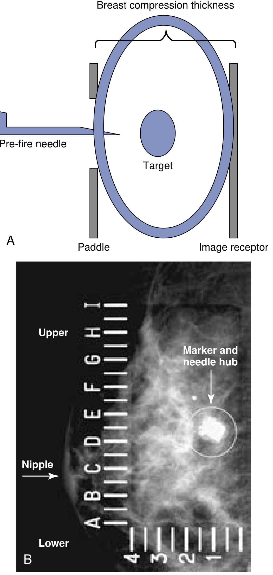
The fundamental principle involves mapping the 3D coordinates of the target lesion in relation to the geometric centre of the compressed breast by acquiring two angled mammographic images.
Step-by-step localisation:
- A scout (0°) image is obtained with the breast compressed between the paddle and the image receptor
- Two additional images are then obtained at +15° and -15° (30° apart)
- The parallax shift of the target lesion between these two angled views allows calculation of its precise X, Y, and Z (depth) coordinates within the compressed breast
- The computer translates these coordinates to position the needle device accurately at the target
Imaging Modalities Used
1. 2D Digital Mammography (Standard)
- The original and most widely used platform
- Acquires two-dimensional images from which 3D coordinates are calculated
- Best for microcalcifications, focal asymmetries, and densities visible on mammography
2. Digital Breast Tomosynthesis (DBT / 3D Mammography)
- FDA-approved; increasingly available
- The X-ray tube moves in an arc around the breast, taking multiple images at 1 mm slice intervals from many angles within seconds
- Computer generates a highly focused 3D image of the breast
- Particularly valuable in dense breast tissue - can distinguish a true distortion or asymmetry from overlapping breast tissue (which standard 2D mammography cannot)
- Reduces recall rates and improves characterisation of lesions
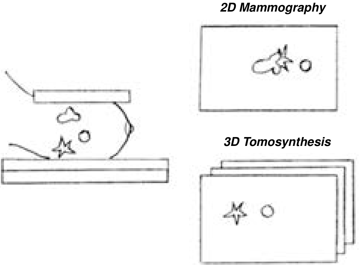
3. MRI-Guided Biopsy
- For lesions visible only on MRI (enhancement without a mammographic or sonographic correlate)
- Uses the same stereotactic coordinate principles but within an MRI suite
4. Ultrasound-Guided Biopsy
- Preferred when the lesion is sonographically visible (palpable masses, solid nodules)
- Real-time visualisation of needle within lesion
- More comfortable patient positioning (no prone requirement)
- Stereotactic guidance is used specifically when the lesion is not seen on ultrasound
BI-RADS Classification and Indications
Breast lesions are classified by the Breast Imaging-Reporting and Data System (BI-RADS):
| BI-RADS Category | Description | Action |
|---|---|---|
| 1 | Negative | Routine screening |
| 2 | Benign finding | Routine screening |
| 3 | Probably benign | Short-term follow-up OR biopsy |
| 4 | Suspicious | Biopsy recommended |
| 5 | Highly suggestive of malignancy | Biopsy mandatory |
| 6 | Known biopsy-proven malignancy | Further tissue sampling if needed |
Indications for Stereotactic Core Biopsy (Box 1 - Current Surgical Therapy 14e)
- BI-RADS 4 - suspicious lesions
- BI-RADS 5 - highly suspicious lesions (confirms diagnosis and facilitates treatment planning)
- BI-RADS 3 - probably benign lesions when:
- Clinical suspicion is elevated
- Patient or physician preference
- Short-term imaging follow-up is not practical
- New suspicious microcalcifications - the classic indication
- Developing asymmetries or architectural distortions on mammography
- Non-palpable asymmetry, focal asymmetry, or density not visible on ultrasound
- Mammographic lesion corresponding to suspicious MRI enhancement
- New mass or abnormal tissue at a previous surgery site
- BI-RADS 6 - known malignancy requiring further sampling for treatment planning (e.g. receptor status before neoadjuvant chemotherapy)
- Multifocal or multicentric lesions - to facilitate treatment planning
- Discordant imaging-pathology assessment after initial biopsy - repeat biopsy needed
Contraindications
Absolute Contraindications
- Pregnancy (radiation exposure)
- Lesion immediately adjacent to a breast implant (risk of implant perforation)
- Patient unable to cooperate (mental disability)
Relative / Technical Contraindications (Box 2 - Current Surgical Therapy 14e)
| Category | Specific Contraindication |
|---|---|
| Patient position | Unable to lie prone; kyphosis preventing adequate positioning |
| Weight | >300 lbs (exceeds weight limit of prone biopsy table) |
| Body habitus | Obesity, pregnancy (also radiation exposure concern) |
| Lesion location | Too posterior (near chest wall) - needle may not reach |
| Lesion location | Too superficial (near skin or nipple) - needle may penetrate through breast |
| Breast size | Too thin/small - inadequate tissue thickness in compression for safe needle travel |
| Breast size | Too large with deep lesion - device may not reach the target |
| Anticoagulation | Must be assessed individually; risk-benefit must be weighed |
Patients who cannot undergo stereotactic biopsy should be referred for open surgical excisional biopsy.
Equipment
A. Core Needle Biopsy (CNB) Device
- Spring-loaded automated 14-gauge core needle
- Appropriate for masses >1 cm
- Retrieves multiple tissue cores in separate insertions
- Provides cores for histology (preserves tissue architecture - unlike FNA which provides only cytology)
B. Vacuum-Assisted Biopsy (VAB) Device - Preferred for Microcalcifications
- 7 to 11 gauge rotating cutter with vacuum suction
- Single-insertion device: vacuum suction pulls tissue into the collecting aperture; rotating cutter excises it
- Can perform up to 12 biopsies in 360° around the lesion with a single insertion
- 9-gauge is most popular - can remove up to 1 cm of tissue
- Allows larger volume sampling - reduces sampling error
- Advantages over 14G CNB:
- Repeat biopsy rate for inadequate microcalcification sampling: 11.6% vs. 23.7% with CNB
- In diagnosing DCIS: only 6% upgraded to invasive carcinoma at surgery vs. 21% with 14G CNB
- Nearly 3x more accurate for diagnosing atypical ductal hyperplasia (ADH) - though underestimation still occurs in 18-25%
- Can completely excise small lesions (<1 cm) percutaneously
C. Marker / Clip Placement
- A radiopaque metallic clip (titanium or other material) should be placed at the biopsy site at the time of the procedure in all suspicious lesions because:
- The lesion may be partially or completely removed during biopsy
- Enables accurate surgical localisation at future operations
- Essential if the patient is expected to undergo neoadjuvant chemotherapy (which causes tumour regression and may eliminate the imaging target)
"A clip or other marking device should be placed at the time of percutaneous biopsy of all suspicious lesions to improve the accuracy of future localisation."
- Current Surgical Therapy 14e
Step-by-Step Procedure
-
Pre-procedure workup: Review complete mammographic examination to confirm the lesion location. Document clinical breast examination. Place radiopaque markers over areas of palpable concern.
-
Informed consent: Discuss the possibility of non-diagnostic results, risks, benefits, and alternatives.
-
Patient positioning:
- Prone biopsy table: Patient lies prone with the breast hanging through a fenestration in the table; the operator works below the table. Optimal stability and patient comfort.
- Upright add-on unit: Stereotactic add-on attached to standard mammography equipment; patient sits or stands.
-
Breast compression: Breast compressed and held between the image receptor and the compression plate throughout the procedure.
-
Target confirmation: Imaging performed to confirm the target lesion lies within the accessible area.
-
Coordinate calculation: Computer generates X, Y, Z coordinates of the target and transfers them to the stereotactic targeting device.
-
Skin preparation and local anaesthesia: Sterile technique; local anaesthetic (usually 1% lidocaine) injected into the skin and breast tissue along the planned needle track.
-
Scout images: Scout view obtained; after target identified, two images at ±15° confirm needle position.
-
Pre-fire images: Images obtained with the needle in pre-fire position to confirm alignment with target.
-
Needle firing / tissue acquisition:
- For CNB: needle is fired and multiple separate passes are made
- For VAB: single insertion, multiple tissue cores obtained by rotation around the lesion
-
Post-fire confirmation: Images obtained after tissue acquisition to confirm adequate sampling (especially that calcifications are present in the specimen).
-
Specimen radiography: Tissue cores are radiographed to confirm calcifications are present in the specimen - this is the definitive evidence of adequate sampling for microcalcification targets.
-
Clip/marker deployment: Radiopaque clip placed at the biopsy site.
-
Post-procedure imaging: Mammogram obtained to document clip position and confirm satisfactory haemostasis.
-
Wound care: Manual pressure, dressing applied. Most patients are discharged within 30-60 minutes.
Accuracy and Performance
- Sensitivity of core needle biopsy performed stereotactically or with ultrasound: 97-99%
- Accuracy increases with larger needle gauge
- Sensitivity is superior to FNA because tissue architecture is preserved
- Specimen radiograph confirming calcifications in the core is mandatory for microcalcification targets
Advantages Over Surgical Excisional Biopsy
| Parameter | Stereotactic Core Biopsy | Open Surgical Biopsy |
|---|---|---|
| Invasiveness | Minimal (percutaneous) | Significant (open operation) |
| Anaesthesia | Local | General or local |
| Scarring | Minimal (needle track) | Significant |
| Cost | Lower | Higher |
| Hospital stay | Outpatient (30-60 min) | Day case or inpatient |
| Sensitivity | 97-99% | Near 100% |
| Tissue architecture | Preserved | Full excision |
| False-negative rate | ~1-3% | Very low |
Radiological-Pathological Concordance
After the biopsy result is received, radiological-pathological concordance must be assessed - i.e. does the pathological result explain the imaging finding?
- Concordant result: pathology explains the imaging lesion (e.g. calcifications in fibrocystic change explain benign-looking microcalcifications; carcinoma explains a BI-RADS 5 mass)
- Discordant result: pathology does NOT explain the imaging appearance (e.g. normal/benign tissue from a BI-RADS 5 lesion) - this requires repeat biopsy or surgical excision
Indications for Surgical Excision After Stereotactic Core Biopsy (Box 3 - Current Surgical Therapy 14e)
Surgical excision is required after SCB when:
- Malignancy (invasive carcinoma or DCIS) confirmed - proceed to definitive surgery
- Discordant radiological-pathological findings - benign/normal result from a suspicious or highly suspicious lesion
- Atypical Ductal Hyperplasia (ADH) - upgrade risk to DCIS or invasive cancer is 18-25% (VAB) to even higher with CNB; surgical excision is standard
- Atypical Lobular Hyperplasia (ALH) and Lobular Carcinoma In Situ (LCIS) - upgrade risk warrants excision
- Flat Epithelial Atypia (FEA) - upgrade risk
- Radial scar / complex sclerosing lesion - may harbour carcinoma not sampled
- Mucocele-like lesion with atypia
- Phyllodes tumour - complete excision needed
- Non-diagnostic specimen (no calcifications in specimen radiograph) with remaining clinical or imaging suspicion
- Lesion completely removed on biopsy without prior clip placement (if clip was not placed and no residual lesion remains for localisation)
- Papillary lesion with atypia or where adequate sampling is uncertain
Complications
Stereotactic core biopsy is a very safe procedure. Complications include:
| Complication | Comment |
|---|---|
| Haematoma | Most common; usually minor and self-limiting; managed with pressure |
| Infection | Rare (<1%); cellulitis; manage with antibiotics |
| Vasovagal reaction | Particularly in upright (sitting) position; managed by lying the patient flat |
| Bleeding | Usually stops with pressure; significant haemorrhage is very rare |
| Pneumothorax | Extremely rare; more risk with posterior lesions near chest wall |
| Implant damage | Rare; contraindication if lesion is adjacent to implant |
| Needle track seeding | Theoretical; clinically not demonstrated to be significant |
| False-negative result | 1-3%; due to sampling error - necessitates radiological-pathological concordance check |
Post-Procedure Follow-up
- Most patients: follow-up mammogram at 6-12 months to confirm stability of the biopsy site and any residual abnormality
- Post-DCIS excision: postoperative mammogram to confirm completeness of calcification removal (beyond specimen radiography and margin assessment)
- Age >35 years: unilateral diagnostic mammogram as a new baseline within 4-6 months
- Clip migration: on post-procedure mammogram, the clip should be confirmed at the correct location; migration may require repeat localisation imaging before any planned surgery
Comparison: CNB vs. VAB vs. FNA
| Feature | FNA | 14G CNB | VAB (9-11G) |
|---|---|---|---|
| Needle gauge | 22-25G | 14G | 7-11G |
| Sample type | Cytology only | Histology (cores) | Large-volume histology |
| Architecture preserved | No | Yes | Yes |
| Marker analysis / IHC | Limited | Yes | Yes |
| Microcalcification adequacy | Poor | Moderate | Best |
| ADH accuracy | Poor | Poor (18-25% upgrade) | Best (18-25% upgrade - still uncertain) |
| Re-biopsy rate (calcifications) | High | 23.7% | 11.6% |
| DCIS upgrade rate at surgery | N/A | 21% | 6% |
| Can remove entire small lesion | No | No | Yes (<1 cm) |
| Preferred for microcalcifications | No | No | Yes - standard |
Summary
Stereotactic core biopsy represents the standard of care for tissue diagnosis of mammographically detected non-palpable breast lesions - particularly microcalcifications - that are not sonographically visible. It has replaced the vast majority of open surgical excisional biopsies for diagnosis, offering 97-99% sensitivity as an outpatient procedure under local anaesthesia. Vacuum-assisted biopsy is the preferred device for microcalcifications, providing larger tissue volumes, lower re-biopsy rates, and more accurate histological characterisation. The fundamental principle of radiology-pathology concordance is central to correct interpretation of results and decision-making about further surgical management.
Source:
- Current Surgical Therapy, 14e - Stereotactic Core Biopsy of Breast Disease (Pastoriza JM, Mallon WC, Jacobs LK), pp. 767-770
Rotation of gut embryology
Gut rotation in embryology involves the midgut herniating into the umbilical cord at week 6, rotating 270° counterclockwise around the superior mesenteric artery axis, and returning to the abdomen by week 10. Abnormal rotation causes malrotation and volvulus.
Searching sources...
Finding Sources
Finding Sources
Finding Sources
Finding Sources
Finding Sources
Finding Sources
Reading File
Reading File
Reading File
Finding Sources
Finding Sources
Reading File
Finding Sources
Loading Image
Reading File
Reading File
Loading Image
Finding Sources
I now have excellent content from multiple authoritative sources. Let me compile the comprehensive note on gut rotation in embryology.
Rotation of the Gut - Embryology
Overview of the Primitive Gut
The primitive gut tube is formed from the endodermal roof of the yolk sac at approximately week 3 of gestation. As lateral folding of the embryo occurs, the endoderm is incorporated into the embryo to form a tubular gut.
The primitive gut is divided into three regions, each supplied by a specific artery:
| Division | Derivatives | Blood Supply |
|---|---|---|
| Foregut | Pharynx → oesophagus → stomach → duodenum (to ampulla of Vater); liver, gallbladder, pancreas | Coeliac artery |
| Midgut | Distal duodenum → jejunum → ileum → caecum → appendix → ascending colon → proximal 2/3 of transverse colon | Superior mesenteric artery (SMA) |
| Hindgut | Distal 1/3 of transverse colon → descending colon → sigmoid → rectum → upper anal canal | Inferior mesenteric artery (IMA) |
I. Foregut Development and Rotation
Stomach
The stomach first appears as a fusiform dilation of the foregut at approximately week 4.
Two rotations occur:
1. Rotation around the longitudinal (craniocaudal) axis - 90° clockwise (as viewed from above):
- Originally the stomach lies in the midline with a dorsal and ventral border
- The dorsal border grows faster → becomes the greater curvature
- The ventral border grows slower → becomes the lesser curvature
- Rotation is 90° clockwise (when viewed from above/front)
- Result: The original left side becomes the anterior (ventral) wall; the original right side becomes the posterior (dorsal) wall
- The left vagus nerve (originally innervating the left side) comes to lie on the anterior surface of the stomach; the right vagus comes to lie posteriorly
2. Rotation around the anteroposterior axis - tilt:
- The pyloric end rotates upward and to the right
- The cardia rotates downward and to the left
- Result: The stomach acquires its oblique orientation, with the pylorus now pointing to the right and upward
Consequences of stomach rotation:
- Formation of the lesser sac (omental bursa) - a space behind the stomach
- Development of the greater omentum from the dorsal mesentery as it enlarges downward
- The lesser omentum connects the lesser curvature to the liver (hepatogastric ligament) and duodenum (hepatoduodenal ligament)
Duodenum
- Develops from the caudal end of the foregut and cranial end of the midgut (junction at ampulla of Vater)
- Initially forms a C-shaped loop
- Rotates with the stomach and becomes retroperitoneal in its 2nd, 3rd, and 4th parts
- The duodenal lumen is temporarily obliterated by epithelial proliferation (weeks 5-6); recanalisation occurs by week 8-9; failure leads to duodenal atresia
- As the midgut rotates (see below), the duodenum traces a C-loop beneath the superior mesenteric artery axis
II. Midgut - Physiological Herniation and Rotation (The Key Event)
This is the most complex and clinically significant aspect of gut rotation.
Stage 1: Physiological Umbilical Herniation (Week 6)
- During weeks 5-6, the midgut elongates rapidly - far faster than the abdominal cavity can accommodate
- The midgut herniates ventrally out through the umbilical ring into the proximal umbilical cord - this is called physiological (umbilical) herniation or the primary intestinal loop
- The loop of herniated midgut is oriented around the axis of the superior mesenteric artery (SMA), which occupies the apex of the loop
- At this stage, the loop has:
- Cranial (pre-arterial) limb: gives rise to the distal duodenum, jejunum, and proximal ileum
- Caudal (post-arterial) limb: gives rise to the distal ileum, caecum, appendix, ascending colon, and proximal two-thirds of transverse colon
- The caecal bud appears as a small swelling on the caudal limb at about week 6
Stage 2: Rotation During Herniation (Weeks 6-10)
While in the umbilical cord, the gut rotates 90° counterclockwise around the SMA axis (as viewed from the front):
- The cranial limb moves from the midline to the right side (downward/clockwise in the patient's view from outside)
- The caudal limb moves to the left side
- Net result: cranial limb now lies to the right; caudal limb (with caecal bud) lies to the left
At this point, 90° of the total 270° counterclockwise rotation has occurred.
Stage 3: Return to the Abdomen (Weeks 10-12)
- By week 10, the abdominal cavity grows large enough to accommodate the intestine
- The herniated gut rapidly returns to the abdominal cavity
- As it does so, it undergoes a further 180° counterclockwise rotation (total = 270° counterclockwise from the original position)
The sequence of return:
- The cranial (pre-arterial) limb returns first, entering to the right of the SMA and then moving to occupy the left side of the abdomen (jejunum and ileum fill the left and central abdomen)
- The caudal (post-arterial) limb returns last - the caecum and ascending colon enter and settle in the upper right/epigastrium initially, then descend to the right lower quadrant
Summary of final positions:
- Duodenojejunal junction: left of midline, below and to the left of the SMA → fixed at the ligament of Treitz (suspensory muscle of duodenum)
- Caecum: right lower quadrant
- Ascending colon: right side, retroperitoneal
- Transverse colon: crosses left to right (cephalad to SMA)
- Duodenum: C-loop, retroperitoneal (2nd, 3rd, 4th parts)
Total rotation = 270° counterclockwise around the SMA axis (when viewed from the front/ventral aspect of the embryo)
Stage 4: Fixation (Week 12 onwards)
After return to the abdomen, the gut undergoes retroperitoneal fixation:
- The duodenum (2nd, 3rd, 4th parts) fuses to the posterior abdominal wall and becomes retroperitoneal; the duodenojejunal junction is anchored by the ligament of Treitz to the left of the midline
- The ascending colon fuses to the right posterior abdominal wall → retroperitoneal
- The descending colon fuses to the left posterior abdominal wall → retroperitoneal
- The caecum becomes fixed to the lateral abdominal wall in the right iliac fossa by peritoneal reflections
- The small bowel mesentery acquires a broad base - its attachment runs from the duodenojejunal flexure (upper left) to the ileocaecal junction (lower right) - this long mesenteric root prevents the gut from twisting
- The transverse colon and sigmoid colon remain intraperitoneal with their mesenteries
"The takeoff of the branches of the SMA elongates and becomes fixed along a line extending from its emergence from the aorta to the cecum in the right lower quadrant."
- Schwartz's Principles of Surgery, 11e
III. Hindgut Development
- Forms the distal 1/3 of transverse colon, descending colon, sigmoid, rectum, and upper anal canal
- Does not herniate into the umbilical cord; remains within the abdominal cavity
- The terminal hindgut opens into the cloaca - a common chamber shared with the urogenital tract
- The urorectal septum (mesoderm) grows caudally to divide the cloaca into:
- Anterior: urogenital sinus
- Posterior: anorectal canal
- The cloacal membrane breaks down, opening the anal and urogenital passages
- The dentate line marks the junction of endoderm-derived hindgut (upper 2/3 of anal canal) and ectoderm-derived proctodeum (lower 1/3)
- The anal sphincters:
- Internal anal sphincter = thickened circular smooth muscle of the rectum (hindgut derived)
- External anal sphincter = posterior part of the cloacal sphincter (ectoderm derived)
IV. Genetic and Molecular Basis of Rotation
The molecular basis of gut rotation remains an area of active research:
- BCL6 gene mutations: absence of left-sided expression of BCL6 transcripts leads to reversed cardiac orientation, defective eye development, and intestinal malrotation - demonstrating the link between left-right axis determination and gut rotation
- FOXF1 transcription factor: essential for formation of the dorsal gut mesentery (the structure that mediates normal intestinal rotation); mutations in FOXF1 cause alveolar capillary dysplasia, consistently associated with intestinal malrotation
- The dorsal gut mesentery plays a central mediating role in left-right asymmetric rotation
- Recent studies in rat embryos challenge the classic 270° rotation concept, suggesting instead that rapid, localised longitudinal growth of the duodenum pushes the duodenojejunal segment beneath the mesenteric root, with the colon and rectum changing position relatively little (mainly the caecum exits and passively returns)
V. Anomalies of Gut Rotation
1. Intestinal Malrotation
Definition: Incomplete rotation and fixation of the midgut after return from the umbilical sac.
Incidence: 2-5 per 1,000 live births (autopsy); clinically apparent in 1 per 6,000 live births; 90% present before 12 months of age; up to 1% present in adults.
Pathophysiology:
- If the duodenojejunal flexure or caecum does not reach its normal final position, the mesenteric root is short (both junctions lie close together in the mid-abdomen)
- This creates a narrow SMA pedicle suspending the entire midgut
- The short mesenteric root predisposes to midgut volvulus (twisting around the SMA)
Consequences:
- Ladd's bands: fibrous peritoneal bands extending from the abnormally placed caecum (which lies in the epigastrium or midabdomen) to the lateral abdominal wall and crossing the duodenum - can cause duodenal obstruction
- Midgut volvulus: twisting of the entire midgut around the SMA → intestinal obstruction and ischaemia
2. Non-Rotation
- The gut returns to the abdomen but no rotation occurs at all
- Small intestine lies entirely on the right; large intestine lies entirely on the left
- Usually asymptomatic but at risk for volvulus
- Often associated with other anomalies: diaphragmatic hernia, gastroschisis, omphalocele
3. Reversed Rotation
- Gut rotates clockwise instead of counterclockwise
- The transverse colon passes posterior to the SMA (instead of anterior)
- Can cause compression of the transverse colon by the SMA
4. Paraduodenal Hernia
- Internal hernia into a peritoneal fossa created by incomplete mesenteric fixation during rotation
- Right paraduodenal hernia: small bowel herniates behind the ascending mesocolon
- Left paraduodenal hernia: small bowel herniates behind the descending mesocolon
- Both can cause acute or intermittent intestinal obstruction
5. Omphalocele (Exomphalos)
- Failure of the physiologically herniated gut to return to the abdominal cavity by week 10-12
- Gut (and often liver) remains outside the abdomen covered only by peritoneum and amnion
- Associated with chromosomal anomalies (trisomy 13, 18, 21) and other defects
6. Gastroschisis
- Herniation of gut through a paraumbilical defect in the anterior abdominal wall (usually to the right of the umbilicus)
- No covering membrane - gut is exposed and bathed in amniotic fluid
- Usually isolated (not chromosomal); incidence is rising
- Gut returns to a normal position but may be matted and oedematous
VI. Clinical Presentation of Malrotation with Midgut Volvulus
| Feature | Detail |
|---|---|
| Classic presentation | Bilious (green) vomiting in a neonate (first few weeks of life) |
| Key warning sign | Any bilious vomiting in a neonate must be assumed to be volvulus until proven otherwise |
| Urgency | True surgical emergency - minutes to hours determine intestinal viability |
| Late signs | Bloody stools, abdominal distension, erythema/oedema of abdominal wall, haemodynamic collapse |
| AXR | Proximal bowel distension, paucity of distal gas; "double bubble" if associated duodenal obstruction |
| Gold standard imaging | Upper GI contrast series (stable patient): shows duodenojejunal junction to the right of midline; "corkscrew" or "bird's beak" deformity of the duodenum |
| Ultrasound | Reversal or abnormal relationship of SMA and SMV (normally: vein to the right of artery); in malrotation, the vein may be to the left or directly anterior) |
VII. Surgical Treatment - Ladd's Procedure (1936)
The Ladd's procedure, described by William Ladd in 1936, remains the standard operation:
- Untwist the volvulus counterclockwise to restore blood flow to the midgut - the first and most urgent step
- Assess bowel viability; warm sponges over ischaemic segments; resect necrotic bowel; borderline segments may be left for second-look laparotomy at 24-36 hours
- Divide Ladd's bands from the caecum/right colon to the lateral abdominal wall and across the duodenum - this relieves duodenal obstruction
- Straighten the duodenum by dividing all peritoneal attachments around it - duodenojejunal junction ends up in the right lower quadrant
- Broaden the mesenteric base by dividing bands across the SMA root - this maximises the distance between the duodenojejunal and ileocaecal junctions and reduces the risk of re-volvulus
- Appendectomy - as the appendix will now lie in the left upper quadrant (abnormal position, potentially confounding future diagnosis of appendicitis)
- Return caecum to the left upper quadrant and small bowel to the right abdomen
Complications:
- Recurrent volvulus in up to 10% after Ladd's procedure
- Short bowel syndrome if extensive bowel resection needed (~18% of paediatric SBS cases are due to midgut volvulus)
- Prolonged postoperative ileus
Summary: Key Timeline of Gut Rotation
| Week | Event |
|---|---|
| Week 3-4 | Primitive gut tube forms from yolk sac endoderm; divided into foregut, midgut, hindgut |
| Week 4 | Caecal bud appears; stomach begins as a fusiform dilation |
| Week 4-6 | Stomach rotates 90° clockwise (longitudinal axis) + tilt; lesser sac forms |
| Week 5-6 | Urorectal septum begins dividing cloaca |
| Week 6 | Midgut herniates into umbilical cord (physiological umbilical hernia); 90° counterclockwise rotation begins |
| Week 6-10 | Gut elongates in the cord; 90° counterclockwise rotation completed |
| Week 10-12 | Gut returns to abdominal cavity; further 180° counterclockwise rotation = total 270° counterclockwise |
| Week 12 onwards | Retroperitoneal fixation: duodenum, ascending and descending colon become fixed; ligament of Treitz forms |
Sources:
- Sabiston Textbook of Surgery - Embryology of the Colon and Rectum, Chapter 95; Intestinal Malrotation and Midgut Volvulus, Chapter 117
- Schwartz's Principles of Surgery (11e) - Malrotation and Midgut Volvulus, Chapter 39
- Gray's Anatomy for Students - Malrotation and Midgut Volvulus, Chapter 4
- Moore KL, Persaud TVN, Torchia MG - The Developing Human, 11e (cited within Sabiston)
This is a shared conversation. Sign in to Orris to start your own chat.