
Papillo edema
papilledema optic disc swelling fundoscopy
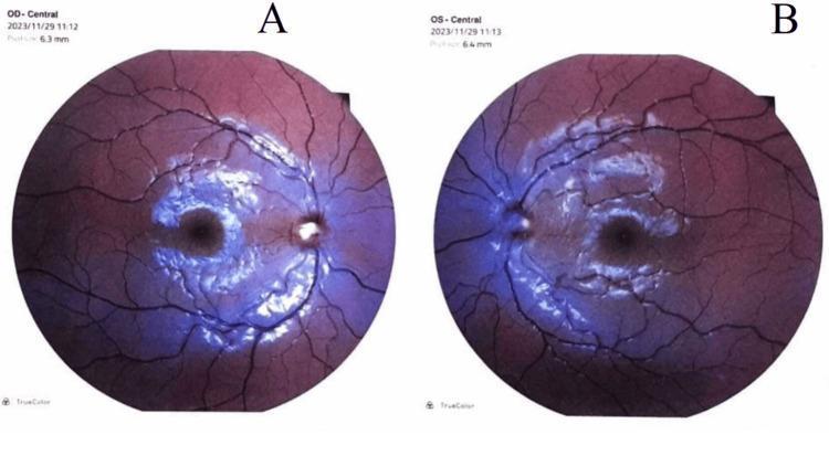
This diagnostic image displays bilateral fundus photographs of the right eye (OD, labeled A) and left eye (OS, labeled B). The images demonstrate severe bilateral papilledema, characterized by significant optic disc swelling with blurred, indistinct disc margins and a peripapillary halo. In both eyes, the optic nerves appear elevated and hyperemic, with a loss of the physiological cup. The retinal vasculature exhibits marked tortuosity and engorgement, with several vessels becoming obscured as they cross the disc margin due to the overlying edema. The surrounding retinal tissue shows evidence of compromised perfusion, with a general hyperemic hue and a lack of clarity in the macula and peripapillary region. These visual findings are consistent with Stage 4 papilledema, often associated with increased intracranial pressure. The modality is fundoscopy (retinal imaging), serving as a critical educational tool for identifying neuro-ophthalmological signs of intracranial hypertension.
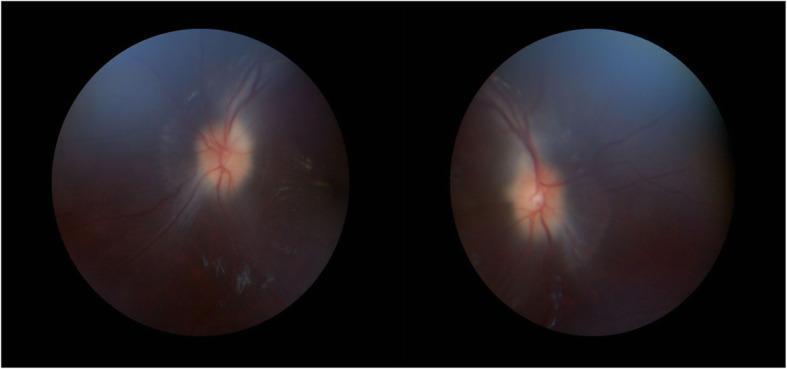
This diagnostic image consists of two fundoscopy circular frames showing the bilateral retinal fundi. The primary clinical finding is bilateral optic disc edema, consistent with grade 2 papilledema. The optic discs demonstrate significant blurring of the margins and visible elevation. Vascular changes include moderate engorgement and increased tortuosity of the retinal veins as they emerge from the disc. The physiological cups appear obscured due to the swelling. The surrounding retinal background maintains a normal red-orange hue, although there is a subtle presence of whitish spots suggestive of exudates in the peripheral field of the left eye. No obvious retinal hemorrhages are visualized. These findings are characteristic of increased intracranial pressure, as seen in conditions like idiopathic intracranial hypertension or space-occupying lesions. This content is intended for medical education in ophthalmology and neurology to demonstrate clinical signs of optic nerve head swelling.
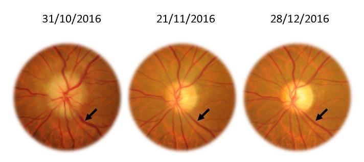
This Comparison Chart consists of three sequential color fundus photographs of the left eye, documenting the progression of ophthalmic findings over a two-month period (31/10/2016 to 28/12/2016). The images demonstrate the resolution of papilledema (optic disc edema) and venous congestion. In the initial photograph, the optic disc exhibits blurred margins, elevation, and obscuration of peripapillary vessels, characteristic of acute swelling. The retinal veins, highlighted by black arrows, appear significantly engorged and tortuous (venous turgescence). Subsequent images show a chronological improvement: the optic disc margins become progressively more distinct and sharp, indicating a reduction in edema. Simultaneously, the caliber of the retinal veins decreases, showing a marked reduction in turgidity and fullness. This timeline serves as a clinical illustration of treatment response or the natural history of intracranial pressure resolution. Key concepts include optic neuropathy, fundoscopy, venous caliber changes, and the morphological stages of papillary edema remission.
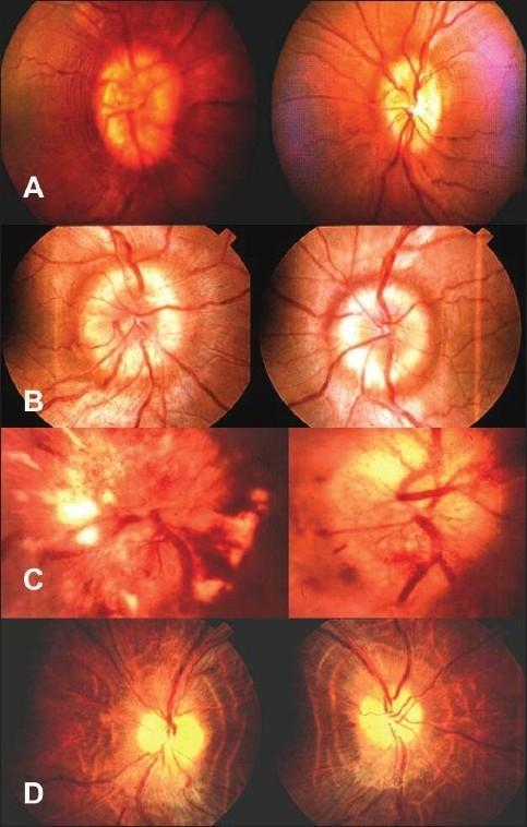
This clinical photograph panel displays four stages of papilledema (A–D) in fundus images, illustrating the progression of optic disc swelling due to increased intracranial pressure. Stage A (Early) shows optic disc hyperemia and mild blurring of the nasal margins with preserved retinal nerve fiber layer (RNFL) striations. Stage B (Established) demonstrates more pronounced circumferential disc elevation and obscuration of major retinal vessels crossing the margin, with increased vascular tortuosity. Stage C (Vintage/Chronic) highlights significant pathological changes including severe edema, cotton-wool spots, and retinal hemorrhages that further mask the disc anatomy. Stage D (Atrophic) shows the late-stage transition to optic atrophy, characterized by a pale, chalky-white optic disc with relatively defined margins compared to acute stages, accompanied by vessel attenuation and thinning of the RNFL. This comparison chart serves as an educational tool for ophthalmologists and neurologists to categorize the severity and duration of papilledema in clinical practice.
papilledema intracranial pressure
Papilledema
- Wills Eye Manual, p. 704
Pathophysiology
- Bradley and Daroff's Neurology in Clinical Practice
Four Stages (Fundoscopic Classification)
| Stage | Key Features |
|---|---|
| A - Early | Minimal disc hyperemia, capillary dilation; mild opacification of nerve fiber layer (loses its linear light reflex); mild disc swelling; absent venous pulsations; peripapillary RNFL hemorrhages |
| B - Fully Developed (Acute) | Disc surface grossly elevated; engorged tortuous retinal veins; splinter hemorrhages at/near disc; surface vessels obscured by opaque NFL; cotton-wool spots; Paton lines (circumferential retinal folds); macular star or exudates |
| C - Chronic | Disc takes on a "champagne cork" appearance; pseudodrusen (extruded axoplasm); pale gliotic disc; hemorrhages resolve; collateral optociliary vessels may develop |
| D - Atrophic | Pale, chalky disc; vessel attenuation; RNFL thinning - end-stage optic atrophy with permanent visual loss |
- Localization in Clinical Neurology, 8e, p. 377
Clinical Features
Symptoms
- Transient visual obscurations - brief (seconds), often bilateral, precipitated by postural change (hallmark symptom)
- Headache (worse in morning, worse with Valsalva)
- Diplopia (VI nerve palsy - false localizing sign)
- Nausea and vomiting
- Visual acuity is typically preserved early - this is a key distinguishing feature from papillitis
Signs
-
Bilateral swollen, hyperemic optic discs (may be asymmetric early)
-
Blurred disc margins - starts at superior and inferior poles (where NFL is thickest)
-
Loss of spontaneous venous pulsations (SVPs)
-
Flame-shaped peripapillary hemorrhages
-
Dilated, tortuous retinal veins
-
Enlarged physiologic blind spot (earliest visual field defect)
-
Normal pupillary reactions and color vision (unless severe asymmetric edema - then APD)
-
Inferonasal arcuate visual field defects, eventually peripheral constriction
-
Wills Eye Manual, p. 704-705; Localization in Clinical Neurology, 8e
Fluorescein Angiography
- Early: absent disc fluorescence (delayed circulation)
- Arteriovenous phase: dilated capillaries and microaneurysms
- Venous phase: dye leakage beyond disc margins
- May be normal in early papilledema
Causes of Raised ICP (Etiology)
| Category | Examples |
|---|---|
| Intracranial mass | Primary or metastatic tumors, brain abscess |
| Hydrocephalus | Obstructive or communicating |
| Idiopathic intracranial hypertension (IIH) | Obese women of childbearing age; also tetracyclines, retinoids, steroids withdrawal, OCP |
| Venous sinus thrombosis | |
| Meningitis/encephalitis | |
| Hypertensive encephalopathy | Malignant hypertension |
| Trauma | Intracranial hematoma |
- Wills Eye Manual, p. 707
Differential Diagnosis of Disc Swelling/Elevation
| Condition | Distinguishing Features |
|---|---|
| Pseudopapilledema (drusen, anomalous disc) | No true disc swelling; vessels not obscured; disc not hyperemic; SVPs often present; drusen on B-scan/autofluorescence/CT |
| Papillitis (optic neuritis) | Usually unilateral; significant visual acuity loss; pain on eye movement; APD present; color vision loss |
| Ischemic optic neuropathy (AION) | Pale or sectoral disc swelling; sudden unilateral visual loss; APD |
| Central retinal vein occlusion | Hemorrhages extend beyond peripapillary region; dilated tortuous veins; usually unilateral |
| Hypertensive optic neuropathy | Very high BP; peripheral hemorrhages; arteriolar narrowing |
| Diabetic papillopathy | Diabetic patient; mild visual loss; spontaneous resolution |
| Leber hereditary optic neuropathy | Young males; peripapillary telangiectasias; rapid sequential visual loss |
| Amiodarone toxicity | Drug history; subacute visual loss |
| Sarcoid/tuberculous infiltration | Other systemic signs |
- Wills Eye Manual, p. 705-706
Investigations
- Urgent neuroimaging (CT or MRI brain with contrast) - rule out mass lesion before LP
- Lumbar puncture - opening pressure >25 cm H₂O (normal <20); CSF analysis
- Visual field testing (Humphrey) - monitor progression
- Fundus photography / OCT - document disc swelling and NFL thickness
- B-scan ultrasound - increased optic nerve sheath diameter (>5 mm), may show buried drusen
- Fluorescein angiography - confirm true papilledema
- MRI venography - if venous sinus thrombosis suspected
Management
Treat the underlying cause
- Intracranial mass: neurosurgery/oncology referral
- Meningitis: antibiotics
- Venous sinus thrombosis: anticoagulation
Idiopathic Intracranial Hypertension (IIH) - specific management:
- Weight loss (10% body weight reduces ICP significantly)
- Acetazolamide (carbonic anhydrase inhibitor) - reduces CSF production; first-line drug
- Topiramate - second-line; also promotes weight loss
- GLP-1 receptor agonists (e.g., semaglutide) - a 2025 meta-analysis (PMID 40937960) confirmed efficacy and safety of GLP-1 and dual GIP/GLP-1 agonists in IIH, making this an emerging pharmacotherapy option
- Therapeutic lumbar puncture - temporary relief
- CSF shunting (ventriculoperitoneal or lumboperitoneal shunt) - for refractory cases
- Optic nerve sheath fenestration - when vision is acutely threatened
Key Clinical Pearls
- Preserved visual acuity in acute papilledema is the hallmark that distinguishes it from papillitis
- Absent venous pulsations on fundoscopy suggest raised ICP (but 20% of normal people lack SVPs)
- Never do LP before neuroimaging - risk of transtentorial herniation if mass lesion present
- The Foster Kennedy syndrome (ipsilateral optic atrophy + contralateral papilledema) occurs with large frontal lobe tumors compressing one optic nerve while raising ICP on the other side
- Unilateral or highly asymmetric papilledema can occur due to anatomical variations in the optic nerve sheath septations
- Terson syndrome: in acute subarachnoid hemorrhage with papilledema, subhyaloid hemorrhages may break into the vitreous
Foster Canada syndrome
Foster Kennedy syndrome optic atrophy papilledema fundus
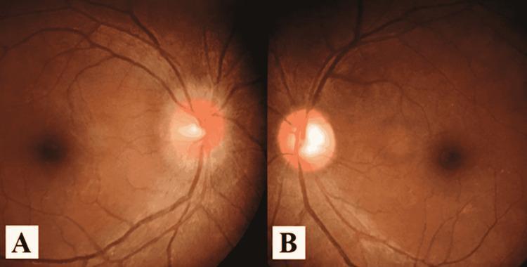
This dual-panel fundus photograph provides a side-by-side comparison of different optic nerve head pathologies in the right and left eyes. Panel A depicts the right eye fundus, showing a markedly hyperemic and swollen optic disc. The disc margins are blurred and indistinct, indicating significant edema, with an absence of a visible physiologic cup. The surrounding retinal vasculature appears slightly engorged. Panel B shows the left eye fundus, characterized by prominent temporal pallor of the optic disc. Unlike the right eye, the disc in the left eye is not elevated, and the nasal margin appears relatively more defined, though the temporal aspect exhibits a stark white coloration indicative of optic atrophy. Both panels show the macula temporally positioned relative to the disc. This comparison is clinically relevant for neuro-ophthalmological evaluation, potentially illustrating conditions such as Foster Kennedy syndrome (though intracranial pressure and imaging context are required for definitive diagnosis) or sequential optic neuropathy involving acute swelling in one eye and chronic atrophy in the other.
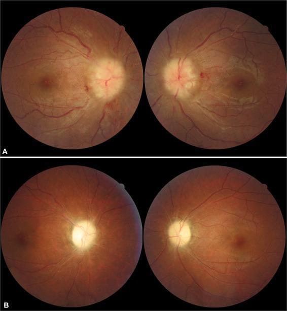
This comparative fundus photograph series demonstrates the progression and resolution of severe bilateral papilledema in a patient with pseudotumor cerebri syndrome. Top row (A): Initial presentation showing grade 5 papilledema in both eyes. The optic discs are severely swollen, elevated, and have blurred margins. Key clinical signs include peripapillary hemorrhages and significant obscuration of the major retinal vessels as they leave the disc. The nerve fiber layer appears thickened and opacified. Bottom row (B): Post-treatment follow-up after 6 months. The acute optic disc edema has completely resolved, but clinical sequelae are visible. The optic discs now exhibit a pale, chalky appearance characteristic of optic atrophy. There is noticeable narrowing (attenuation) of the retinal arterioles and a reduction in the previous vascular engorgement. The image highlights the critical transition from acute hypertensive disc swelling to chronic secondary optic atrophy, emphasizing the importance of monitoring vascular changes and disc pallor in neuro-ophthalmological conditions.
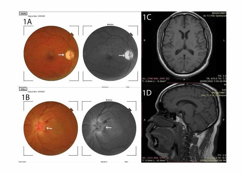
This composite medical image presents diagnostic findings for a case of pseudo-Foster Kennedy syndrome, specifically diabetic papillopathy. Panes 1A and 1B display fundus photography and red-free imaging of the right and left eyes, respectively. Pane 1A (OD) demonstrates a markedly pale, atrophic optic disc (indicated by a white arrow). Pane 1B (OS) shows a significantly edematous and elevated optic disc with blurring of the disc margins and peripapillary retinal hemorrhages, characteristic of acute disc swelling. Panes 1C and 1D provide neuroradiological correlation via T1-weighted Fast Spin Echo (FSE) MRI. Pane 1C shows an axial view at the level of the lateral ventricles, and Pane 1D shows a mid-sagittal view of the brain and orbit. The MRI demonstrates normal intracranial anatomy with no evidence of space-occupying lesions, masses, or increased intracranial pressure, which is critical for differentiating pseudo-Foster Kennedy syndrome from true Foster Kennedy syndrome (typically caused by a frontal lobe tumor). Educational focus includes ophthalmological manifestations of diabetes and neuro-imaging in the differential diagnosis of optic disc edema.
Foster Kennedy Syndrome
Definition
The Classic Triad
| Feature | Side | Mechanism |
|---|---|---|
| Anosmia | Ipsilateral to lesion | Direct compression of olfactory bulb or tract |
| Optic atrophy | Ipsilateral to lesion | Direct injury/compression of the ipsilateral optic nerve |
| Papilledema | Contralateral eye | Raised intracranial pressure secondary to the mass |
- Localization in Clinical Neurology, 8e, p. 310-311
Classic Causes
- Olfactory groove meningioma - the prototypical cause
- Sphenoid ridge/wing meningioma
- Frontal lobe glioma or abscess
- Giant anterior communicating artery aneurysm
- Pituitary adenoma with suprasellar extension (rare)
- Intracranial meningiomatosis compressing optic nerve + blocking superior sagittal sinus
Fundoscopic Appearance
Mechanism in More Detail
- Localization in Clinical Neurology, 8e, p. 379-380
Pseudo-Foster Kennedy Syndrome
| True Foster Kennedy | Pseudo-Foster Kennedy | |
|---|---|---|
| Cause | Frontobasal mass lesion | Sequential bilateral disease affecting optic nerves at different times |
| Anosmia | Present (olfactory involvement) | Usually absent |
| Neuroimaging | Mass lesion present | No intracranial mass; normal ICP |
| Common etiology | Olfactory groove/sphenoid meningioma | Sequential NAION, optic neuritis, syphilis, arachnoiditis, occult trauma |
-
Sequential NAION (non-arteritic anterior ischemic optic neuropathy) - active disc edema in one eye + old atrophy in the other
-
Sequential optic neuritis
-
Diabetic papillopathy affecting each eye at different times
-
Localization in Clinical Neurology, 8e; Kanski's Clinical Ophthalmology, 10e
Associated Features
- Frontal lobe signs: personality change, disinhibition, contralateral hemiparesis (late)
- Raised ICP symptoms: morning headache, nausea/vomiting, diplopia (VI nerve palsy)
- Visual field defects: central scotoma or arcuate defects on the side of optic atrophy; enlarged blind spot contralaterally
- Optociliary shunt vessels may develop at the atrophic disc margin - these represent anastomotic channels between the central retinal vein and choroidal circulation, bypassing compressed venous channels
Investigations
- MRI brain with contrast - identifies the frontobasal mass
- Formal visual fields (Humphrey perimetry) - document defects in both eyes
- Fundus photography / OCT - document disc atrophy vs papilledema
- Olfactory testing - confirm anosmia
- Lumbar puncture - only after neuroimaging excludes mass; to measure ICP
Key Exam Points
- Ipsilateral optic atrophy + contralateral papilledema = Foster Kennedy syndrome until proven otherwise
- The atrophic disc cannot swell - this is the physiological basis of the syndrome
- Anosmia is the third component - often tested in exams but clinically overlooked
- Pseudo-Foster Kennedy (sequential NAION) is actually more common in clinical practice than true Foster Kennedy
- Always do neuroimaging first before LP