
Floating shoulder in detail
Reading File
Reading File
Finding Sources
Finding Sources
Searching PubMed
"floating shoulder"
Reading File
Reading File
Searching PubMed
"floating shoulder" AND (fixation OR outcomes)
Part of the chapter was truncated, so I'll read the remaining sections.
Reading File
Reading File
Reading File
Reading Article
PMID: 36283563
Reading File
Floating shoulder
Summary
Floating shoulder is the classic term for ipsilateral fractures of the clavicle and the scapular (glenoid) neck. Rockwood now treats it as a controversial label. Its core meaning is a glenoid that has lost its osseoligamentous connection to the rest of the scapula and the axial skeleton. A clavicle fracture plus a scapular neck fracture does not always produce that state.
1. Definition and history
-
Ganz and Noesberger first described it: a fracture of the scapular column combined with a clavicle fracture and rupture of the coracoclavicular (CC) and coracoacromial (CA) ligaments.
-
Herscovici et al. coined "floating shoulder" for ipsilateral midshaft clavicle and scapular neck fractures. The clavicle fracture lies medial to the CC ligament insertion.
-
Goss introduced the superior shoulder suspensory complex (SSSC). This is an osseofibrous ring made of the glenoid, coracoid, CC ligament, lateral clavicle, AC joint, AC ligament and acromion. Two breaks in the ring, or one break plus a strut fracture (clavicle or scapular), can leave it unstable. Instability risks delayed union, nonunion or malunion.
-
Later authors reduced the definition to "any clavicle fracture plus any scapular neck or body fracture" and ignored the CC and CA ligaments. This inflated the number of reported cases.
-
Source: Rockwood and Green's Fractures in Adults, 10th ed 2025, pp. 1179-1182 (Ch. 33).
2. Anatomy and why it can be unstable
- The SSSC is the ring above. Together with the clavicle (the superior strut) and the scapular body and spine (the inferior strut), it links the glenoid to the axial skeleton.
- Rockwood's critique:
- A midshaft clavicle fracture alone does not displace the scapular body much. The trapezius and other scapuloaxial muscles hold the scapular position.
- A clavicle fracture plus a surgical neck fracture alone cannot produce a true floating shoulder. Williams et al. argued that CC and CA ligament disruption is also needed.
- Only a fracture of the anatomical neck or surgical neck can separate the glenoid from the body.
- Displaced anatomical neck fractures are primarily unstable, usually with distal and valgus displacement. Arts and Louette consider them a variant of floating shoulder even without a clavicle fracture.
- Surgical neck stability depends on the CC and AC ligaments, or on an intact coracoid base. If both ligaments rupture, the muscles on the coracoid pull the glenoid distally and medially, and the coracoid-to-clavicle distance widens.
- Many "scapular neck fractures" in the floating shoulder literature were really transverse two-part infraspinous fractures of the scapular body (the so-called infraglenoid neck fractures). The fracture line runs below the glenoid, so it does not destabilise the glenoid. The apparent medial glenoid displacement is really lateral displacement of the infraspinous part, pulled by the intact rotator cuff.
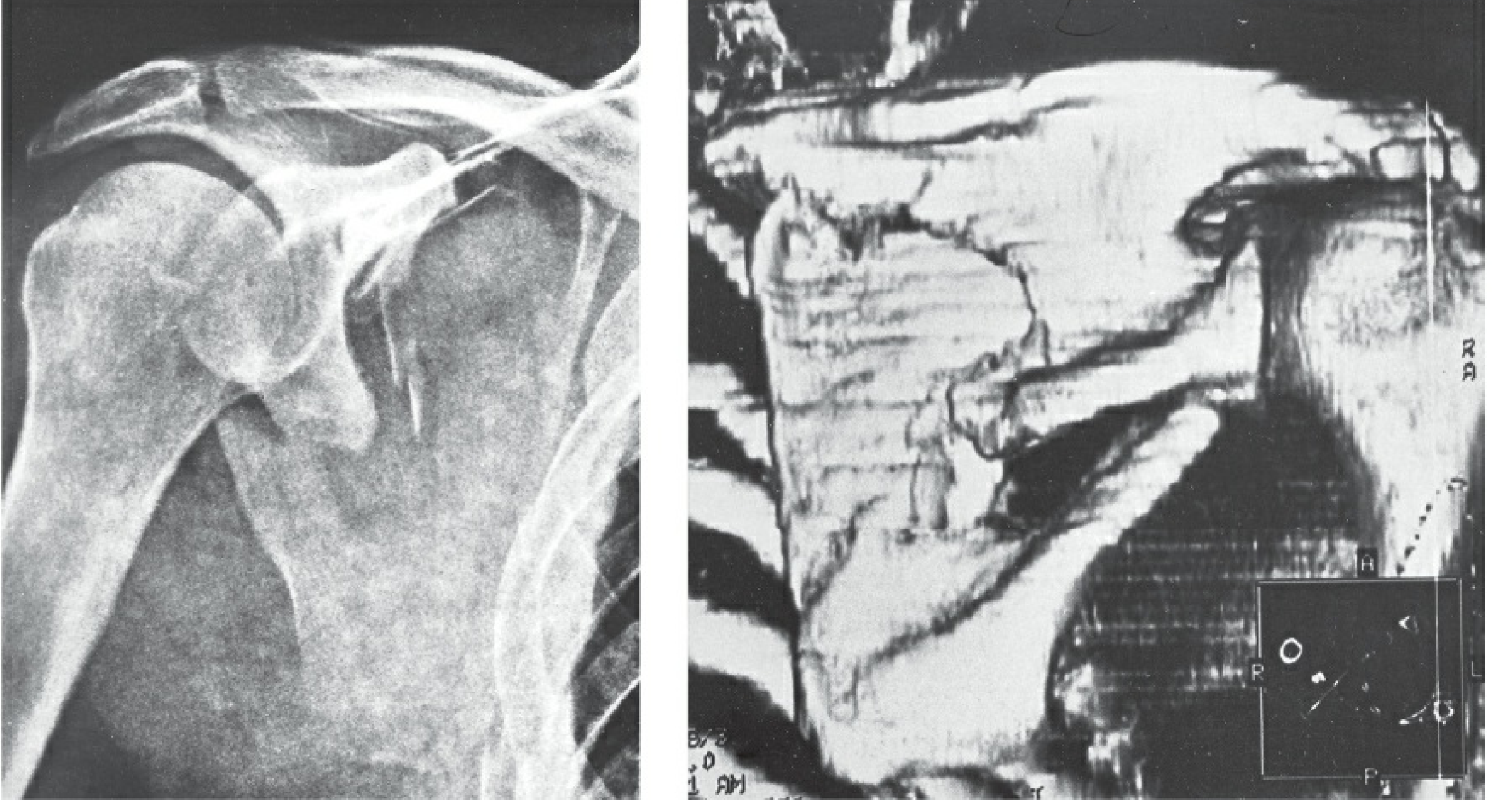
Figure 33-26 from Rockwood and Green's, p. 1181. The AP film is read as a neck fracture, but the CC distance is normal. The 3D CT shows a two-part scapular body fracture.
3. Epidemiology
In the chapter author's series of 519 scapular fractures:
- Surgical neck fractures: 13 (about 2.5% of scapular fractures).
- Anatomical neck fractures: 6.
- Only one surgical neck fracture was totally unstable, with CC and CA rupture.
- Midshaft clavicle fractures occurred in 16% of all scapular fractures, 24% of scapular body fractures and 32% of two-part infraspinous body fractures.
This overlap explains the "seemingly high number" of floating shoulder cases in the literature.
- Source: Rockwood and Green's, 10th ed, p. 1181.
4. Mechanism and associated injuries
These come from my own knowledge, not the retrieved text:
- It usually follows high-energy blunt trauma such as a motor vehicle crash, a fall from height or a direct blow to the shoulder.
- Associated injuries are common: rib fractures, pneumothorax or haemothorax, pulmonary contusion, head and spine injury, and brachial plexus or vascular injury. Always rule out scapulothoracic dissociation.
- Scapulothoracic dissociation is shown in Figure 33-27 of the chapter. It has an enlarged thoracoscapular space and can include neurovascular injury.
5. Diagnosis
- Plain films: a trauma series (true AP of the scapula, scapular Y, axillary) plus a clavicle view. Scapular neck fractures, especially surgical neck, are hard or impossible to diagnose reliably on plain films.
- CT with 3D reconstruction: recommended (DeFranco and Owens) to identify the true fracture pattern, including neck versus infraspinous body fracture.
- CC ligament status: check the coracoclavicular distance. In many published "floating shoulder" films the distance was normal, meaning the CC ligaments were intact. No study stated radiologic criteria for CC ligament injury.
- Glenopolar angle (GPA): a common measure of glenoid medialisation. A GPA of less than about 20 to 30 degrees suggests significant displacement. This threshold is from my own knowledge. Rockwood notes GPA on plain films cannot capture 3D displacement.
6. Treatment
Controversy
Studies are heterogeneous. Some treated both fractures nonoperatively, some fixed only the clavicle, and some fixed both. Outcomes did not differ significantly except in a few studies.
Clavicle-only fixation
- Rationale: reducing the clavicle indirectly reduces and stabilises the glenoid. This avoids a difficult glenoid or scapular exposure with a high complication rate, especially for less experienced surgeons (Rockwood and Green's, Ch. 34, p. 4411 of the block file).
- Caveats from Ch. 33:
- Better results after clavicle fixation may partly reflect earlier rehabilitation, not better reduction of the scapula.
- Persistent scapular body malposition after isolated clavicle fixation has been seen.
- Oh et al. reported two failures of isolated clavicle plating in floating shoulder, so implants should match the higher loads.
- Open reduction of scapular neck fractures is harder after a few days because of rapid callus formation.
- Suggested sequence in Ch. 34:
- Anatomically reduce and fix the clavicle first.
- Re-image the shoulder.
- If the glenoid alignment is acceptable, stop and follow closely.
- If the glenoid is still unacceptable, fix the glenoid or scapular neck, typically through a posterior approach.
Fixation of both bones
Labler et al. and Leung et al. reported very good results with simultaneous clavicle and scapular fixation. The chapter author uses open reduction and internal fixation (ORIF) via the Judet approach for displaced anatomical or surgical neck fractures.
The author's view
- A true floating shoulder is a displaced anatomical neck fracture or a totally unstable surgical neck fracture. This is where the glenoid has lost all osseoligamentous connection.
- The midshaft clavicle fracture is largely irrelevant to glenoid stability. Clavicle fixation has little effect on the glenoid fragment.
- SSSC injury without an anatomical or surgical neck fracture is a separate problem. Double or triple SSSC lesions are potentially unstable and are treated operatively.
- Page numbers for this section are 1181 to 1182.
Commonly cited operative indications
This is from my own knowledge and was not in the retrieved text. Values vary between sources.
- Glenoid or scapular neck displacement of more than about 1 cm (some use more than 5 mm).
- Angulation of more than about 40 degrees.
- GPA below about 20 to 30 degrees.
- Open fracture, neurovascular injury, or a polytrauma patient who needs early mobilisation.
Fixation notes
- Clavicle: plate fixation, superior or anteroinferior.
- Scapula: posterior (Judet) or modified approach, with fixation along the lateral border.
- If the CC and AC ligaments are disrupted, consider CC ligament repair or reconstruction as well.
7. Recent evidence
- A 2023 cadaveric study in J Shoulder Elbow Surg supports the ligament-based view.
- Cutting the CC ligaments reduced the GPA and increased displacement under load.
- With intact ligaments, clavicle plating alone improved the GPA but not displacement.
- With AC, CA and CC disruption, clavicle fixation plus CC repair improved both GPA and displacement.
- It concludes that treatment should account for ligament integrity.
- My search for recent systematic reviews or meta-analyses on floating shoulder returned only frozen shoulder papers, which are unrelated. I found no recent high-level evidence. The guidance above rests on retrospective series, case reports and biomechanical data.
- Recent case reports describe a modified direct lateral approach for lateral border and scapular spine fixation. I have not read these in full.
8. Illustrative case
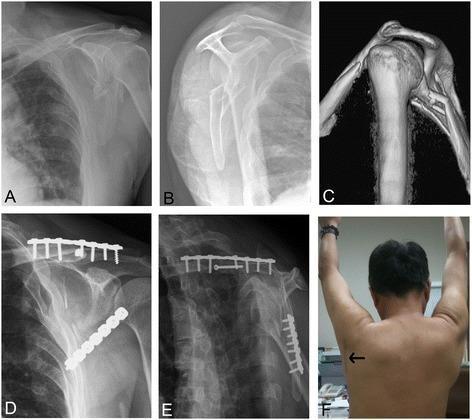
The figure shows pre-op radiographs and 3D CT with a GPA of 3 degrees and 43 degrees of neck angulation. Post-op films show clavicle reconstruction plate and lateral border scapular plate fixation, with the GPA improved to 36 degrees. A posterior incision was used.
Key takeaways
- The classic definition is ipsilateral clavicle and scapular neck fractures. The modern view is a disrupted SSSC with a glenoid unlinked from the scapula and axial skeleton.
- Use CT to distinguish a true neck fracture from an infraspinous body fracture, and check CC ligament integrity.
- Many cases can be managed nonoperatively or with clavicle fixation alone. Re-image after clavicle fixation and add glenoid or scapular fixation if alignment remains unacceptable.
- Unstable anatomical or surgical neck fractures need ORIF. Treat associated chest, spine and neurovascular injuries first.
This is a shared conversation. Sign in to Orris to start your own chat.