
Ligaments of knee joint for 6m Short notes
Reading File
The knee has several major ligaments: the ACL and PCL (cruciate ligaments inside the joint), the MCL and LCL (collateral ligaments on the sides), and the popliteal ligaments posteriorly. Let me pull detailed textbook content.
Searching sources...
Finding Sources
Finding Sources
Reading File
Finding Sources
Reading File
Reading File
Reading File
Finding Sources
Reading File
Loading Image
Reading File
Loading Image
I now have all the information needed for a thorough short note. Let me compile the answer.
Ligaments of the Knee Joint
The knee joint is a synovial hinge joint between the femur and tibia. Because the bony surfaces are not closely apposed, the joint relies heavily on strong ligaments for stability. The ligaments are divided into extrinsic (outside the capsule) and intrinsic (within the capsule) groups.
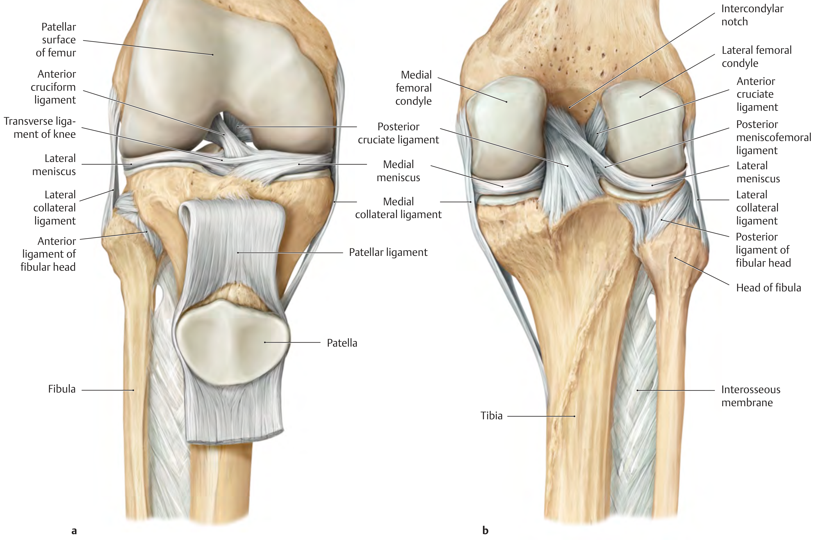
Classification
| Group | Ligaments |
|---|---|
| Extrinsic - Anterior | Patellar ligament, Medial & Lateral patellar retinacula |
| Extrinsic - Medial/Lateral | Medial collateral (tibial collateral), Lateral collateral (fibular collateral) |
| Extrinsic - Posterior | Oblique popliteal ligament, Arcuate popliteal ligament |
| Intrinsic (intra-articular) | Anterior cruciate, Posterior cruciate, Transverse ligament of knee, Posterior meniscofemoral ligament |
1. Patellar Ligament (Anterior)
- The distal continuation of the quadriceps femoris tendon.
- Runs from the apex and adjacent margins of the patella to the tibial tuberosity.
- It is the strongest ligament anteriorly and acts as part of the extensor mechanism.
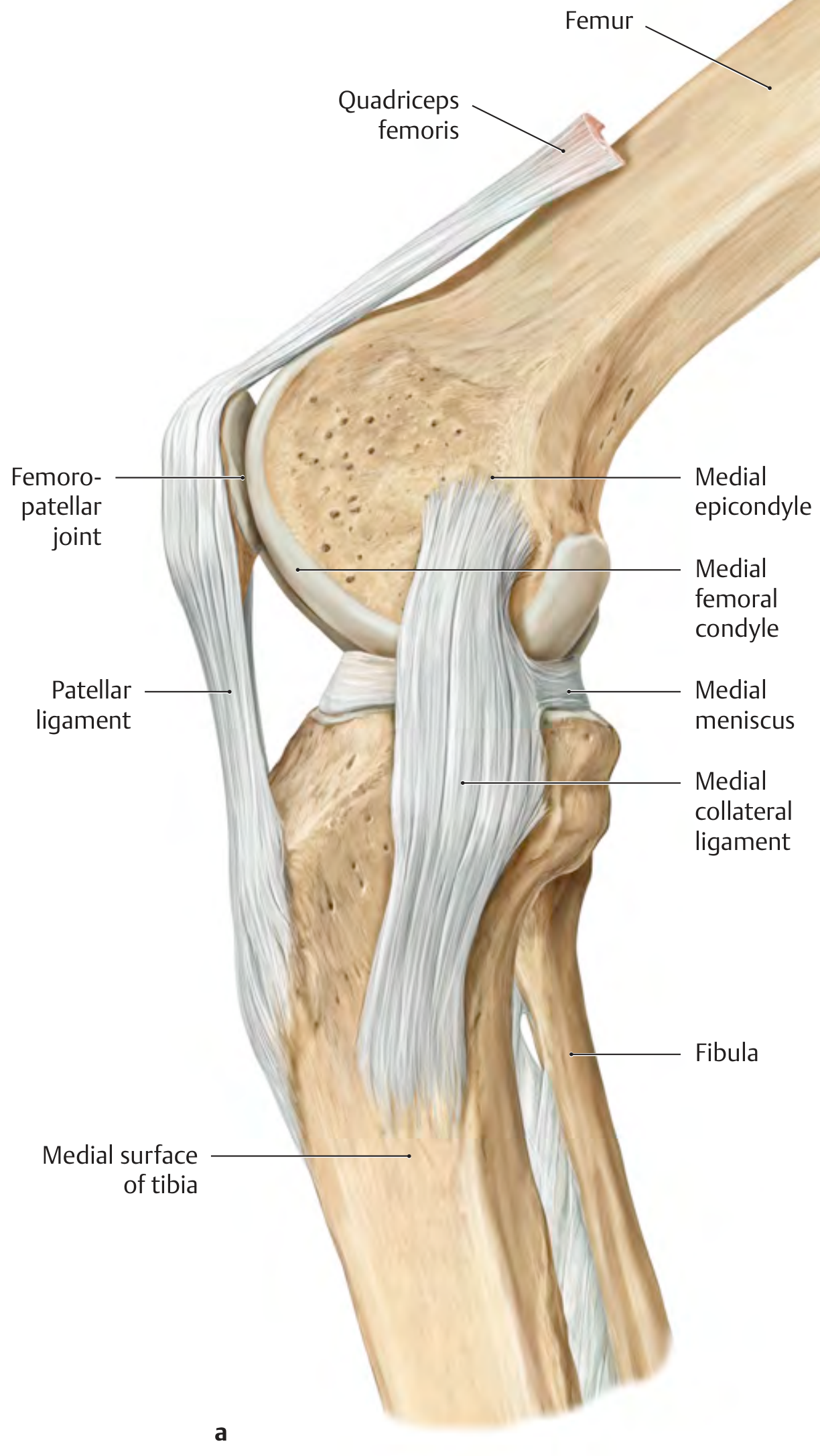
2. Medial Collateral Ligament (MCL / Tibial Collateral Ligament)
- Shape: Broad, flat; has superficial and deep layers separated by loose connective tissue.
- Attachments: Superficial layer - medial epicondyle of femur → medial surface of proximal tibia, ~7-8 cm below the tibial plateau. Deep layer - inserts into the medial meniscus just above the joint line.
- Function: Resists valgus stress.
- Key point: The deep layer is firmly attached to the medial meniscus, which is why MCL injuries frequently co-occur with medial meniscus tears.
- Tension: Taut in extension; lax in flexion.
3. Lateral Collateral Ligament (LCL / Fibular Collateral Ligament)
- Shape: Small, rounded cord (unlike the flat MCL).
- Attachments: Lateral epicondyle of femur → head of fibula (runs obliquely downward and backward).
- Function: Resists varus stress.
- Key point: Has no attachment to the lateral meniscus (unlike MCL and medial meniscus), hence isolated LCL injury does not tear the lateral meniscus.
- Tension: Taut in extension; lax in flexion.
4. Anterior Cruciate Ligament (ACL)
- Location: Intra-articular but extrasynovial (covered anteriorly and laterally by synovial membrane).
- Attachments: Anterior intercondylar area of tibia → medial surface of the lateral femoral condyle (runs upward, backward, and laterally).
- Fiber bundles: Two bundles - anteromedial (AM) and posteromedial (PM), allowing some fibers to be taut in every joint position.
- Function: Prevents anterior subluxation of the tibia on the femur. Also limits internal rotation and hyperextension.
- Clinical test: Anterior drawer test; Lachman test.
- Blood supply: Middle genicular artery (from popliteal artery).
- Common injury: Pivoting/twisting trauma; part of the unhappy triad (ACL + MCL + medial meniscus).
5. Posterior Cruciate Ligament (PCL)
- Attachments: Posterior intercondylar area of tibia → lateral surface of the medial femoral condyle (runs upward, forward, and medially).
- Fiber bundles: Anterolateral (larger/stouter) and posteromedial (more slender).
- Size: Thicker and stronger than the ACL; oriented approximately at a right angle to it.
- Function: Prevents posterior subluxation of the tibia on the femur. The primary restraint to posterior tibial displacement.
- Clinical test: Posterior drawer test; posterior sag sign.
The two cruciate ligaments cross each other, keep the articular surfaces in contact, and stabilize the knee primarily in the sagittal plane.
6. Oblique Popliteal Ligament (Posterior)
- A broad, fibrous band derived from the semimembranosus tendon.
- Runs obliquely upward and laterally across the posterior capsule.
- Reinforces the posterior capsule.
7. Arcuate Popliteal Ligament (Posterior)
- An inverted Y-shaped ligament arising from the head of the fibula.
- Arches over the popliteus muscle to attach to the posterior capsule.
- Along with the oblique popliteal ligament, forms the posterolateral corner stabilizing complex.
8. Transverse Ligament of the Knee (Intrinsic)
- Connects the anterior horns of the medial and lateral menisci.
9. Posterior Meniscofemoral Ligament (Ligament of Wrisberg) (Intrinsic)
- Runs from the posterior horn of the lateral meniscus to the lateral surface of the medial femoral condyle.
Summary Table - Clinical Correlations
| Ligament | Tests | Common Injury Mechanism |
|---|---|---|
| ACL | Anterior drawer, Lachman | Pivot/twist, non-contact deceleration |
| PCL | Posterior drawer, Posterior sag | Dashboard injury, fall on flexed knee |
| MCL | Valgus stress test | Valgus force (blow to lateral knee) |
| LCL | Varus stress test | Varus force (blow to medial knee) |
Sources: THIEME Atlas of General Anatomy & Musculoskeletal System; Rheumatology 2-Volume Set (Elsevier, 2022)
This is a shared conversation. Sign in to Orris to start your own chat.