
Is measels affect adult
measles adults clinical presentation complications
measles rash koplik spots adult
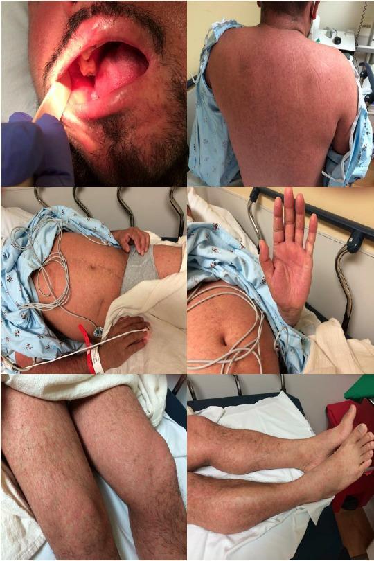
A series of clinical photographs documenting the exanthem and enanthem of measles (rubeola) in an adult male. The oral cavity image demonstrates pathognomonic Koplik spots: tiny, bluish-white or whitish-gray macules resembling grains of sand on an erythematous base, located on the buccal mucosa. The remaining images show a generalized, blanching, erythematous maculopapular rash. This exanthem exhibits a cephalocaudal progression, appearing as numerous discrete macules and papules that have coalesced into larger patches on the back, abdomen, and extremities, including the palms and feet. The presentation illustrates the classic clinical manifestations of a primary measles infection, highlighting the diagnostic importance of the enanthem during the prodromal phase and the characteristic distribution of the subsequent skin eruption for emergency and infectious disease medicine.
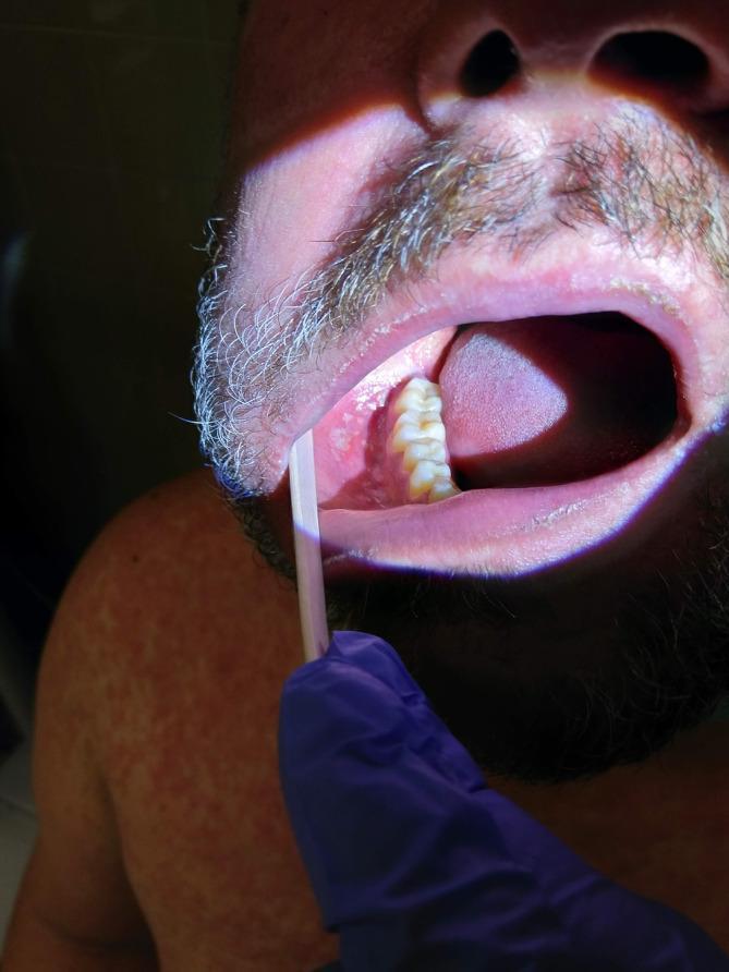
This clinical photograph shows the intraoral cavity and shoulder of an adult male, highlighting pathognomonic signs of a viral exanthem. The primary focus is the buccal mucosa, which is retracted to reveal Koplik's spots: multiple small, 1–2 mm, irregular, bluish-white or gray-white grain-like elevations on an erythematous base. These spots are located opposite the mandibular molars. In the background, the patient's shoulder and chest display a faint, widespread, red maculopapular rash. The image demonstrates key diagnostic features of the prodromal phase of measles (rubeola), where intraoral lesions precede the full systemic rash. This visual is suitable for medical education regarding infectious diseases, dermatology, and clinical diagnosis of childhood and adult exanthems.
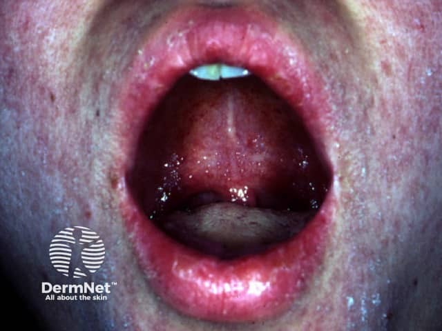
Clinical photograph of the oral cavity showing Koplik spots, a classic mucosal manifestation of measles. Modality: Clinical photography; technique: intraoral close‑up of the buccal mucosa. Location: buccal mucosa near the molar teeth, on an erythematous background. Visual features: numerous small, discrete, bluish‑white to white centers surrounded by red mucosa; lesions measure approximately 1–3 mm in diameter and may appear vesicular or punctate. The mucosa surrounding the spots is inflamed and moist, with mild hyperemia and glossed surfaces in this region. Significance: Koplik spots are highly suggestive of acute measles infection, often appearing during the prodromal phase before the maculopapular rash; they are considered pathognomonic when present. Pathology: viral cytopathic changes in the oropharyngeal epithelium with focal desquamation; clinically, the finding helps confirm a diagnosis of measles in the appropriate clinical context. Diagnostic significance: supports presumptive diagnosis of measles; prompts public health actions, isolation, and antiviral/public health precautions. Differential considerations include aphthous ulcers, herpangina, other viral exanthems, or mucosal lesions in systemic illness, though Koplik spots possess a distinctive bluish center on erythematous mucosa. Clinical correlation: correlate with fever, cough, coryza, conjunctivitis, and rash onset. Utility: educational for clinicians, nurses, medical/dental students; aids recognition and differential diagnosis of febrile rash illness.
Measles in Adults
Who Gets It?
Classic Symptoms in Adults
| Stage | Features |
|---|---|
| Prodrome (days 1-4) | High fever, cough, runny nose (coryza), sneezing, red eyes (conjunctivitis), photophobia |
| Koplik spots | Pathognomonic small bluish-white spots on the buccal (inner cheek) mucosa - appear BEFORE the rash |
| Rash (day 3-5) | Maculopapular rash starting at the hairline/behind ears, spreads down the face, trunk, then limbs; lesions coalesce |
| Resolution | Rash fades with brownish discoloration and mild skin peeling after 6-7 days |
Complications - Adults at Higher Risk
-
Pneumonia - The most dangerous complication. Viral pneumonia occurs in 3-15% of adults with measles. Multilobar reticulonodular opacity is the most common X-ray finding in young adults. Secondary bacterial pneumonia (from H. influenzae, S. pneumoniae, N. meningitidis) develops 5-10 days after rash onset and causes >90% of measles deaths. - Murray & Nadel's Respiratory Medicine
-
Giant cell pneumonia - A rare but lethal complication in adults with deficient cell-mediated immunity (e.g., HIV, cancer). Mortality in HIV patients ~17%, in oncology patients ~70%.
-
Encephalitis - Occurs in ~1 in 1000 cases. Mortality 10-20%; most survivors have lasting neurological damage. - Jawetz Medical Microbiology
-
SSPE (Subacute Sclerosing Panencephalitis) - A very rare late complication (1 in 10,000-100,000 cases) occurring 5-15 years after infection; progressive mental deterioration, rigidity, coma - usually fatal in 1-3 years.
-
Otitis media - Most common complication overall (5-9% of cases).
-
Thrombocytopenic purpura
-
In pregnancy - Associated with fetal death and severe pneumonia. - Creasy & Resnik's Maternal-Fetal Medicine
Atypical Measles in Adults
- High fever, headache, myalgias, vomiting, abdominal pain
- Rash starts on palms, soles, wrists, and ankles (opposite direction from classic measles - no cephalocaudal spread)
- Lesions may be vesicular, petechial, or purpuric
- No Koplik spots
- Pulmonary involvement in most cases; can cause acute respiratory failure
- Residual nodular lung opacities may persist for years
- Murray & Nadel's Respiratory Medicine; Harrison's Principles of Internal Medicine 22E
Diagnosis
- Measles-specific IgM antibody - detectable from day 3 after rash onset (most common method)
- RT-PCR - throat, nasal, urine samples (most sensitive, sent to CDC)
- Fourfold rise in IgG between acute and convalescent samples
- Multinucleated giant cells on stained respiratory/urine sediment specimens
Treatment
- No specific antiviral exists for measles
- Supportive care: fluids, fever control, treat secondary bacterial infections promptly
- Vitamin A reduces morbidity/mortality (especially in children, but also used in severe adult cases)
- Ribavirin, interferon-alpha, and immunoglobulin have been used for severe measles pneumonia
- Respiratory isolation is required
Prevention
- MMR vaccine (live attenuated): Two doses are 97% effective
- Adults who are unvaccinated or have uncertain vaccination history should receive 1-2 doses of MMR
- 92-95% population coverage is needed for herd immunity