
Anorectal malformation 10 marks question answer for surgery pg resident university exam
"anorectal malformations" AND classification
Anorectal Malformations (Imperforate Anus) — 10 Mark Answer
1. Definition
2. Embryology
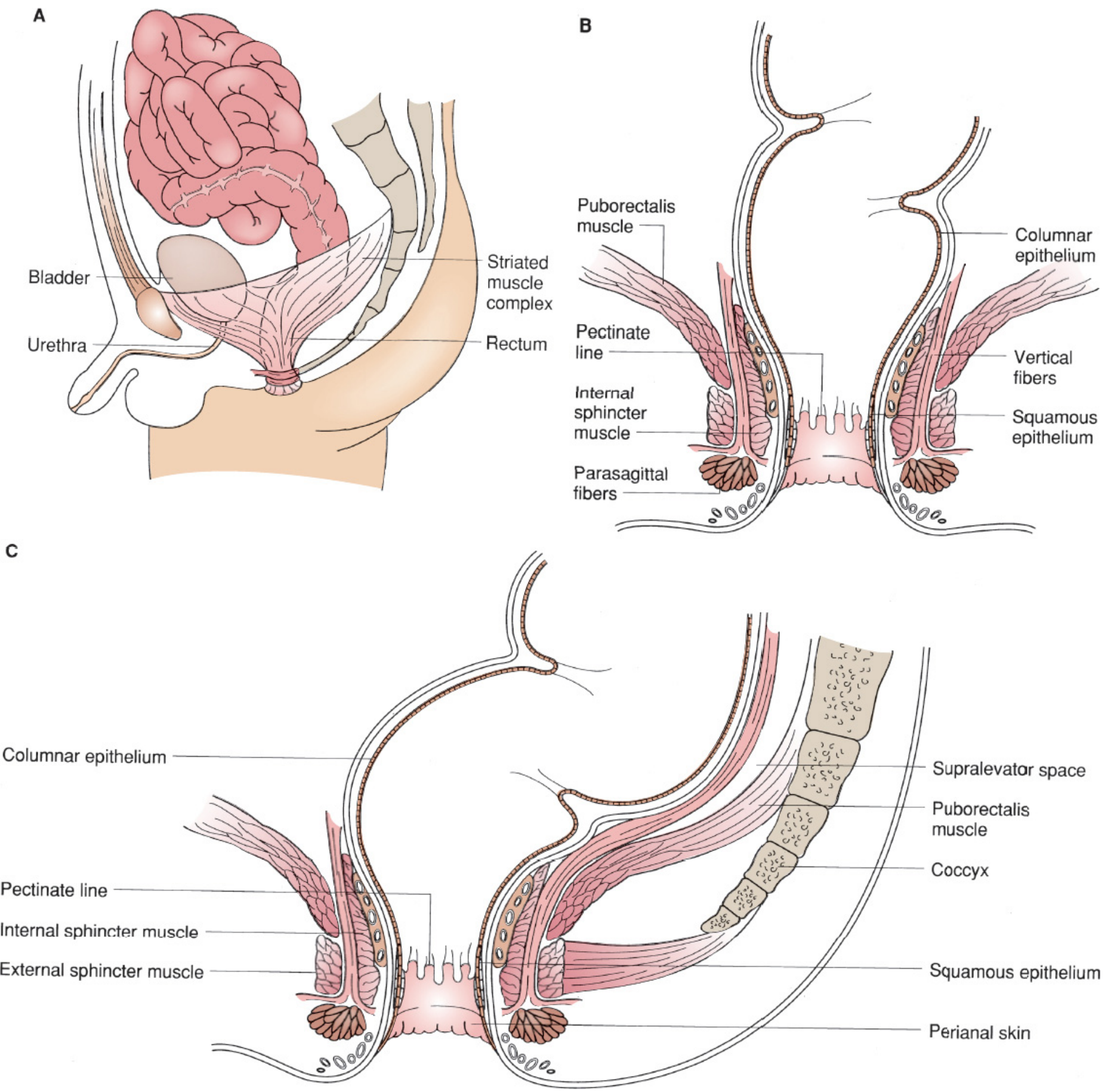
- The cloaca is a common chamber into which hindgut and allantois open, lined by endoderm and separated from surface ectoderm by the cloacal membrane.
- The urorectal septum grows caudally and, with fusion of the lateral cloacal ridges, divides the cloaca into a dorsal rectum and ventral urogenital sinus by about week 5-6.
- The caudal end of the septum forms the perineal body.
- The anal membrane normally ruptures by the 8th week, establishing anal patency.
- Arrest or deviation of this process at different stages produces the wide anatomical spectrum of ARM, explaining the frequent fistulous communication with the urinary or genital tract (Mulholland and Greenfield's Surgery, p. 5485-86).
3. Classification
| Level | Female | Male |
|---|---|---|
| High | Anorectal agenesis ± rectovaginal fistula; rectal atresia | Anorectal agenesis ± rectoprostatic/rectovesical fistula; rectal atresia |
| Intermediate | Rectovestibular fistula; rectovaginal fistula; anal agenesis without fistula | Rectobulbar urethral fistula; anal agenesis without fistula |
| Low | Anovestibular fistula; anovulvar/anocutaneous fistula; anal stenosis | Anocutaneous (perineal) fistula; anal stenosis |
- Perineal (cutaneous) fistula
- Rectourethral fistula (bulbar/prostatic)
- Rectovesical fistula
- Vestibular fistula
- Cloaca
- No fistula
- Anal stenosis
- Rare variants (rectal atresia/stenosis, pouch colon, rectovaginal fistula, H-fistula)
4. Associated Anomalies (VACTERL)
- Vertebral (sacral agenesis, hemivertebrae — assess sacral ratio)
- Anorectal malformation itself
- Cardiac defects
- Tracheo-Esophageal fistula/atresia
- Renal and urinary tract anomalies (vesicoureteric reflux, renal agenesis)
- Limb anomalies (radial ray defects)
5. Clinical Features
- Failure to pass meconium within 24-48 hours of birth
- Absent or abnormally sited anal opening on perineal inspection
- Meconium passed per urethra/vagina in fistulous variants, or meconium-stained "pearls" along a perineal fistula track (bucket-handle deformity)
- Abdominal distension, bilious vomiting if untreated
- Flat/absent perineum, sacral dimple/scoliosis suggesting associated anomalies
6. Investigations
- Clinical perineal examination at 24 hours - single most important step to look for a visible fistula (do not rush to imaging before this).
- Invertogram / cross-table lateral prone shoot-through X-ray (Wangensteen-Rice) at 24 hours of life with a radio-opaque marker on the anal dimple, infant prone with hips elevated, to estimate the distance of the gas-filled rectal pouch from the perineal skin.
- Abdominal and perineal ultrasound to assess pouch distance and bladder filling.
- MCU / distal colostography (after colostomy) - gold standard to delineate fistula anatomy before definitive repair.
- Renal ultrasound, spinal ultrasound/MRI, echocardiography, spinal X-ray (sacral ratio) to screen for associated VACTERL anomalies.
- Karyotype if ambiguous genitalia/cloacal anomaly suspected.
7. Management
- Can undergo primary neonatal anoplasty / minimal PSARP without a covering colostomy.
- Stage 1: Diverting colostomy (usually descending colostomy, or transverse) shortly after birth to decompress the bowel and protect the urinary tract from fecal contamination.
- Stage 2: Posterior Sagittal Anorectoplasty (PSARP) - described by Alberto Peña - performed at around 3 months of age (or when the infant reaches ~10 kg). The midline posterior sagittal incision splits the sphincter complex precisely under electrical muscle stimulation, mobilizes the rectal pouch, divides the fistula, and places the neorectum accurately within the striated muscle (levator ani, puborectalis, external sphincter) complex. For high anomalies or cloaca, a laparoscopic-assisted approach may be combined (LAARP).
- Stage 3: Colostomy closure after anal calibration/dilatation program confirms an adequately healed, appropriately sized neoanus (typically 6-8 weeks post-PSARP).
8. Complications / Prognosis
- Anal stricture, mucosal prolapse
- Constipation (very common, especially with low lesions or rectal dilation)
- Fecal incontinence - correlates with the level of the original lesion, sacral development, and quality of the sphincter/striated muscle complex; low lesions have the best continence prognosis, high lesions and cloaca the worst.
- Urinary incontinence, recurrent UTIs
- Missed or recurrent fistula
- Long-term, multidisciplinary follow-up (colorectal, urology, spine) is required into adulthood as outcomes such as continence and quality of life evolve with growth (APSA 2025 systematic review on functional outcomes, PMID 40023107, and the ARMOUR project, PMID 40664458, both emphasize standardized long-term outcome reporting for these patients).
Diagram
1.Feeding jejunostomy due to acid ingestion 2.Ileal conduit due to repeated bladder stone after trauma 35 years ago... Give case presentation of these two cases in surgery pg level to write and present in front of surgery professor in pg university practical exam... Mention details about the point to read..and what are the questions they will ask..and full detailed 30 mark answer for feeding jejunostomy and ileal conduit
Which case should I prepare in full first?
Anorectal Malformations (Imperforate Anus) — 10 Mark Answer
1. Definition
2. Embryology
- The cloaca is a common chamber into which hindgut and allantois open, lined by endoderm and separated from surface ectoderm by the cloacal membrane.
- The urorectal septum grows caudally and, with fusion of the lateral cloacal ridges, divides the cloaca into a dorsal rectum and ventral urogenital sinus by about week 5-6.
- The caudal end of the septum forms the perineal body.
- The anal membrane normally ruptures by the 8th week, establishing anal patency.
- Arrest or deviation of this process at different stages produces the wide anatomical spectrum of ARM, explaining the frequent fistulous communication with the urinary or genital tract (Mulholland and Greenfield's Surgery, p. 5485-86).
3. Classification
| Level | Female | Male |
|---|---|---|
| High | Anorectal agenesis ± rectovaginal fistula; rectal atresia | Anorectal agenesis ± rectoprostatic/rectovesical fistula; rectal atresia |
| Intermediate | Rectovestibular fistula; rectovaginal fistula; anal agenesis without fistula | Rectobulbar urethral fistula; anal agenesis without fistula |
| Low | Anovestibular fistula; anovulvar/anocutaneous fistula; anal stenosis | Anocutaneous (perineal) fistula; anal stenosis |
- Perineal (cutaneous) fistula
- Rectourethral fistula (bulbar/prostatic)
- Rectovesical fistula
- Vestibular fistula
- Cloaca
- No fistula
- Anal stenosis
- Rare variants (rectal atresia/stenosis, pouch colon, rectovaginal fistula, H-fistula)
4. Associated Anomalies (VACTERL)
- Vertebral (sacral agenesis, hemivertebrae — assess sacral ratio)
- Anorectal malformation itself
- Cardiac defects
- Tracheo-Esophageal fistula/atresia
- Renal and urinary tract anomalies (vesicoureteric reflux, renal agenesis)
- Limb anomalies (radial ray defects)
5. Clinical Features
- Failure to pass meconium within 24-48 hours of birth
- Absent or abnormally sited anal opening on perineal inspection
- Meconium passed per urethra/vagina in fistulous variants, or meconium-stained "pearls" along a perineal fistula track (bucket-handle deformity)
- Abdominal distension, bilious vomiting if untreated
- Flat/absent perineum, sacral dimple/scoliosis suggesting associated anomalies
6. Investigations
- Clinical perineal examination at 24 hours - single most important step to look for a visible fistula (do not rush to imaging before this).
- Invertogram / cross-table lateral prone shoot-through X-ray (Wangensteen-Rice) at 24 hours of life with a radio-opaque marker on the anal dimple, infant prone with hips elevated, to estimate the distance of the gas-filled rectal pouch from the perineal skin.
- Abdominal and perineal ultrasound to assess pouch distance and bladder filling.
- MCU / distal colostography (after colostomy) - gold standard to delineate fistula anatomy before definitive repair.
- Renal ultrasound, spinal ultrasound/MRI, echocardiography, spinal X-ray (sacral ratio) to screen for associated VACTERL anomalies.
- Karyotype if ambiguous genitalia/cloacal anomaly suspected.
7. Management
- Can undergo primary neonatal anoplasty / minimal PSARP without a covering colostomy.
- Stage 1: Diverting colostomy (usually descending colostomy, or transverse) shortly after birth to decompress the bowel and protect the urinary tract from fecal contamination.
- Stage 2: Posterior Sagittal Anorectoplasty (PSARP) - described by Alberto Peña - performed at around 3 months of age (or when the infant reaches ~10 kg). The midline posterior sagittal incision splits the sphincter complex precisely under electrical muscle stimulation, mobilizes the rectal pouch, divides the fistula, and places the neorectum accurately within the striated muscle (levator ani, puborectalis, external sphincter) complex. For high anomalies or cloaca, a laparoscopic-assisted approach may be combined (LAARP).
- Stage 3: Colostomy closure after anal calibration/dilatation program confirms an adequately healed, appropriately sized neoanus (typically 6-8 weeks post-PSARP).
8. Complications / Prognosis
- Anal stricture, mucosal prolapse
- Constipation (very common, especially with low lesions or rectal dilation)
- Fecal incontinence - correlates with the level of the original lesion, sacral development, and quality of the sphincter/striated muscle complex; low lesions have the best continence prognosis, high lesions and cloaca the worst.
- Urinary incontinence, recurrent UTIs
- Missed or recurrent fistula
- Long-term, multidisciplinary follow-up (colorectal, urology, spine) is required into adulthood as outcomes such as continence and quality of life evolve with growth (APSA 2025 systematic review on functional outcomes, PMID 40023107, and the ARMOUR project, PMID 40664458, both emphasize standardized long-term outcome reporting for these patients).
Diagram
feeding jejunostomy complications
ileal conduit complications
ESPEN practical guideline clinical nutrition surgery jejunostomy feeding indications
EAU guidelines urinary diversion ileal conduit complications follow up 2026
1. Feeding Jejunostomy in a Patient With Acid Ingestion
A. How to present the case in practical examination
Opening case presentation
“This is a ___-year-old male/female, who was admitted with alleged history of ingestion of a corrosive acid ___ days/months ago. The patient subsequently developed progressive dysphagia, initially to solids and later to liquids, associated with vomiting/regurgitation after feeds and significant weight loss.In view of inadequate oral intake due to corrosive upper gastrointestinal injury/oesophageal stricture, a feeding jejunostomy was performed on ___ date for enteral nutritional rehabilitation.At present, the patient is receiving ___ mL of ___ feed every ___ hours/continuous feeding through the jejunostomy. There is/there is no pain, leakage, skin excoriation, blockage, dislodgement, fever, abdominal distension, vomiting or diarrhoea.”
“The patient has no history suggestive of hematemesis, melena, aspiration, or features of intestinal obstruction. I would specifically enquire regarding the nature and quantity of acid ingested, intent of ingestion, immediate treatment received, endoscopy findings, prior dilatation, and psychiatric history.”
Probable diagnosis
“This is a case of corrosive acid ingestion with upper gastrointestinal injury, most likely a corrosive oesophageal stricture/gastric outlet involvement, status post feeding jejunostomy for enteral nutritional support.”
B. Important history to elicit
1. Corrosive ingestion history
- Substance: acid or alkali, name of agent, concentration if available.
- Amount, time since ingestion, accidental versus suicidal ingestion.
- Was vomiting induced? Was gastric lavage done? Any antidote administered?
- Early symptoms: oral burns, drooling, odynophagia, dysphagia, retrosternal/epigastric pain, hematemesis, stridor, breathing difficulty.
- ICU admission, perforation, mediastinitis/peritonitis, emergency surgery.
- Endoscopy report and grade of injury, if available.
- Psychiatric evaluation in intentional ingestion.
2. Symptoms of stricture or obstruction
- Dysphagia: solids first, then liquids.
- Odynophagia and regurgitation.
- Postprandial vomiting, early satiety, epigastric fullness: suggests corrosive gastric outlet obstruction.
- Weight loss and dehydration.
- Aspiration cough or recurrent lower respiratory infection.
3. Feeding jejunostomy history
- Date and indication for insertion.
- Operative/laparoscopic/endoscopic placement.
- Type and size of tube, if records available.
- Nature of feed, frequency, volume and water flushes.
- Whether oral feeding is allowed.
- Problems: blockage, accidental tube removal, peritubal leak, pain, skin excoriation, diarrhoea, abdominal distension, fever.
4. Nutritional history
- Pre-ingestion and current weight.
- Percentage weight loss over 1-6 months.
- Appetite and daily calorie/protein intake.
- Features of micronutrient deficiency.
- Diabetes, renal failure, liver disease and medications.
C. Examination points
General examination
- Nutritional state: weight, BMI, temporal wasting, loss of subcutaneous fat, muscle wasting.
- Pallor, dehydration, pedal oedema.
- Oral cavity scars, dental injury, oral burns.
- Cervical scars if cervical oesophagostomy was done.
- Respiratory examination for aspiration-related chest infection.
- Mental health status and self-harm risk where appropriate.
Abdominal examination
Inspection
- Midline laparotomy/laparoscopy scars.
- Feeding jejunostomy tube, usually in the left upper abdomen.
- Note whether tube is securely fixed and whether a marking at skin level is documented.
- Peristomal erythema, induration, leakage, granulation tissue, cellulitis, excoriation.
- Distension, visible peristalsis or hernia.
Palpation
- Local tenderness or warmth around the jejunostomy site.
- Guarding/rigidity: consider leakage or peritonitis.
- Organomegaly, mass and ascites.
Auscultation
- Bowel sounds.
Demonstration at the tube site
“I will inspect the tube for patency, fixation, external length, leak and skin condition. I will not forcibly flush a blocked tube or replace a dislodged tube blindly, especially if it is recently inserted, due to the risk of intraperitoneal placement and peritonitis.”
D. What to read before the practical
-
Corrosive ingestion
- Difference between acid and alkali injury.
- Acute management: airway first, resuscitation, early upper GI endoscopy where indicated.
- Contraindications: no emesis induction, neutralisation, blind nasogastric tube insertion or routine gastric lavage.
- Late complications: oesophageal stricture, gastric outlet obstruction, perforation, carcinoma risk.
-
Enteral nutrition
- Indications and contraindications.
- Feeding routes: nasogastric, nasojejunal, PEG, PEG-J, surgical jejunostomy and parenteral nutrition.
- Refeeding syndrome and its prevention.
-
Feeding jejunostomy
- Indications, methods, procedure, feeding regimen, complications and management.
- Witzel technique and needle catheter jejunostomy.
-
Future reconstruction
- Endoscopic dilatation for oesophageal strictures.
- Gastric outlet obstruction management.
- Oesophageal replacement options: gastric pull-up, colonic interposition and jejunal interposition, depending on anatomy.
Feeding Jejunostomy: 30-Mark Theory Answer
Definition
Indications
General indications
- Inability to maintain adequate oral intake despite a functioning intestine.
- Upper GI obstruction:
- Corrosive oesophageal stricture.
- Corrosive gastric outlet obstruction.
- Oesophageal or gastric malignancy.
- Following major upper GI surgery:
- Oesophagectomy.
- Gastrectomy.
- Pancreaticoduodenectomy in selected patients.
- Severe pancreatitis when gastric feeding is not tolerated.
- Neurological dysphagia with high aspiration risk when gastric feeding is unsuitable.
- Enterocutaneous fistula where feeding distal to the fistula is possible.
Specific indications in corrosive ingestion
- Severe oesophageal injury with inability to swallow.
- Associated gastric injury or gastric outlet obstruction.
- Nutritional rehabilitation before oesophageal reconstruction.
- After emergency oesophagogastrectomy for transmural necrosis.
- Anticipated prolonged need for enteral access.
Contraindications
Absolute
- Distal intestinal obstruction.
- Diffuse peritonitis.
- Bowel ischaemia.
- Severe uncorrectable coagulopathy.
- Non-functional gut with need for total parenteral nutrition.
Relative
- Massive ascites.
- Severe haemodynamic instability.
- Extensive adhesions.
- Inflammatory bowel disease involving the proposed segment.
- Severe malabsorption/short bowel syndrome.
Advantages
- Provides physiological nutrition through the gut.
- Maintains intestinal mucosal integrity and reduces bacterial translocation.
- Avoids the need for central venous access and its septic/thrombotic complications.
- Avoids passage of feed through injured oesophagus or stomach.
- Suitable for prolonged feeding.
- Can be placed at laparotomy, laparoscopy, or by endoscopic/radiological methods.
Disadvantages
- Requires an invasive procedure.
- Requires careful tube care and feeding supervision.
- Jejunal feeds need slow, controlled administration because the stomach reservoir is bypassed.
- Mechanical, infectious, metabolic and bowel complications may occur.
Types and techniques
-
Open surgical feeding jejunostomy
- Witzel tunnel technique.
- Stamm jejunostomy.
- Needle catheter jejunostomy.
-
Laparoscopic feeding jejunostomy
-
Percutaneous endoscopic jejunostomy
- Direct percutaneous endoscopic jejunostomy.
- PEG with jejunal extension, PEG-J.
Witzel Feeding Jejunostomy: Operative Steps
- Under general anaesthesia, perform upper midline laparotomy or use a laparoscopic approach.
- Identify the duodenojejunal flexure and select a mobile jejunal loop approximately 20-30 cm distal to the ligament of Treitz.
- Choose the antimesenteric border.
- Place a purse-string seromuscular suture.
- Make a small enterotomy and introduce a suitable feeding tube into the jejunum, directing it distally.
- Advance the tube approximately 20-30 cm beyond the entry site.
- Tighten the purse-string around the tube.
- Create a serosal tunnel by placing interrupted Lembert sutures over the tube, usually 5-7 cm long. This is the Witzel tunnel, which reduces leakage.
- Fix the jejunum to the parietal peritoneum with a few sutures to prevent torsion and leakage.
- Bring the tube out through a separate stab incision in the abdominal wall, avoiding the main wound.
- Fix the tube to the skin, document external length, and start feeds after confirming clinical stability.
Postoperative Feeding Protocol
- Initially keep tube on drainage or clamp as per operative protocol.
- Confirm clinical stability and absence of leak/peritonitis.
- Start sterile water or isotonic feed at a low rate, usually continuous pump feeding.
- Gradually increase rate and concentration as tolerated.
- Use polymeric formula when possible. Semi-elemental feed may be used in malabsorption or poor tolerance.
- Flush tube with water before and after feeds/medications, and intermittently during continuous feeding.
- Monitor:
- Abdominal distension, vomiting, diarrhoea.
- Tube-site leak.
- Fluid balance.
- Blood glucose.
- Electrolytes, magnesium and phosphate.
- Weight and nitrogen/protein targets.
Refeeding syndrome
- Correcting potassium, magnesium and phosphate deficits.
- Giving thiamine before and during initiation.
- Starting feed slowly.
- Monitoring electrolytes and fluid status closely.
Complications
Early complications
- Bleeding.
- Peritoneal contamination and peritonitis.
- Jejunal leak.
- Tube malposition.
- Wound infection and cellulitis.
- Ileus.
- Small bowel obstruction.
- Volvulus around fixation point.
- Aspiration is less likely than with gastric feeding but still possible due to reflux/vomiting.
Late complications
- Tube blockage.
- Tube dislodgement or migration.
- Peritubal leakage and skin excoriation.
- Granulation tissue.
- Jejunal fistula following removal.
- Small bowel obstruction/intussusception.
- Buried bumper-type problems are uncommon in surgical tubes.
- Diarrhoea, cramps, dumping-type symptoms.
- Dehydration, electrolyte imbalance and hyperglycaemia.
Management principles
- Blocked tube: attempt gentle warm-water flush. Do not use excessive force or sharp instruments.
- Leaking tube: assess fixation, tube size, skin infection and intraluminal obstruction. Protect skin.
- Recent accidental dislodgement: emergency surgical/radiological evaluation. Do not blindly reinsert.
- Late dislodgement after mature tract formation: replace under appropriate guidance according to local protocol.
- Peritonitis/acute abdomen: resuscitation, imaging and urgent surgical review.
Prognosis and follow-up in corrosive injury
- Endoscopic dilatation for oesophageal stricture.
- Definitive surgery for refractory stricture or gastric outlet obstruction.
- Nutritional optimisation before oesophageal replacement.
Likely examiner questions and short answers
| Question | Model answer |
|---|---|
| Why jejunostomy and not gastrostomy? | Gastric feeding may be unsafe or ineffective when stomach is injured, gastric outlet is obstructed, or reflux/aspiration risk is high. Jejunostomy bypasses the oesophagus and stomach. |
| Where is the jejunostomy made? | Usually on a proximal mobile jejunal loop, around 20-30 cm distal to the ligament of Treitz. |
| What is Witzel jejunostomy? | A tube jejunostomy in which a serosal tunnel is created over the catheter with Lembert sutures to reduce leak. |
| When do you start feeding? | After ensuring haemodynamic stability and no evidence of leak or peritonitis, commonly within 12-24 hours depending on intraoperative findings and protocol. |
| What is the commonest tube problem? | Blockage, dislodgement and peritubal leakage are common practical problems. |
| How do you prevent tube blockage? | Use liquid medications where possible, give each drug separately, and flush before/after feeds and medications. |
| What is refeeding syndrome? | Metabolic shifts after restarting nutrition in a starved patient, especially hypophosphataemia, hypokalaemia and hypomagnesaemia, causing arrhythmias, fluid overload and neurological complications. |
| What should never be done in corrosive ingestion? | Do not induce vomiting, neutralise the agent, perform routine gastric lavage, or blindly insert an NG tube. |
| What are late effects of corrosive ingestion? | Oesophageal stricture, gastric outlet obstruction, malnutrition, aspiration, and increased long-term risk of oesophageal carcinoma. |
2. Ileal Conduit in a Patient With Repeated Bladder Stones Following Old Pelvic Trauma
Important clinical clarification
“I will review the operative records to establish whether the patient had cystectomy with ileal conduit, or supravesical diversion with the native bladder left in situ. In this patient, repeated bladder calculi were likely a consequence of chronic urinary stasis, infection, foreign material, outlet obstruction or neurogenic/post-traumatic dysfunction. The indication for diversion was probably an end-stage dysfunctional lower urinary tract rather than recurrent calculi alone.”
A. Practical case presentation
Opening case presentation
“This is a ___-year-old male/female, who sustained pelvic/bladder/urethral trauma in a road traffic accident ___ years ago. The patient subsequently developed lower urinary tract dysfunction with recurrent urinary tract infections and recurrent vesical calculi, requiring ___ procedures such as cystolitholapaxy/open cystolithotomy/urethral dilatation.In view of a severely dysfunctional bladder/outlet, recurrent calculi and infections, with or without upper tract deterioration, the patient underwent urinary diversion by ileal conduit on ___ date.The patient currently passes urine continuously through a right iliac fossa urostomy into an external appliance. The stoma output is approximately ___ mL/day, clear/cloudy, with/without mucus. There is no fever, loin pain, reduced output, peristomal skin excoriation, haematuria, colicky pain or appliance leakage.”
Summary and diagnosis
“This is a case of urinary diversion by ileal conduit following a long-standing post-traumatic dysfunctional bladder/urethra, complicated by recurrent vesical calculi and urinary tract infection.”
B. History to take
1. Original trauma
- Mechanism: road traffic accident, crush injury, pelvic fracture, penetrating trauma.
- Bladder rupture: intraperitoneal or extraperitoneal.
- Urethral injury: blood at meatus, suprapubic cystostomy, urethroplasty, recurrent stricture.
- Previous operative details.
2. Lower urinary tract dysfunction
- Retention, incontinence, dysuria, frequency.
- Poor stream, straining, incomplete emptying.
- Clean intermittent catheterisation or long-term catheter use.
- Recurrent UTI, pyelonephritis.
- Stone history, number and type of stone procedures.
- Haematuria and suprapubic pain.
- Vesicocutaneous/urethrocutaneous fistula.
3. Diversion history
- Was cystectomy performed?
- Is there a retained bladder?
- Date and indication for ileal conduit.
- Any perioperative complications.
- Stoma output and character of urine.
- Stoma appliance: type, frequency of changing, leakage.
- Passage of mucus: small amounts can occur because bowel mucosa secretes mucus.
- Recurrent upper tract infection, stones, flank pain or reduced urine output.
4. Renal and metabolic symptoms
- Fever, chills and loin pain.
- Polyuria, dehydration.
- Weakness, fatigue, altered sensorium: consider metabolic acidosis.
- Bone pain/fractures in long-standing acidosis.
- Renal function test trends.
C. Examination of an ileal conduit
General examination
- Nutrition, pallor, hydration.
- Fever.
- Oedema and hypertension suggesting renal impairment.
- Signs of chronic kidney disease.
- Examine scars from pelvic trauma, laparotomy, suprapubic cystostomy and urethral procedures.
Local abdominal examination
Inspection
- Usually a right iliac fossa stoma, though site may vary.
- Stoma should be:
- Pink/red, moist, viable.
- Protruding slightly from skin.
- Continuously draining urine.
- Look for:
- Ischaemia/necrosis: dusky black stoma.
- Retraction.
- Prolapse.
- Stenosis.
- Peristomal dermatitis/excoriation.
- Parastomal hernia.
- Leakage around appliance.
- Crystals/encrustation.
- Papillary lesions or bleeding.
Palpation
- Tenderness, parastomal hernia.
- Suprapubic fullness/mass if bladder remains in situ.
- Renal ballotability or loin tenderness.
- Abdominal masses/scars.
Stoma assessment statement
“The stoma is situated in the right iliac fossa. It is pink, moist and viable, with continuous clear urine drainage into a properly fitting appliance. I will assess for stenosis, retraction, prolapse, parastomal hernia and peristomal skin complications.”
D. Investigations to mention
- Urinalysis and urine culture, preferably specimen from a freshly changed appliance or catheterised conduit sample, not from an old bag.
- Serum creatinine, urea, electrolytes, bicarbonate and chloride.
- CBC and inflammatory markers if infection is suspected.
- Ultrasound KUB for hydronephrosis, stones and renal size.
- CT urography/CT KUB when stone, obstruction or upper tract pathology is suspected.
- Loopogram/conduitogram to assess conduit, reflux and obstruction.
- Nuclear renal scan if differential renal function or obstruction requires assessment.
- Cystoscopy of retained bladder if haematuria, pyocystis, pain or suspicion of retained bladder pathology.
- Stone analysis and metabolic evaluation in recurrent stone disease.
Ileal Conduit: 30-Mark Theory Answer
Definition
Indications
Common indications
- After radical cystectomy for bladder cancer.
- Non-functioning or end-stage bladder due to:
- Neurogenic bladder with upper tract damage.
- Severe post-traumatic bladder/urethral destruction.
- Refractory incontinence with irreparable outlet.
- Severe radiation cystitis.
- Interstitial cystitis or contracted bladder in selected cases.
- Intractable bleeding.
- Complex genitourinary fistula.
- Diversion before renal transplantation in a non-compliant, unsafe bladder.
- Failed previous reconstructive surgery.
- Severe recurrent infection and stones in a dysfunctional lower tract, when bladder-preserving methods are no longer suitable.
Contraindications
Absolute/major
- Severe short bowel syndrome.
- Extensive ileal Crohn disease.
- Previously irradiated or unusable ileum.
- Severe mesenteric vascular insufficiency.
- Inability to tolerate major surgery.
Relative
- Advanced renal dysfunction.
- Severe hepatic dysfunction.
- Extensive intra-abdominal adhesions.
- Poor manual/social ability to manage an appliance, although this is generally manageable with stoma-care support.
Advantages
- Technically simpler than continent diversion or orthotopic neobladder.
- Shorter operative time.
- Lower demand for patient dexterity than self-catheterisable reservoirs.
- Reliable continuous low-pressure drainage.
- Suitable for elderly, frail, impaired renal function, impaired sphincter function, or patients unsuitable for continent diversion.
- Does not depend on urethral integrity.
Disadvantages
- Permanent external appliance.
- Continuous urinary drainage.
- Stomal complications.
- Mucus in urine.
- Infection, stone formation and upper tract deterioration.
- Metabolic complications, although lower than with continent reservoirs because of shorter urine-bowel contact time.
Preoperative preparation
- Explain all diversion options: ileal conduit, continent cutaneous diversion and orthotopic neobladder where relevant.
- Assess renal function, electrolytes, nutritional status, comorbidities and bowel disease.
- Treat UTI and optimise anaemia/nutrition.
- Imaging of kidneys, ureters and bladder.
- Identify and correct obstruction where feasible.
- Review previous trauma/reconstruction records.
- Mark stoma site preoperatively with a stoma therapist:
- Right lower abdomen.
- Away from scars, skin creases, belt line, bony prominences and umbilicus.
- In a location visible to the patient while sitting and standing.
Operative technique
Essential principles
- Isolate a well-vascularised, tension-free ileal segment.
- Preserve mesenteric blood supply.
- Achieve tension-free ureteroileal anastomoses.
- Protect anastomoses with temporary ureteric stents where used.
- Construct a viable, non-twisted, protruding stoma.
Steps
- Lower midline incision and abdominal exploration.
- If indicated, perform cystectomy or manage the diseased bladder according to pathology.
- Mobilise ureters carefully, preserving periureteric adventitia and blood supply.
- Select a 10-15 cm segment of distal ileum, generally about 10-15 cm proximal to the ileocaecal valve.
- Preserve a broad mesenteric vascular pedicle.
- Divide bowel at both ends and restore intestinal continuity by ileoileal anastomosis.
- Close mesenteric defects to prevent internal hernia.
- Bring the isolated ileal segment to the right iliac fossa through an appropriately sized abdominal wall opening.
- Implant the ureters into the proximal conduit:
- Bricker technique: separate ureteroileal anastomoses.
- Wallace technique: ureters joined together, then implanted as a common anastomosis.
- Place ureteric stents according to technique/practice.
- Mature the distal end as a spout stoma, commonly 2-3 cm above skin to reduce urine contact with peristomal skin.
- Confirm viability and absence of twisting.
Ureteroileal anastomotic techniques
Bricker technique
- Each ureter is implanted separately into the ileal conduit.
- Advantage: separate access to each ureter.
- Disadvantage: two anastomoses.
Wallace technique
- Ureters are spatulated and joined to create a common ureteral plate, then anastomosed to the conduit.
- In some series, it has lower anastomotic complication rates, but the consequence of a common stricture can affect both upper tracts.
Postoperative care
- Monitor urine output from conduit hourly initially.
- Examine stoma colour, viability, oedema and output.
- Fluid balance, daily weight, renal function, chloride and bicarbonate.
- Early mobilisation, DVT prophylaxis and respiratory physiotherapy.
- Stoma education before discharge.
- Remove ureteric stents according to operative protocol.
- Watch for ileus, leak, sepsis, obstruction, worsening renal function and metabolic acidosis.
Complications
Early complications
- Paralytic ileus.
- Wound infection, intra-abdominal collection and sepsis.
- Anastomotic urinary leak.
- Ureteroileal anastomotic leak.
- Bowel leak or bowel obstruction.
- Stomal ischaemia or necrosis.
- Pyelonephritis/UTI.
- Thromboembolism.
Late complications
-
Stomal complications
- Parastomal hernia.
- Stenosis.
- Retraction.
- Prolapse.
- Peristomal dermatitis.
- Appliance leakage.
-
Upper urinary tract complications
- Reflux.
- Hydroureteronephrosis.
- Ureteroileal stricture.
- Recurrent pyelonephritis.
- Progressive renal impairment.
-
Stone disease
- Conduit stone, renal/ureteric stones.
- Causes: chronic bacteriuria, mucus, stasis, foreign bodies/stents, metabolic abnormalities and obstruction.
-
Metabolic
- Hyperchloraemic metabolic acidosis due to chloride absorption and bicarbonate loss through bowel mucosa exposed to urine.
- Risk rises with renal dysfunction, dehydration, obstruction and prolonged urine contact.
- Hypokalaemia may occur.
- Vitamin B12 deficiency can occur years later if a sufficiently long terminal ileal segment has been resected, though this is less likely with a short conduit.
-
Other
- Sexual dysfunction and infertility depending on associated pelvic surgery.
- Retained bladder complications if bladder is left in situ: pyocystis, pain, bleeding and rarely malignancy.
Follow-up
- Stoma and peristomal skin examination.
- Serum creatinine, electrolytes, bicarbonate and chloride.
- Urinalysis/culture only when clinically indicated, as asymptomatic bacteriuria is common.
- Ultrasound KUB for hydronephrosis and stones.
- CT urography or functional imaging where indicated.
- Monitoring for recurrent infection, obstruction and stone disease.
- Stoma nurse support and appliance review.
Likely examiner questions and model answers
| Question | Model answer |
|---|---|
| Define ileal conduit. | An incontinent urinary diversion using a short isolated ileal segment implanted with ureters proximally and brought as a cutaneous stoma distally. |
| Why is the stoma commonly on the right side? | The distal ileum lies naturally in the right lower abdomen and can reach the skin with a safe mesenteric pedicle. |
| What length of ileum is used? | Usually 10-15 cm. |
| How far from ileocaecal junction is it selected? | Usually about 10-15 cm proximal to the ileocaecal valve. |
| What are Bricker and Wallace techniques? | Bricker is separate implantation of each ureter; Wallace is a common ureteral plate implanted into the conduit. |
| Why is there mucus in the urine? | The conduit is living bowel mucosa and continues to produce mucus. |
| Does bacteriuria always need treatment? | No. Treat symptomatic UTI, pyelonephritis or before instrumentation; asymptomatic bacteriuria is common. |
| Why does metabolic acidosis occur? | Intestinal exposure to urine causes chloride/ammonium absorption and bicarbonate loss, producing hyperchloraemic normal-anion-gap metabolic acidosis. |
| Why can this patient get recurrent stones? | Urinary stasis, chronic infection with urease-producing organisms, mucus, foreign bodies, metabolic abnormalities, outlet/conduit obstruction and upper tract dilatation. |
| What are stoma complications? | Stenosis, retraction, prolapse, parastomal hernia, ischaemia and peristomal dermatitis. |
| What are the long-term concerns? | Renal function decline, hydronephrosis, ureteroileal stricture, UTI, stones, stomal complications and metabolic acidosis. |
| Why not perform an ileal conduit for a routine bladder stone? | Bladder stones should first be treated by removing the stone and correcting the underlying cause. Diversion is reserved for an irreparably damaged or unsafe lower urinary tract. |