
Non alcoholic fatty liver disease
"nonalcoholic fatty liver disease"[MeSH Terms] AND treatment
nonalcoholic fatty liver disease NAFLD histology steatosis steatohepatitis
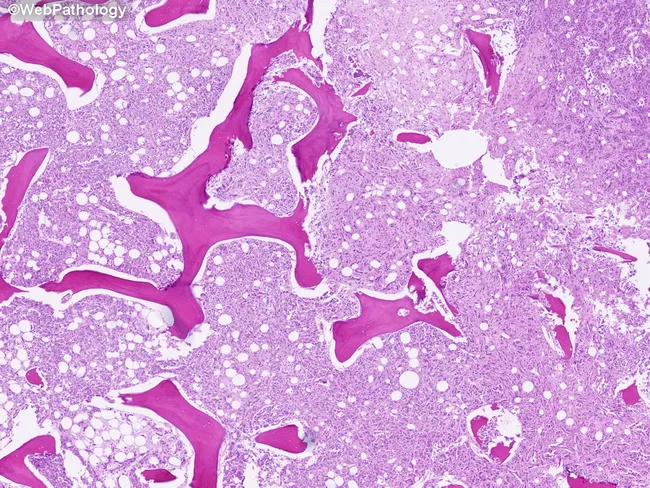
Light-microscopy histology of liver tissue demonstrates macrovesicular steatosis. Imaging modality: Histology using Hematoxylin and Eosin (H&E) stain on paraffin-embedded sections. Anatomical localization: hepatic parenchyma of the liver, with hepatocytes arranged in plates separated by sinusoids and preserved lobular architecture visible at low magnification. Visual features: numerous intracellular lipid vacuoles produce large clear droplets that displace hepatocyte nuclei to the periphery; cytoplasm otherwise eosinophilic; occasional subtle ballooning is not evident; portal tracts and central veins are discernible but not markedly inflamed. Pathological interpretation: macrovesicular steatosis consistent with fatty liver changes; most compatible with nonalcoholic fatty liver disease (NAFLD) or alcoholic fatty liver disease (AFLD) depending on patient history. In this image, steatosis is the predominant finding with minimal inflammatory infiltrate and no definitive fibrosis at this level of magnification. Clinical significance: fatty change indicates hepatocellular stress from metabolic syndrome or hepatotoxic exposure; may precede steatohepatitis, fibrosis, cirrhosis, and increased risk of hepatocellular carcinoma with ongoing injury. Potential clinical use cases: assessment of steatosis burden in NAFLD/NASH studies, biopsy-guided staging, therapeutic monitoring, and education for trainees. The image serves as an educational reference for recognizing hepatocellular lipid accumulation and distinguishing it from other hepatic pathologies such as steatohepatitis and cholestatic injury.
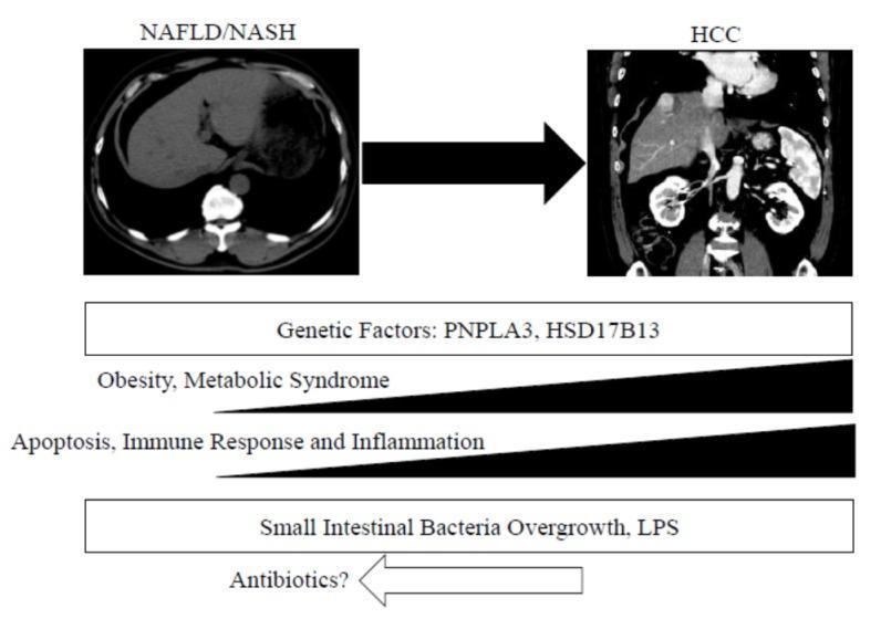
This medical infographic illustrates the pathophysiology and progression of liver disease from Nonalcoholic Fatty Liver Disease/Nonalcoholic Steatohepatitis (NAFLD/NASH) to Hepatocellular Carcinoma (HCC). The upper portion features two diagnostic CT images: an axial section of a liver with NAFLD/NASH showing diffuse decreased attenuation (steatosis) and a coronal section demonstrating HCC with multiple heterogeneously enhancing masses distorting the hepatic contour. Below the imaging, horizontal bars and arrows correlate clinical progression with underlying molecular and environmental drivers. Key factors identified include genetic predispositions (PNPLA3, HSD17B13), the increasing influence of obesity and metabolic syndrome, and escalating levels of apoptosis, immune response, and inflammation. The diagram also highlights the role of the gut-liver axis, specifically Small Intestinal Bacterial Overgrowth (SIBO) and Lipopolysaccharides (LPS), with a potential therapeutic intersection for antibiotics. The graphic serves as a summary of hepatocarcinogenesis in the context of metabolic dysfunction-associated liver disease.
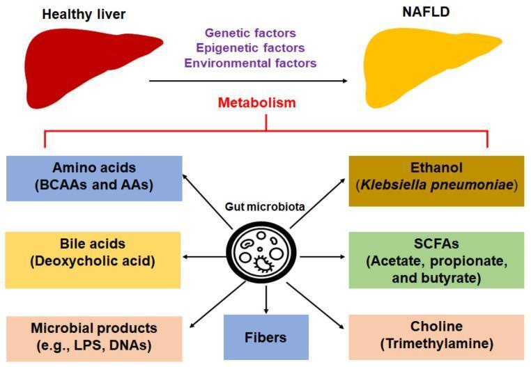
This pathophysiology diagram illustrates the metabolic cross-talk between the gut microbiota and the development of nonalcoholic fatty liver disease (NAFLD). At the top, a 'Healthy liver' depicted in red transitions via an arrow towards a yellow-colored liver labeled 'NAFLD,' signifying steatosis. This progression is influenced by genetic, epigenetic, and environmental factors. Centrally, a 'Gut microbiota' icon radiates arrows toward various metabolites and microbial compounds categorized by colored boxes. These include amino acids (BCAAs and AAs), bile acids (deoxycholic acid), microbial products (LPS, DNAs), dietary fibers, short-chain fatty acids (SCFAs like acetate, propionate, and butyrate), ethanol (specifically mentioning Klebsiella pneumoniae), and choline/trimethylamine. A red bracket connects these metabolic influences to the core transition arrow labeled 'Metabolism,' highlighting how gut-derived signals modulate liver health and the progression toward NAFLD and potentially nonalcoholic steatohepatitis (NASH). This educational infographic is designed to explain the biochemical pathways and systemic interactions involved in metabolic liver disease.
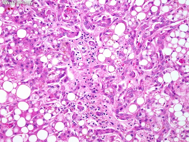
Alcoholic steatohepatitis manifested on liver biopsy as macrovesicular steatosis with hepatocellular injury, inflammatory infiltrates, and early fibrosis. This bright-field histology image, obtained from a paraffin-embedded liver biopsy and stained with Hematoxylin and Eosin, highlights lobular and portal changes. Large fat vacuoles displace hepatocyte nuclei; scattered ballooning degeneration accompanies hepatocellular damage. In the portal tracts, mixed inflammatory cells—neutrophils and lymphocytes including CD4+ and CD8+ T cells—are evident, often forming a neutrophilic infiltrate around ductular structures. A ductular reaction is visible as proliferating biliary-like cells at the limiting plate and portal-periportal interface. The surrounding parenchyma shows cytoplasmic rarefaction and mild-to-moderate inflammation, with early fibrotic bands encroaching on the portal triad region. Collectively, these features support an active alcoholic injury pattern rather than pure steatosis, differentiating from nonalcoholic fatty liver disease. Clinically, this image underpins diagnoses of alcoholic hepatitis/ASH in the appropriate history and contributes to assessments of disease activity and prognosis via fibrosis staging. It is relevant for medical education, histopathology libraries, and research on inflammatory liver diseases, fibrosis progression, and the hepatobiliary response to alcohol exposure. This image supports differential diagnosis with NAFLD, alcoholic hepatitis, DILI; useful for pathology education, case conferences, radiology-pathology correlation, and AI dataset curation, and clinical teaching.
"nonalcoholic fatty liver disease"[MeSH Terms] AND "diagnosis"
Non-Alcoholic Fatty Liver Disease (NAFLD)
Definition & Nomenclature
-
NAFL (Non-Alcoholic Fatty Liver): isolated steatosis, low risk of progression
-
NASH (Non-Alcoholic Steatohepatitis): steatosis + liver injury + inflammation ± fibrosis
-
NASH Cirrhosis: end-stage, with risk of HCC
-
Robbins & Kumar Basic Pathology, p. 1910
Epidemiology
-
Global prevalence of NAFLD: ~25% of the population; NASH: 1.5-6%
-
NAFLD is the most prevalent chronic liver disease worldwide
-
Expected to increase 60% by 2030 alongside the obesity epidemic
-
~20% of patients with NASH develop cirrhosis
-
Obese patients have a fourfold higher risk of developing NAFLD
-
NAFLD has become one of the leading causes of liver transplantation in the US
-
With declining HCV treatment burden, NASH is likely to overtake HCV as the leading risk factor for HCC
-
Bailey and Love's Short Practice of Surgery 28th Ed., p. 2599; Washington Manual of Medical Therapeutics
Risk Factors / Associated Conditions
| Metabolic Factor | Notes |
|---|---|
| Obesity (primarily central) | Most common; obese patients 4x more likely to develop NAFLD |
| Insulin resistance / Type 2 Diabetes | Key driver; family history also a risk |
| Dyslipidemia | Hypertriglyceridemia, low HDL, high LDL |
| Hypertension | Part of metabolic syndrome triad |
| Metabolic syndrome | All components increase risk |
- Robbins & Kumar, p. 1912-1918
Pathogenesis
- Obesity → insulin resistance → increased adipose lipolysis → excess free fatty acids (FFAs) delivered to liver
- Reduced adiponectin → decreased FFA oxidation in skeletal muscle → increased FFA uptake into hepatocytes → triglyceride accumulation
- De novo lipogenesis promoted in the liver
- Accumulated lipids cause lipotoxicity: saturated FFAs (e.g. palmitate) and lysophosphatidylcholine → hepatocyte apoptosis
- ER stress, lysosomal dysfunction, mitochondrial dysfunction
- Reactive oxygen species (ROS) generation
- Inflammasome activation → IL-1 release → local inflammation
- Gut microbiome dysbiosis → increased gut-derived LPS (endotoxin) → amplifies hepatic inflammation
- SIBO (small intestinal bacterial overgrowth) and gut-liver axis disruption
- Hepatocyte injury activates hepatic stellate cells → collagen deposition → fibrosis → cirrhosis
- Yamada's Textbook of Gastroenterology 7th Ed., p. 1899; Robbins & Kumar, p. 1920
Histopathology
| Feature | Description |
|---|---|
| Macrovesicular steatosis | Large lipid vacuoles displacing hepatocyte nucleus to periphery (≥5% hepatocytes) |
| Hepatocyte ballooning | Cytoplasmic rarefaction, key marker of injury |
| Lobular inflammation | Neutrophilic and mononuclear infiltrates |
| Mallory-Denk bodies | Less common than in alcoholic hepatitis |
| Fibrosis staging | F0=none; F1=perisinusoidal/periportal; F2=both; F3=bridging; F4=cirrhosis |
- Yamada's Textbook of Gastroenterology, p. 1886-1897; Robbins & Kumar, p. 1910
Fibrosis Progression
-
A proportion of patients show improvement in NAS spontaneously (even in placebo arms of clinical trials)
-
~25% of patients with simple steatosis will progress to NASH
-
Progression to NASH cirrhosis reported at ~11% over 15 years
-
A small proportion can progress to advanced fibrosis within 5-7 years
-
Obesity and weight gain are the strongest drivers of fibrosis progression
-
Washington Manual of Medical Therapeutics, p. 6037-6039
Clinical Features
- Most patients are asymptomatic (most common cause of incidentally elevated transaminases)
- Symptoms when present: fatigue, malaise, right upper quadrant discomfort
- Severe disease: symptoms of chronic liver disease, portal hypertension
- AST:ALT ratio < 1 (contrast: alcoholic liver disease typically >2)
- Elevated ALT and AST, but can be normal even in advanced NASH
-
Increased incidence of coronary artery disease (shared metabolic risk factors)
-
Cardiovascular disease is the most common cause of death in NAFLD patients
-
HCC risk, even in non-cirrhotic NASH
-
Robbins & Kumar, p. 1922
Diagnosis
Clinical Diagnosis
- Significant alcohol use (>21 units/week men, >14 women)
- Medications causing steatosis (methotrexate, tamoxifen, amiodarone, corticosteroids)
- Other causes of chronic liver disease (viral hepatitis, Wilson's, autoimmune)
Imaging
- Ultrasound: First-line; detects steatosis but cannot grade inflammation or fibrosis
- CT/MRI: More quantitative assessment of fat; CT shows decreased attenuation
- MR Elastography (MRE) and Vibration-Controlled Transient Elastography (VCTE/FibroScan): Useful non-invasive tools to assess liver fibrosis
Non-Invasive Biomarkers
- FIB-4 score (age × AST / [platelet count × √ALT]): widely used for fibrosis risk stratification
- NAFLD Fibrosis Score: uses age, BMI, glucose, AST/ALT ratio, platelet count, albumin
- Serum biomarkers (ELF panel, Pro-C3, etc.) increasingly used
Liver Biopsy
-
Gold standard for diagnosing NASH and staging fibrosis
-
Required to distinguish NASH from uncomplicated steatosis
-
Recommended in: high-risk for steatohepatitis, advanced fibrosis, or unclear diagnosis
-
Washington Manual of Medical Therapeutics, p. 6000-6008
Treatment
1. Lifestyle Modification (First-Line)
| Weight Loss Target | Expected Benefit |
|---|---|
| ≥3% body weight | Improvement of steatosis |
| ≥7% body weight | Resolution of NASH |
| ≥10% body weight | Fibrosis regression |
- Diet + exercise is the most effective treatment
- Fewer than 20% of patients can maintain the lower body weight long-term
2. Pharmacotherapy
| Drug | Evidence | Notes |
|---|---|---|
| Vitamin E (800 IU/day) | Reduces liver inflammation in non-diabetic NASH | Not recommended in diabetics, males with prostate risk |
| Pioglitazone (thiazolidinedione) | Improves histology in NASH (including diabetics) | Weight gain, heart failure risk |
| GLP-1 agonists (semaglutide) | Emerging evidence; weight loss + hepatic benefit | Not yet formal NASH indication in older guidelines |
| Resmetirom (thyroid hormone receptor-β agonist) | FDA-approved March 2024 for NASH with fibrosis - first drug specifically approved for MASH | Significant milestone |
3. Bariatric Surgery
- Considered for eligible obese patients with NASH
- Prospective studies show significant improvement or resolution of NASH post-bariatric surgery
- Not yet a standalone specific treatment for NASH
4. Liver Transplantation
-
For NASH-related ESLD (end-stage liver disease)
-
NASH is now one of the leading indications for liver transplant in the US
-
Washington Manual of Medical Therapeutics, p. 6011-6032
Chemotherapy-Associated Hepatitis (CASH)
- Methotrexate, 5-fluorouracil, irinotecan, tamoxifen, L-asparaginase
- Bailey and Love's Short Practice of Surgery 28th Ed., p. 2602-2611
Prognosis Summary
| Parameter | Value |
|---|---|
| Steatosis → NASH progression | ~25% |
| NASH → cirrhosis (15-year period) | ~11% |
| Most common cause of death | Cardiovascular disease |
| HCC risk | Present even without cirrhosis in NASH |
| Expected NAFLD prevalence increase by 2030 | +60% |
Key Differentials
- Alcoholic Fatty Liver Disease (AFLD): Clinically indistinguishable histologically; history is key. AFLD typically has AST:ALT >2, Mallory-Denk bodies more prominent, neutrophilic infiltrate more intense
- Drug-induced steatosis (see CASH above)
- Wilson's disease, autoimmune hepatitis, viral hepatitis - must be excluded