
Please teach me this : Late deceleration — In most cases, a late deceleration is a reflex fetal response to transient hypoxemia during a uterine contraction (table 1 and waveform 2 and waveform 3 and waveform 4 and waveform 5 and waveform 6) [33]. When uterine contractions compress maternal blood vessels traversing the uterine wall, maternal perfusion of the intervillous space is reduced; myometrial diastolic blood flow may cease temporarily when intrauterine pressure exceeds 35 mmHg [34,35]. Reduced delivery of oxygenated blood to the intervillous space due to strong, excessively frequent, and/or prolonged contractions can reduce diffusion of oxygen into the fetal capillary blood in the chorionic villi, leading to a decline in fetal PO2. The fetus can compensate for reductions in oxygen delivery by the high oxygen affinity of fetal red cells (high hemoglobin F) and the high oxygen-carrying capacity of fetal blood (high hemoglobin level) [36]. When fetal PO2 falls below the normal range (approximately 15 to 25 mmHg in the umbilical artery), chemoreceptors initiate an autonomic reflex response. Initially, sympathetic outflow causes peripheral vasoconstriction, shunting oxygenated blood flow away from non-vital vascular beds and toward vital organs such as the brain, heart and adrenal glands. The resulting increase in fetal blood pressure is detected by baroreceptors, which trigger a parasympathetic reflex and slow the heart rate, reduce cardiac output, and return blood pressure to normal. After the contraction, fetal oxygenation is restored, autonomic reflexes subside, and the FHR gradually returns to baseline. This combined sympathetic-parasympathetic reflex response to transient interruption of fetal oxygenation has been confirmed in animal studies [33,37-45]. Interruption of the oxygen pathway to the fetus can occur at multiple maternal levels in addition to uterine contractions, such as the lungs (eg, maternal hypoxemia), heart (eg, poor cardiac output) or vasculature (eg, hypotension). Rarely, fetal oxygenation is interrupted sufficiently to result in both severe hypoxemia and metabolic acidemia and, in turn, direct myocardial depression and late decelerations [33]. Late decelerations related to severe hypoxemia, metabolic acidemia, and myocardial depression increase the risk of adverse neonatal outcome. Late decelerations resulting from a reflex response to transient hypoxemia can be distinguished from more concerning late decelerations by the presence of moderate baseline variability or accelerations, which reliably exclude the presence of damaging degrees of hypoxia-induced metabolic acidemia [23-25]. Recurrent late decelerations with absent/minimal variability and no accelerations require prompt attention
late deceleration fetal heart rate physiology mechanism uterine contraction hypoxemia
Late Decelerations - A Complete Teaching Guide
What Is a Late Deceleration?
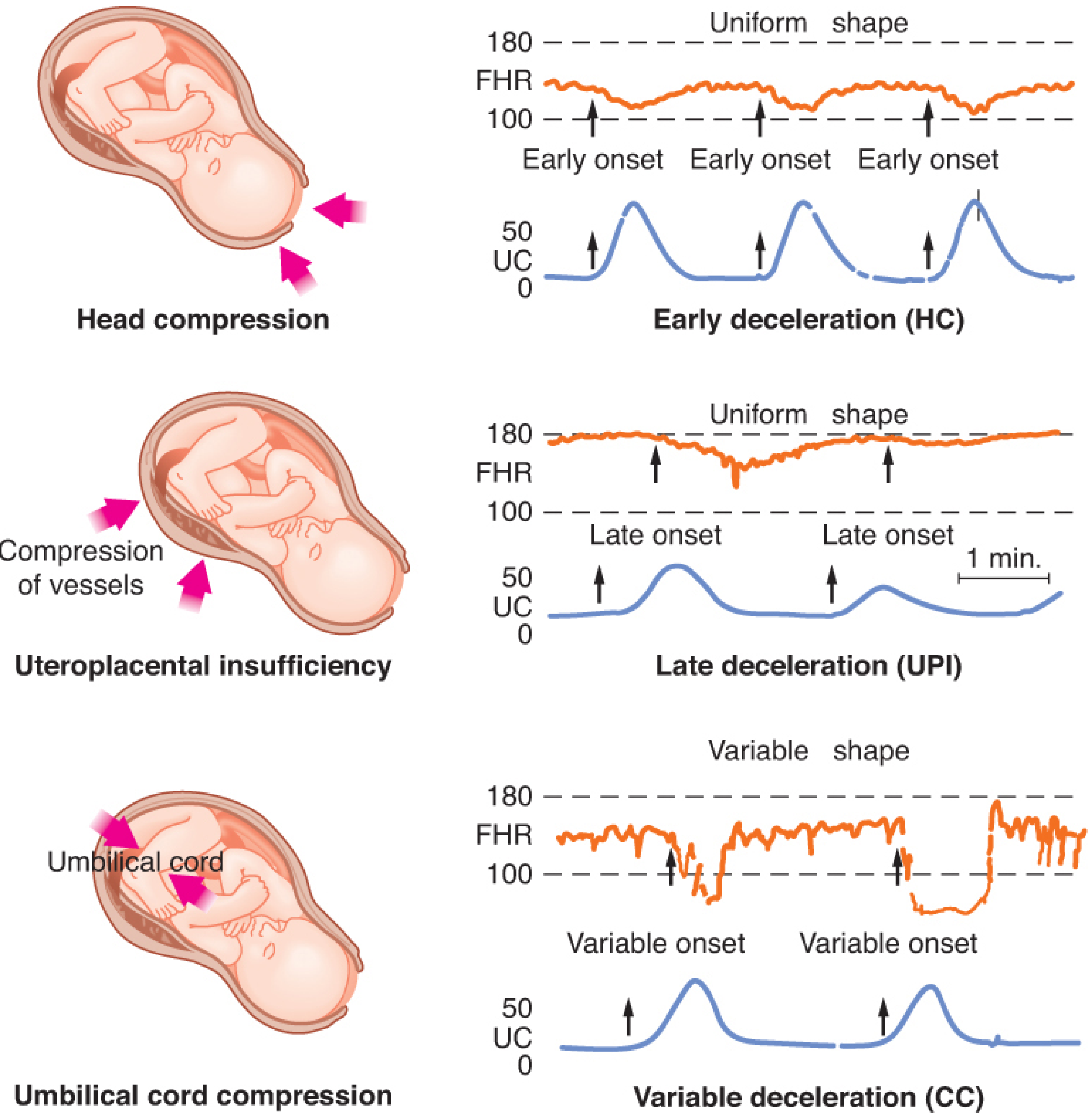
- Top panel (early deceleration): The FHR dip mirrors the contraction peak - caused by fetal head compression increasing vagal tone. Benign.
- Middle panel (late deceleration): The FHR dip comes after the contraction peak - the "late onset" arrows show this clearly. This is the pattern to worry about.
- Bottom panel (variable deceleration): Abrupt, unpredictable - caused by umbilical cord compression.
Why Does It Happen? The Two Mechanisms
Mechanism 1: Reflex Late Deceleration (more common, less dangerous)
- The fetus normally compensates well because it has high-affinity fetal hemoglobin (HbF) and high hemoglobin levels overall, both of which maximize oxygen-carrying capacity. But strong or prolonged contractions can overwhelm this.
| Step | What happens |
|---|---|
| Chemoreceptor activation | Senses low PO₂ |
| Sympathetic outflow | Peripheral vasoconstriction - blood is shunted away from non-vital tissues toward brain, heart, adrenals |
| Blood pressure rises | Because of vasoconstriction |
| Baroreceptors activated | Detect the BP rise |
| Parasympathetic (vagal) reflex | Slows the heart rate, reduces cardiac output, normalizes BP |
| FHR dips = the late deceleration | This is the heart rate drop you see on the monitor |
- Deoxygenated blood to travel from the placenta through the umbilical vein to the fetal heart and chemoreceptors
- The PO₂ must fall below a threshold before vagal activity is triggered
Mechanism 2: Non-Reflex Late Deceleration (less common, more dangerous)
- Prolonged or severe hypoxemia causes metabolic acidemia
- The deoxygenated blood reaching the heart is insufficient to maintain normal myocardial function
- There is direct myocardial depression - the heart slows because it is failing, not because of a protective vagal reflex
- Increased vagal activity also contributes
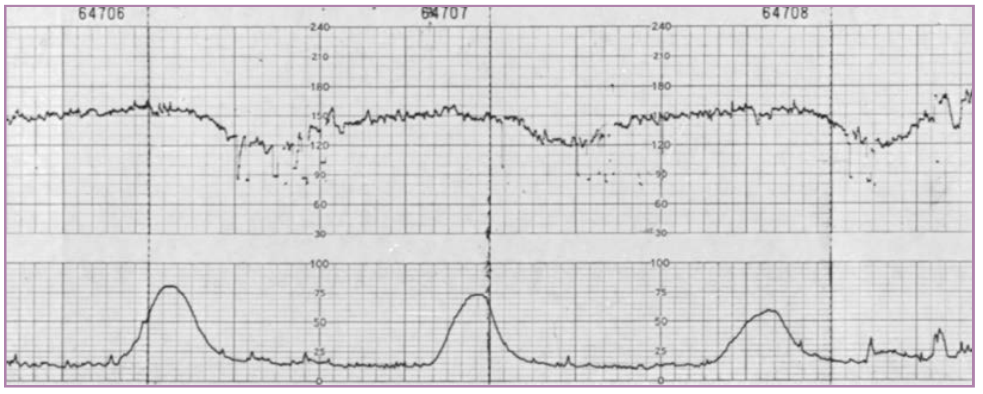
The Key Clinical Distinction: Can I Tell Them Apart?
| Feature | Reflex Late Decel | Non-Reflex Late Decel |
|---|---|---|
| Mechanism | Chemoreceptor/baroreceptor vagal reflex | Direct myocardial depression + acidemia |
| FHR variability | Moderate (normal) | Absent or minimal |
| Accelerations | May be present | Absent |
| CNS status | Intact - brain is oxygenated | Compromised |
| Metabolic acidemia | Not present | Present |
| Risk | Lower - transient hypoxemia | Higher - fetal decompensation |
| Action | Optimize blood flow, monitor | Prompt intervention |
Where Else Can the Oxygen Pathway Be Interrupted?
Maternal lungs → Maternal circulation → Intervillous space → Placental membrane → Fetal blood → Fetal heart/brain
- Lungs: Maternal hypoxemia (e.g., pulmonary edema, asthma exacerbation)
- Heart: Poor maternal cardiac output
- Vasculature: Maternal hypotension (e.g., from epidural/spinal, hemorrhage, aortocaval compression)
- Uterus: Excessive/prolonged contractions (hyperstimulation from oxytocin), placental abruption
- Placenta itself: Preeclampsia, intrauterine growth restriction (reduced placental reserve)
Summary Flow Diagram
Uterine contraction
        ↓
Compression of uterine vessels
        ↓
Reduced intervillous space perfusion
        ↓
Fetal PO₂ drops below ~15-25 mmHg
        ↓
        ┌─────────────────────────────────────┐
        ↓                                     ↓
 Chemoreceptors activated              (if severe/prolonged)
        ↓                              Direct myocardial depression
 Sympathetic: vasoconstriction              ↓
        ↓                              NON-REFLEX late deceleration
 BP rises → Baroreceptors activated    (absent variability, no accels)
        ↓                              → FETAL DECOMPENSATION
 Vagal: HR slows, CO decreases
        ↓
 REFLEX late deceleration
 (normal variability ± accels present)
 → CNS intact, manageable
        ↓
 Contraction ends → O₂ restored → FHR returns to baseline
Clinical Takeaways
- Late = delayed relative to contraction peak - onset-to-nadir ≥30 seconds, and the dip doesn't start until after the contraction begins.
- The most common mechanism is a reflex - chemoreceptors → sympathetic → baroreceptors → vagal slowing. This is physiologically protective.
- Variability is your most important clue - moderate variability = CNS intact = reflex deceleration = fetus is compensating. Absent/minimal variability = fetal decompensation = act now.
- Recurrent late decelerations + absent variability + no accelerations = urgent assessment and intervention.
- Think beyond the uterus - maternal hypotension, hypoxemia, or poor cardiac output can all cause the same pattern.