
Styphalocovus scaled skin syndrome
Reading File
Reading File
Finding Sources
Searching PubMed
"staphylococcal scalded skin syndrome"
Searching the Web
Staphylococcal scalded skin syndrome CDC clinical overview treatment
Searching Images
staphylococcal scalded skin syndrome clinical skin rash flaccid bullae
You likely mean staphylococcal scalded skin syndrome (SSSS).
It is a serious skin infection-related emergency, usually in infants and young children, caused by toxin-producing Staphylococcus aureus. The toxin makes the top layer of skin peel off, so the skin can look red, tender, blistered, or “scalded.”
Typical signs
- Fever, irritability, poor feeding
- Painful, widespread red skin, often starting around the mouth, neck, armpits, or groin
- Fragile/flaccid blisters and peeling skin
- Skin may rub off easily with gentle pressure
- The mouth and other mucous membranes are usually spared, unlike Stevens-Johnson syndrome/TEN
It needs urgent hospital assessment, IV anti-staphylococcal antibiotics, fluids, pain control, and skin/wound care. DermNet's SSSS overview describes it as a dermatologic emergency requiring prompt hospital treatment.
If you or a child currently has painful red peeling or blistering skin, fever, or looks unwell, seek emergency care now. Do not try to peel skin or apply home remedies.
Find recent clinical guidelines for SSSS management and full detail about this in detail
Reading File
Reading File
Reading File
Finding Sources
Finding Sources
Finding Sources
Searching PubMed
"staphylococcal scalded skin syndrome" AND management
Searching the Web
staphylococcal scalded skin syndrome clinical guideline management pediatric 2024 2025
Searching Images
Staphylococcal scalded skin syndrome infant erythema superficial skin peeling clinical photograph
Reading File
Reading File
Reading File
Reading Article
PMID: 40650480
Reading Article
PMID: 33283348
Reading Article
PMID: 36671239
Reading Web Page
https://pch.health.wa.gov.au/For-health-professionals/Emerge…
Staphylococcal scalded skin syndrome (SSSS): current management
SSSS is a medical emergency, particularly in neonates and young children. It is caused by toxin-producing Staphylococcus aureus, not by the bacteria directly invading every area of peeling skin. Exfoliative toxins A and B enter the circulation and cleave desmoglein-1 in the superficial epidermis. This causes fragile blisters and sheet-like peeling, while the mouth, eyes, and genital mucosa are usually spared.
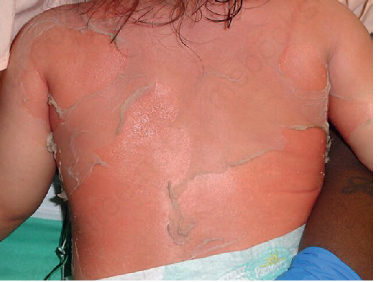
SSSS occurs mainly in infants and children younger than 5 years. In adults, it is uncommon and should prompt assessment for renal impairment, immunosuppression, malignancy, or another major underlying illness.
What recent guidance is available?
There is no single globally adopted SSSS guideline equivalent to a major IDSA or AAP guideline dedicated solely to SSSS. Management is therefore based on pediatric emergency/dermatology pathways, local antimicrobial-resistance patterns, and the best recent evidence.
The most useful current sources are:
-
Perth Children’s Hospital Emergency Department guideline, endorsed October 2023 and scheduled for review in October 2026. It is a practical pediatric assessment-and-management pathway, although it explicitly requires adaptation to local policy and senior clinical judgment. See the PCH SSSS guideline.
-
2025 systematic review of non-neonatal pediatric SSSS. It reviewed 48 studies and supports a “less is more” approach: targeted cultures, limited nonspecific blood testing, beta-lactam therapy as first-line where MSSA is likely, IV fluids only when oral intake is inadequate, and bland emollient skin care. Importantly, it found no evidence that routine adjunctive clindamycin improved outcomes. Gray et al. (2025), Pediatric Dermatology, PMID 40650480.
-
Practical current clinical summaries from Merck Manual Professional and DermNet align with this approach.
Clinical presentation
Typical progression is rapid:
- Fever, irritability, lethargy, reduced feeding, or malaise
- Tender, diffuse, sunburn-like erythema, often beginning around the eyes, nose, mouth, neck, axillae, and groin
- Wrinkling of skin followed by superficial flaccid bullae
- Positive Nikolsky sign: gentle pressure causes superficial epidermal separation
- Sheet-like superficial peeling over hours to 1 to 2 days
- Mucosal sparing is an important clue
- Palms and soles are often spared
The infection source can be distant from the rash, such as the nose, nasopharynx, conjunctiva, ear, throat, umbilicus in neonates, wound, or localized impetigo. The blister roof itself is often sterile because the disease is toxin-mediated.
Immediate assessment and disposition
Admit to hospital
Most children with suspected SSSS should be admitted because they may require:
- IV antibiotics
- Hydration and electrolyte monitoring
- Analgesia
- Temperature support
- Wound/skin care
- Monitoring for progression, secondary infection, and sepsis
Consider high-dependency, ICU, or burn-unit input when there is extensive skin loss, poor perfusion, hypotension, marked dehydration, electrolyte disturbance, inability to feed, sepsis concern, or an adult with SSSS.
Immediate priorities
- ABCDE assessment
- Assess airway, breathing, circulation, hydration, mental state, temperature, and urine output.
- Assess extent and severity
- Percentage of body surface involved
- Degree of pain and skin tenderness
- Feeding/oral intake and urine output
- Signs of sepsis or shock
- Start empiric anti-staphylococcal treatment promptly
- Do not wait for culture results in clinically convincing disease.
- Seek pediatric, dermatology, infectious-disease, and burn/wound-care input as appropriate.
Investigations
SSSS is mainly a clinical diagnosis. Tests should aid treatment or exclude dangerous alternatives, not delay antibiotics.
Recommended microbiology
Obtain cultures before antibiotics if this does not delay treatment:
- Swab/culture from a suspected primary focus:
- Nasopharynx or nose
- Conjunctiva
- Umbilicus in neonates
- Ear, throat, wound, impetigo lesion, or other focal infection
- Blood culture if febrile, systemically unwell, neonate, immunocompromised, or sepsis is suspected
- Urine culture may be appropriate in neonates or where a urinary source is possible
Do not rely on blister-fluid culture. Bullae are commonly sterile in SSSS, unlike bullous impetigo, where blister fluid often grows S. aureus. Targeted cultures from periorificial sites are more useful.
Laboratory tests
Use selectively:
- CBC, electrolytes, glucose, urea/creatinine
- CRP or other inflammatory markers when sepsis is considered or the diagnosis is uncertain
- Blood gas/lactate in an ill or poorly perfused child
- Strict fluid balance and urine-output monitoring if skin loss is extensive
The 2025 systematic review found that routine blood counts, chemistry panels, and inflammatory markers are generally nonspecific and do not establish the diagnosis or substantially guide routine care.
Skin biopsy
A biopsy is not routinely needed. Obtain urgent dermatology input and consider frozen-section histology if the main concern is SJS/TEN, autoimmune blistering disease, or another uncertain diagnosis. SSSS shows superficial, non-inflammatory intraepidermal splitting at the granular layer.
Antimicrobial management
First-line treatment
Start an IV anti-staphylococcal beta-lactam for presumed methicillin-susceptible S. aureus (MSSA), according to the local pediatric formulary and susceptibility patterns. Common options include:
- Flucloxacillin or cloxacillin
- Nafcillin or oxacillin
- Cefazolin
The exact drug, dose, and interval must be selected by the treating pediatric team, especially in neonates and in renal impairment.
When to cover MRSA
Use an MRSA-active agent, commonly vancomycin, when any of the following applies:
- Known MRSA colonization or previous MRSA infection
- High local prevalence of MRSA
- Severe illness or septic presentation
- A culture suggesting MRSA
- Failure to improve on appropriate first-line MSSA therapy
- Relevant outbreak or healthcare exposure
Once culture and susceptibility findings are available, narrow therapy to the most appropriate agent.
Clindamycin: current evidence
Historically, clindamycin was often added because it may suppress toxin production. However, the strongest recent pediatric evidence does not show improved outcomes from routine addition:
- The 2025 systematic review found no outcome benefit supporting routine clindamycin addition.
- An 84-patient pediatric cohort similarly found no reduction in hospital stay with clindamycin and advised against skin debridement. The 2021 cohort study supports culture-directed therapy instead.
Therefore, do not add clindamycin routinely just for toxin suppression unless a local guideline, microbiology advice, resistance pattern, or individual clinical circumstance supports it.
IV-to-oral transition and duration
Reassess daily. Transition to oral antistaphylococcal therapy when the patient:
- Is clearly improving
- Is afebrile or clinically stable
- Is drinking/feeding adequately
- Has no sepsis concern
- Can tolerate oral medication
The total duration should be individualized according to disease severity, source control, culture results, and clinical response. Do not use a fixed duration without local pediatric or infectious-disease guidance.
Supportive care
Supportive care is as important as antibiotics.
Fluids and electrolytes
Superficial epidermal loss can cause fluid loss and poor oral intake.
- Assess hydration, capillary refill, heart rate, blood pressure, urine output, and weight.
- Give IV fluids if oral intake is inadequate, dehydration is present, or there is extensive denudation.
- Monitor electrolytes in moderate to severe disease.
- Avoid both under-resuscitation and unnecessary fluid overload.
Pain management
Skin tenderness can be substantial.
- Give age-appropriate analgesia early.
- Use opioid analgesia if pain is severe, with monitored clinical care.
- Minimize repeated dressing changes and adhesive tapes, which may worsen epidermal loss.
Skin and wound care
- Handle skin gently.
- Use bland emollients such as petrolatum where appropriate.
- Use non-adherent dressings for denuded areas.
- Avoid routine surgical debridement or aggressive removal of skin. It can worsen pain, increase complications, and prolong hospitalization.
- Avoid potentially irritating topical agents unless advised by dermatology or wound-care specialists.
- Maintain a warm environment to prevent hypothermia.
Nutrition and monitoring
- Encourage feeding and oral fluids when safe.
- Monitor input/output closely.
- Watch for secondary skin infection, cellulitis, pneumonia, bacteremia, sepsis, hypothermia, and electrolyte abnormalities.
Key differential diagnoses
| Condition | Features favoring it over SSSS |
|---|---|
| SJS/TEN | Prominent mucosal erosions, targetoid lesions, drug exposure, deeper full-thickness epidermal loss, typically more common in older children/adults |
| Bullous impetigo | Localized flaccid bullae, usually no fever or systemic illness, blister fluid more likely culture-positive |
| Staphylococcal toxic shock syndrome | Hypotension, shock, multiorgan involvement, diffuse rash followed by later desquamation |
| Scarlet fever | Sandpaper-like rash, pharyngitis, strawberry tongue, lacks superficial flaccid bullae |
| Epidermolysis bullosa / inherited blistering disorders | Recurrent or early-life blistering, trauma-related lesions, family history |
| Autoimmune bullous disease | Different distribution, may have mucosal involvement, requires dermatologic testing |
| Thermal burn or chemical injury | Exposure history and distribution pattern |
The most urgent distinction is from SJS/TEN. In SSSS, mucosa is generally spared and cleavage is superficial. In SJS/TEN, mucosal disease is common and skin injury is deeper and more dangerous.
Prognosis
With prompt antimicrobial and supportive treatment, children usually improve rapidly and heal without scarring, often within about 5 to 7 days after treatment begins. Adults have a substantially higher risk of severe disease and death because SSSS in adults is often associated with renal dysfunction, immunosuppression, or serious comorbidity.
Infection prevention and outbreak control
For neonatal-unit, nursery, or daycare clusters:
- Notify infection prevention and public health teams according to local requirements.
- Reinforce hand hygiene and contact precautions.
- Culture possible sources or carriers when directed by outbreak-control teams.
- Consider staff, caregiver, or parental carriage evaluation in persistent clusters.
- Use molecular typing if available for outbreak investigation.
A neonatal outbreak review emphasizes source identification, hygienic measures, and limiting exposure through physical barriers and infection-control practices. Nusman et al. (2022), PMID 36671239.
Practical clinical algorithm
- Suspect SSSS in a febrile, irritable infant/young child with painful erythema, superficial peeling, positive Nikolsky sign, and mucosal sparing.
- Admit and assess severity. Stabilize circulation, hydration, pain, and temperature.
- Obtain targeted cultures from the suspected source and blood cultures if systemically unwell. Do not depend on blister culture.
- Start IV anti-staphylococcal beta-lactam promptly if MSSA is likely. Add MRSA-active therapy only when risk or local epidemiology justifies it.
- Provide gentle skin care, non-adherent dressings, emollient, analgesia, and fluid support. Avoid debridement.
- Review daily with culture results and clinical progression. Narrow antibiotics and convert to oral therapy when stable.
- Escalate care for extensive skin loss, poor intake, dehydration, shock, sepsis, or diagnostic uncertainty, especially possible SJS/TEN.
This is educational information, not an individualized prescription. Any person with widespread tender red skin, blistering/peeling, fever, reduced feeding, lethargy, or signs of dehydration should receive urgent in-person medical assessment.
This is a shared conversation. Sign in to Orris to start your own chat.