
Types of ameloblastoma
ameloblastoma types histology
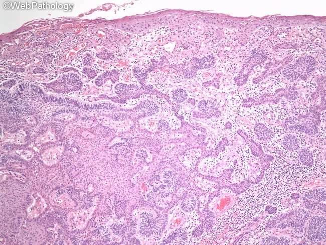
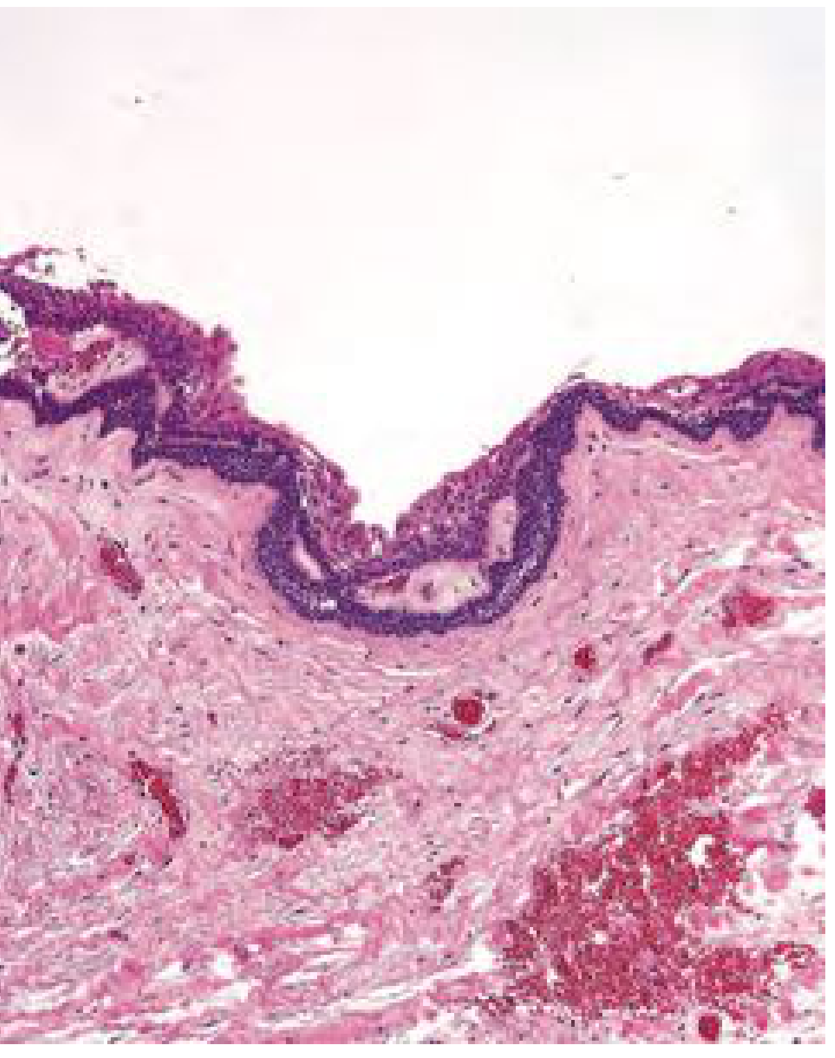
Comprehensive Description: Histology image of a peripheral (extraosseous) ameloblastoma involving oral mucosa of the mandible. Modality: Light microscopy of formalin-fixed, paraffin-embedded tissue; staining: Hematoxylin and Eosin (H&E). The surface mucosa appears intact with underlying lamina propria populated by odontogenic epithelium forming islands and cords. In the follicular pattern, nests of ameloblastic epithelium display peripheral palisading of columnar cells with reverse polarity against a basement membrane, and a central stellate reticulum–like zone resembling developing enamel organ. The nests are embedded in a mature, densely fibrous to loosely arranged connective tissue stroma. In the plexiform pattern, interconnecting thin cords or sheets of basaloid cells traverse the stroma, frequently lacking classic peripheral palisading and reverse nuclear polarity, with more loosely arranged cells within the cords. The surrounding stroma is vascularized loose connective tissue. Features mimic the intraosseous counterpart seen in jaw lesions but arise in soft tissue. Clinically, peripheral ameloblastoma is benign but locally aggressive with potential for recurrence after incomplete excision. This image supports recognition of characteristic histologic hallmarks, aids differential diagnosis from basal cell lesions and other odontogenic tumors, and informs surgical planning and prognostic assessment. Educational relevance includes teaching follicular vs plexiform morphologies and enamel organ–like architecture in peripheral contexts.
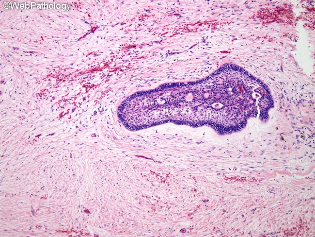
Histopathology of ameloblastoma, desmoplastic variant, on a Hematoxylin and Eosin stained slide from jaw bone tissue. The lesion shows dense, sclerotic stroma with compressed bone trabeculae and scattered islands and thin cords of odontogenic epithelium embedded in the fibrous matrix. Epithelial components consist of cuboidal to columnar cells, with partial reverse polarity, and areas reminiscent of stellate reticulum are minimal or absent. Peripheral palisading is often not evident in this variant. The stroma demonstrates marked desmoplasia with abundant type I collagen and sparse inflammatory infiltrate. Keratinization is uncommon, and mitotic activity is typically low. The tumor is locally infiltrative despite its benign designation, and the desmoplastic pattern can mimic fibrous or desmoplastic fibromatous lesions radiographically. Diagnostic significance rests on identifying odontogenic epithelium within a densely fibrous milieu, consistent with desmoplastic ameloblastoma, a distinct histologic subtype of ameloblastoma. Differential considerations include conventional ameloblastoma subtypes (follicular, plexiform), fibrous dysplasia, odontogenic fibroma, and fibromatosis. Clinically, lesions tend to occur in the anterior jaws and may present as swelling. Surgical management requires complete excision due to recurrence risk. This image is educational for pathology training, histology atlases, and radiology–histology correlation studies. Interpretation should integrate clinical context, radiographs, and immunohistochemical confirmation when available, guiding management.
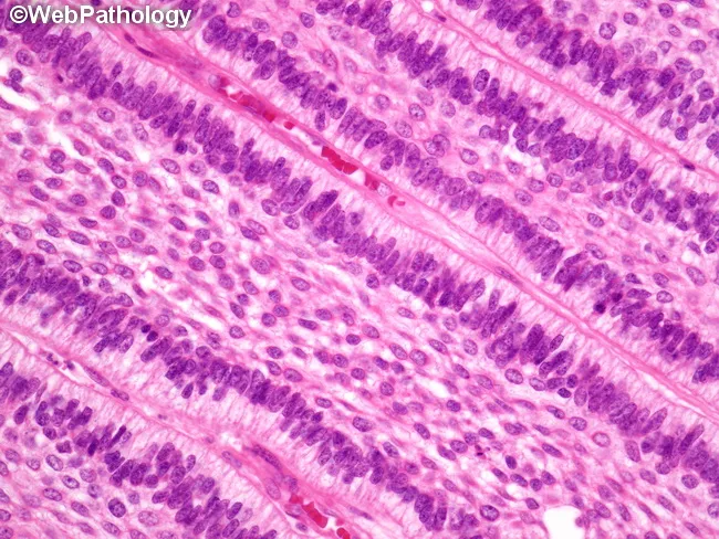
This histology image depicts ameloblastoma of the jaw, captured on a hematoxylin and eosin–stained biopsy section examined under light microscopy. The dominant pattern shown is follicular ameloblastoma, with oval to rounded islands of odontogenic epithelium embedded in a dense fibrous stroma. Peripheral columnar cells exhibit classic reverse polarity and peripheral palisading, while the central cells resemble the stellate reticulum of the developing tooth germ. In some regions, the epithelial nests are triangular or elongated and dispersed, consistent with alternate patterns that may coexist in the same tumor, including plexiform, acanthomatous, basaloid, granular cell, or desmoplastic variants. The nests are separated by a mature, hyalinized connective tissue stroma without prominent inflammatory infiltrate. The myxoid or hyalinized stroma and highly organized epithelial architecture help distinguish ameloblastoma from other odontogenic lesions. Clinically, these histologic features correlate with locally aggressive behavior despite benign cytology, underscoring the need for complete surgical excision with clean margins to minimize recurrence risk. The image emphasizes the diagnostically relevant hallmarks—pigmented? No, not pigmented; but rather cohesive islands with peripheral palisading and stellate reticulum core. This slide serves educational value for surgical planning, differential diagnosis, and radiologic-pathologic correlation in jaw lesions. It is suitable for teaching, research, and case documentation.
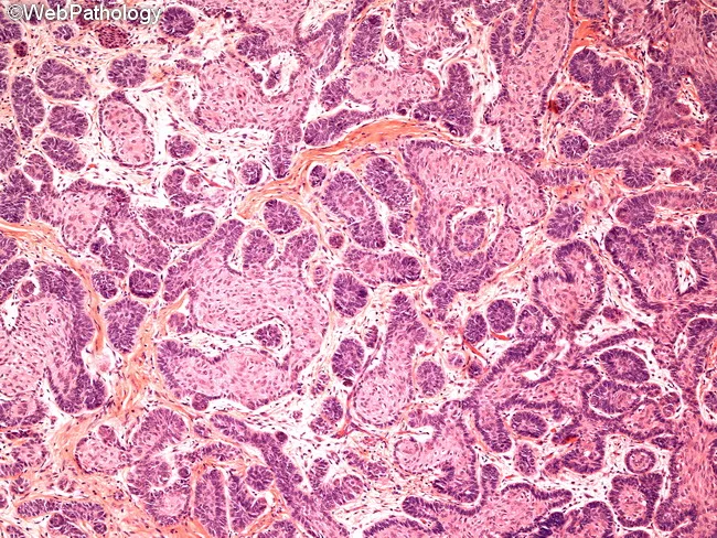
Histology: Light microscopy of a jaw lesion shows classic ameloblastoma morphology on hematoxylin and eosin stained tissue. The neoplasm is composed of multiple epithelial islands and cords embedded in a fibrous connective tissue stroma. Follicular pattern predominates, with nests of odontogenic epithelium displaying peripheral palisading of columnar cells and reversal of polarity, nuclei oriented away from the basement membrane. Central cells resemble stellate reticulum with loosely arranged, angular morphology. Some islands exhibit a loosely arranged, anastomosing arrangement compatible with plexiform architecture. Overall cellularity is moderate, with minimal pleomorphism and no overt mitotic activity in the field, consistent with benign but locally aggressive behavior. Basal layer cells are darkly basophilic with prominent nucleoli, while surrounding stroma is densely fibrous and occasionally myxoid, producing a subtle desmoplastic impression in portions of the section. The tumor borders appear infiltrative into adjacent connective tissue, reflecting its known propensity for local invasion despite benign cytology. There is an absence of keratinization, dysplasia, or metastatic features. The image highlights the diagnostic hallmarks: enamel organ–like islands, reverse polarity, stellate reticulum–like center, and a fibrous stroma. These histologic features underline confirmation of diagnosis and guide surgical planning in odontogenic tumors and jaw neoplasms. For educational and diagnostic purposes.
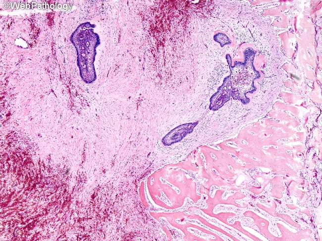
Light microscopic histopathology of a desmoplastic ameloblastoma. Imaging modality: Brightfield hematoxylin-eosin stained tissue section, scanned at low-to-intermediate magnification to reveal structural context within the jaw bone. The tumor shows islands, cords, and narrow strands of odontogenic epithelium embedded in a densely collagenized, fibrous stroma (desmoplasia). Epithelial nests consist of cuboidal to columnar cells with scant cytoplasm and hyperchromatic nuclei; peripheral basaloid cells may exhibit loose alignment rather than classic palisading. Central regions lack a prominent stellate reticulum, consistent with desmoplastic morphology. Surrounding stroma is markedly hyalinized and densely fibrous, often with wedge-shaped or kite-like stromal condensations that compress adjacent osseous trabeculae. Occasional chondro-osseous metaplasia or calcifications can be seen, though not obligatory. The lesion is typically benign, slow growing, and locally invasive, with no malignant cytology evident in this field. Immunoprofile is supportive but not decisive; transforming growth factor beta driven desmoplasia contributes to the sclerotic background. Clinically, desmoplastic ameloblastoma may present as a mixed radiolucent-radiopaque jaw lesion, frequently in anterior mandible. Recurrent behavior is possible without wide local excision. This histology helps distinguish the desmoplastic variant from follicular or plexiform patterns and guides surgical planning and prognosis. Correlative radiology and clinicopathologic data enhance diagnostic confidence and treatment planning for jaw lesions.
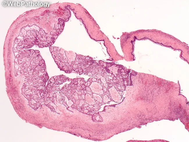
Histology: Light microscopy; Hematoxylin and Eosin (H&E) stained section of a unicystic ameloblastoma arising in the mandible. The specimen is a cystic lesion, typically associated with an unerupted third molar, showing a well circumscribed cavity lined by ameloblastomatous epithelium. The lining displays basal palisading of columnar cells with reversed nuclear polarity and an overlying zone resembling stellate reticulum. In many fields there are intraluminal or mural tumor nests within the cyst wall, representing the proliferative component. The surrounding stroma is usually fibrous and vascular, sometimes with mild inflammation. Clinically and radiographically, the lesion can mimic a dentigerous cyst, primordial cyst, radicular cyst, or residual cyst; histopathology confirms ameloblastic differentiation and classifies it as unicystic variant. Pathologic diagnosis has prognostic significance because involvement limited to the cyst lining (luminal or simple unicystic) has a favorable prognosis with conservative enucleation, while invasion of the cyst wall (mural unicystic) or solid nodules increases recurrence risk and may require more aggressive resection. Potential clinical uses include surgical planning, radiologic-pathologic correlation, and educational differentiation of odontogenic cystic lesions. This image exemplifies the diagnostic interface between histology and radiology in juvenile jaw lesions and odontogenic tumors.
Types of Ameloblastoma
Overview
- ~85% occur in the mandible (molar/ramus region most common)
- Slow, painless jaw expansion
- MAPK pathway mutations (especially BRAF V600E) found in ~90% of conventional lesions
- Radiographically: radiolucent, well-demarcated, unilocular or multilocular
1. Conventional (Solid/Multicystic) Ameloblastoma
| Subtype | Key Feature |
|---|---|
| Follicular | Islands of odontogenic epithelium with peripheral palisading + central stellate reticulum - most common |
| Plexiform | Interconnecting cords/sheets of basaloid cells in loose stroma |
| Acanthomatous | Squamous metaplasia with keratin pearls in the center of epithelial islands |
| Granular cell | Central cells replaced by eosinophilic granular cytoplasm |
| Desmoplastic | Dense fibrous/collagenized stroma, often in anterior jaw; mixed radiolucent-radiopaque on imaging |
| Basal cell | Resembles basal cell carcinoma; least common histologic variant |
2. Unicystic Ameloblastoma
- Columnar basilar cells with nuclear palisading
- Nuclear polarization away from the basement membrane
- Hyperchromatism of basal cell nuclei
- Subnuclear vacuolization
- Overlying loosely aggregated stellate cells (resembling stellate reticulum)
| Subtype | Description | Treatment | Recurrence |
|---|---|---|---|
| Luminal | Ameloblastic epithelium lining lumen only | Enucleation/conservative excision | Low (~10%) |
| Intraluminal | Nodule(s) projecting into lumen; no wall invasion | Conservative excision | Low-moderate |
| Mural | Tumor invades fibrous cyst wall | Resection with clear margins (more aggressive) | Higher |
3. Peripheral (Extraosseous) Ameloblastoma
- Typically in middle-aged adults
- Presents as a sessile or pedunculated gingival mass
- Radiographic features: limited utility since the lesion is extraosseous
- Histologically: ameloblastic epithelium below the lamina propria, with same patterns as conventional type
- Treatment: Surgical excision (conservative, since bone not involved)
- Recurrence rate: 15-20%
4. Metastasizing Ameloblastoma
- Histologically benign-appearing tumor that nevertheless metastasizes
- Most common metastatic sites: Lungs and cervical lymph nodes
- No cytologic atypia distinguishing it from conventional ameloblastoma
- Radiographic features identical to conventional type
- Treatment: Depends on stage
- Prognosis: Fair to poor
Summary Table
| Type | Location | Age | Key Feature | Treatment | Recurrence |
|---|---|---|---|---|---|
| Conventional | Intraosseous (mandible >> maxilla) | >20 yrs | Multilocular radiolucency; MAPK mutations | Resection with 1 cm margin | Moderate-high |
| Unicystic | Intraosseous (posterior mandible) | 2nd-3rd decade | Unilocular, mimics dentigerous cyst | Depends on subtype (luminal vs. mural) | 10-20% |
| Peripheral | Extraosseous (gingiva/mucosa) | Middle age | Soft tissue mass; no bone involvement | Local excision | 15-20% |
| Metastasizing | Intraosseous | Variable | Benign histology + metastasis (lung/LN) | Stage-dependent | Poor |
- K. J. Lee's Essential Otolaryngology, pp. 757-758
- Cummings Otolaryngology Head and Neck Surgery, Chap. 88