
List all the possible causes for thehairfall with its treatment
causes of hair loss alopecia types treatment 2024
"hair loss" OR "alopecia" treatment causes
types of alopecia hair loss causes diagram
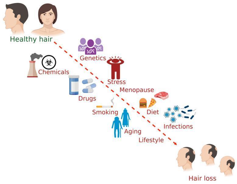
This medical infographic illustrates the multifactorial etiology of hair loss in humans, depicting a progression from 'Healthy hair' to 'Hair loss.' The diagram utilizes a descending dashed red arrow to represent the transition between these two clinical states. Positioned along this pathway are various internal and external contributing factors, each represented by a descriptive icon and text label. These factors include: Chemicals (environmental/occupational exposure), Genetics (hereditary predisposition), Drugs (pharmacological side effects), Stress (psychosocial impact), Smoking, Menopause (hormonal changes), Aging (senescence), Diet (nutritional status/lifestyle), and Infections (pathogenic microorganisms). The top-left corner features healthy male and female profiles with full scalp coverage, while the bottom-right corner displays three male profiles exhibiting progressive stages of androgenetic alopecia-like patterns, including bitemporal recession and vertex thinning. This visual aid is designed for medical education to summarize the complex interactions between lifestyle, environmental, and biological factors that influence follicular health and the progression of alopecia.
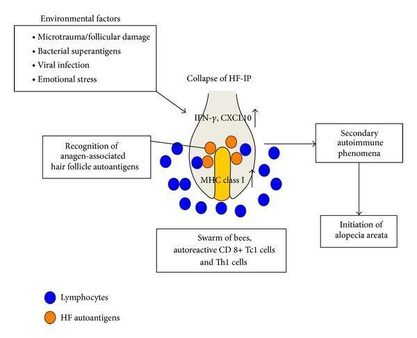
This pathophysiology diagram illustrates the development of alopecia areata (AA) through a multi-step autoimmune process. The central anatomical focus is an anagen-phase hair follicle (HF) bulb. A flow diagram depicts 'Environmental factors' (e.g., viral infection, stress, microtrauma) triggering the 'Collapse of HF-immune privilege (IP)'. This leads to increased expression of IFN-γ and CXCL10, as well as MHC class I within the dermal papilla/matrix area (yellow region). Visually, blue circles (representing autoreactive CD8+ Tc1 and Th1 cells) accumulate around the follicle base in a 'swarm of bees' pattern. These lymphocytes recognize anagen-associated HF autoantigens (orange circles). The resulting secondary autoimmune phenomena culminate in the final box labeled 'Initiation of alopecia areata'. The diagram serves as an educational tool for dermatology students to understand how the loss of hair follicle immune privilege and subsequent T-cell infiltration drive inflammatory non-scarring hair loss.
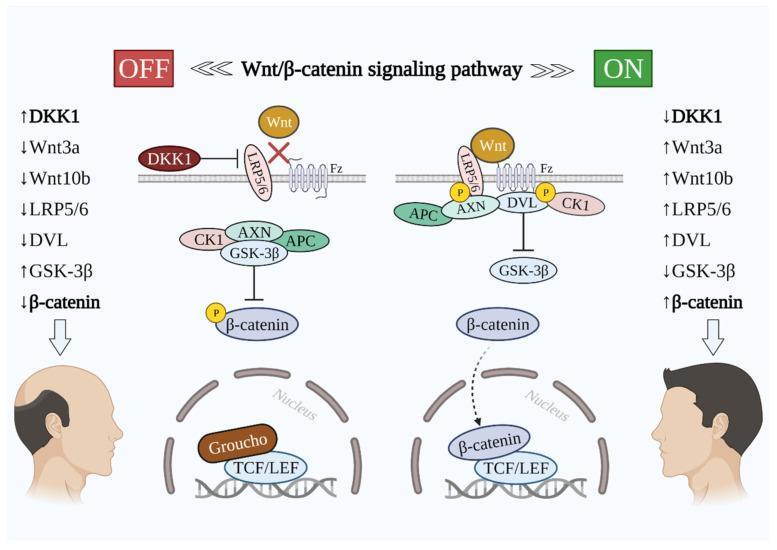
A comparative medical illustration detailing the Wnt/β-catenin signaling pathway in the context of hair growth and loss (androgenetic alopecia). The diagram is split into two states: 'OFF' (left) and 'ON' (right). In the 'OFF' state, DKK1 is upregulated, binding to and inhibiting the LRP5/6 co-receptor. This prevents Wnt ligands (Wnt3a, Wnt10b) from activating the Frizzled (Fz) receptor. Consequently, a cytoplasmic destruction complex (composed of CK1, AXN, GSK-3β, and APC) phosphorylates β-catenin, leading to its degradation. Within the nucleus, the repressor Groucho remains bound to TCF/LEF, suppressing gene transcription. This state is visually correlated with a male profile showing significant hair thinning and a receding hairline. In the 'ON' state, DKK1 is downregulated, allowing Wnt ligands to bind the Fz-LRP5/6 complex. This recruits Dishevelled (DVL), inactivating the destruction complex and inhibiting GSK-3β. Stabilized β-catenin translocates into the nucleus, displaces Groucho, and binds TCF/LEF to initiate hair growth-related gene expression. This state is correlated with a male profile showing full hair density.
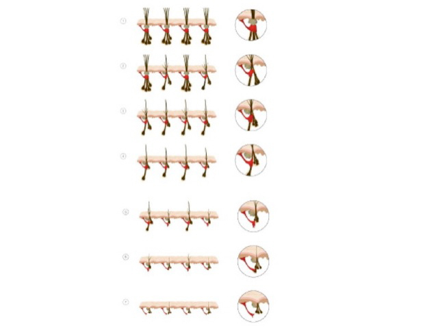
This diagram depicts hair follicle miniaturization in the scalp, a key feature of androgenetic alopecia. Using an illustrative cross-sectional montage, the image shows a series of hair follicles along the dermal–epidermal junction, each progressively smaller and producing thinner, shorter shafts. The leftmost follicles illustrate terminal hair with robust shaft diameter and deep dermal hair bulbs; adjacent panels display incremental reduction in follicle size, shortened anagen phases, and transition toward vellus-like hair with diminished density. The right-hand circles present schematic cross-sections highlighting reduced follicle depth, diminished sebaceous gland prominence, and a shallower bulge region in advanced stages. Overall, the sequence conveys the morphologic spectrum from normal hair morphology to pronounced miniaturization, including decreased follicular lumen size, altered hair shaft keratinization, and increased fibrous stroma in late stages. Clinically, this representation supports understanding of pattern hair loss, explains why hairs become finer and less pigmented, and underpins diagnostic considerations such as androgen-driven alopecia versus other diffuse hair loss processes. It also serves as an educational resource for clinicians, researchers, and students to recognize follicular remodeling, anticipate responses to anti-minoxidil or anti-androgen therapies, and plan histological or genetic investigations as needed for reference.
Causes of Hair Fall (Alopecia) and Their Treatments
PART 1: NON-SCARRING (NON-CICATRICIAL) ALOPECIA
1. Androgenetic Alopecia (Male/Female Pattern Hair Loss)
- The most common type. Androgens - especially dihydrotestosterone (DHT), produced from testosterone by the enzyme 5α-reductase - cause progressive miniaturization of terminal hair follicles into fine, hypopigmented vellus hairs.
- Polygenic inheritance (both maternal and paternal). The androgen receptor gene (AR) on chromosome Xq11-12 is a major determinant.
- Men: bitemporal recession then vertex thinning (Hamilton-Norwood scale).
- Women: diffuse thinning along the midline/crown, frontal hairline usually preserved (Ludwig scale).
- Topical minoxidil (2% women, 5% men): first-line; prolongs anagen phase
- Oral finasteride (1 mg/day in men): 5α-reductase inhibitor, reduces DHT; shown to work better in men with fewer AR-CAG repeats
- Low-dose oral minoxidil: increasingly used for both sexes
- Spironolactone (women): anti-androgen
- Hair transplant surgery: follicular unit transplantation (FUT/FUE) for advanced cases
- Platelet-rich plasma (PRP): adjunctive - Andrews' Diseases of the Skin
- Dutasteride: inhibits both type 1 and 2 5α-reductase; emerging evidence supports topical form (per 2025 review in Ther Deliv, PMID 39641480)
- Micronutrient correction: vitamin D, iron, zinc deficiencies linked to worsening AGA (per systematic review, PMID 39440586)
2. Alopecia Areata (AA)
- Autoimmune: collapse of hair follicle immune privilege, leading to CD8+ T-lymphocyte infiltration around the hair bulb in a "swarm of bees" pattern.
- Environmental triggers: viral infections, stress, microtrauma.
- Variants: patchy AA, alopecia totalis (entire scalp), alopecia universalis (whole body).
- Intralesional corticosteroids (triamcinolone acetonide): mainstay for patchy disease
- Topical corticosteroids, minoxidil, anthralin, tazarotene
- Topical contact sensitizers (DPCP, SADBE): induce immune deviation
- JAK inhibitors - newer FDA-approved options:
- Baricitinib (Olumiant): FDA-approved 2022
- Ritlecitinib (Litfulo): FDA-approved 2023
- Deuruxolitinib (Leqselvi): FDA-approved July 2024 - ~1/3 of patients with >50% hair loss achieved 80% regrowth at 24 weeks
- Systemic corticosteroids: short courses for rapidly progressing disease
- Observation for limited, stable disease (often spontaneously resolves)
3. Telogen Effluvium
- A major physiological or psychological stress shifts large numbers of hair follicles from anagen (growth) into telogen (resting/shedding) phase simultaneously, causing diffuse shedding 2-3 months after the trigger.
- Triggers include:
- High fever or severe infection
- Major surgery or illness
- Postpartum hormonal change (very common)
- Crash dieting / severe protein deficiency
- Thyroid disease (hypothyroidism or hyperthyroidism)
- Iron deficiency anemia
- Medications (see below)
- Psychosocial stress
- Usually self-limiting and reversible within 3-6 months
- Identify and correct the underlying cause
- Thyroid function tests, CBC, ferritin, zinc, vitamin D - correct deficiencies
- Discontinue offending drugs
- Topical minoxidil can accelerate recovery
- Observe for at least 6 months before considering surgical treatment - Cummings Otolaryngology
4. Anagen Effluvium
- Hair shaft fracture during the anagen (growth) phase, not at the root.
- Most commonly caused by chemotherapy agents (doxorubicin, cyclophosphamide, nitrosoureas, antimetabolites, mitotic inhibitors) - causes Pohl-Pinkus constrictions.
- Other causes: isoniazid (INH), thallium poisoning, boron toxicity.
- Can also occur in alopecia areata and secondary syphilis due to inflammation of the hair bulb.
- Loss appears 1-2 months after exposure; usually reversible.
- Usually reverses after stopping the offending agent
- Scalp cooling (hypothermia) during chemotherapy can reduce severity (though there is concern for scalp metastasis protection)
- Topical minoxidil shortens the period of baldness by an average of ~50 days - Andrews' Diseases of the Skin
5. Tinea Capitis (Scalp Ringworm)
- Dermatophyte fungal infection (most commonly Trichophyton tonsurans, Microsporum canis).
- Common in children; causes scaly, itchy patches with broken-off hairs ("black dot" pattern).
- Can cause kerion (suppurative, inflammatory mass) which may lead to scarring.
- Oral griseofulvin: first-line in children
- Oral terbinafine or itraconazole: effective alternatives
- Topical antifungals alone are insufficient for scalp infections
- Selenium sulfide or ketoconazole shampoo as adjunct (reduces spore shedding)
6. Traction Alopecia
- Chronic mechanical tension on hair from tight hairstyles: braids, cornrows, tight ponytails, extensions.
- Common in African and South Asian women.
- Initially non-scarring but can progress to permanent scarring if chronic.
- Stop the offending hairstyle - primary intervention
- Topical minoxidil for early non-scarring cases
- No effective treatment once scarring has occurred
7. Trichotillomania
- Obsessive-compulsive spectrum disorder: compulsive hair pulling by the patient, often unconsciously.
- Irregular patches with hairs of different lengths; mainly on scalp, eyebrows, eyelashes.
- Cognitive behavioral therapy (CBT) and habit reversal training - primary treatment
- Clomipramine or SSRIs (e.g., fluoxetine) for the OCD component
- N-acetylcysteine has some evidence
8. Drug-Induced Hair Loss
- Chemotherapy agents (anagen effluvium)
- Anticoagulants: heparin, warfarin
- Retinoids (isotretinoin, acitretin)
- Beta-blockers (propranolol, metoprolol)
- Anticonvulsants (valproate, carbamazepine)
- Antithyroid drugs (propylthiouracil, carbimazole)
- Lithium
- Oral contraceptives (on cessation - "pill-induced telogen effluvium")
- Hormone therapy (androgens, anabolic steroids)
- Discontinue or switch the offending drug when clinically safe
- Most cases reverse within 3-6 months
9. Hormonal and Endocrine Causes
- Hypothyroidism: dry, coarse hair; diffuse telogen effluvium - outer third of eyebrow loss (Hertoghe's sign)
- Hyperthyroidism: fine, diffuse hair thinning
- Polycystic ovary syndrome (PCOS): hyperandrogenism drives female pattern loss
- Adrenal tumors/hyperplasia: excess androgens
- Hypopituitarism / panhypopituitarism
- Postpartum: estrogen drop after delivery triggers telogen effluvium
- Menopause: declining estrogen increases androgen effect
- Treat the underlying endocrine disorder (thyroid replacement, PCOS management with metformin/OCP/spironolactone)
10. Nutritional Deficiencies
- Iron deficiency (most common worldwide)
- Zinc deficiency
- Vitamin D deficiency
- Biotin deficiency (rare except in parenteral nutrition or raw egg consumption)
- Protein/calorie malnutrition (kwashiorkor, anorexia nervosa)
- Correct the specific deficiency via diet and supplementation
- Iron, zinc, vitamin D supplementation where deficient - especially relevant to AGA (per PMID 39440586)
11. Systemic Diseases Causing Hair Loss
| Disease | Pattern |
|---|---|
| Systemic lupus erythematosus (SLE) | Diffuse loss; "lupus hair" (brittle, front hairline) |
| Secondary syphilis | "Moth-eaten" patchy alopecia |
| Dermatomyositis | Diffuse or patterned loss |
| Hypothyroidism / Hyperthyroidism | Diffuse telogen effluvium |
| HIV/AIDS | Diffuse telogen effluvium or AA |
| Diabetes mellitus | Diffuse thinning |
PART 2: SCARRING (CICATRICIAL) ALOPECIA
| Type | Cause | Treatment |
|---|---|---|
| Lichen planopilaris (LPP) | Autoimmune destruction of the follicle | Hydroxychloroquine, topical/intralesional steroids, cyclosporine, pioglitazone |
| Discoid lupus erythematosus (DLE) | Autoimmune scarring on scalp | Topical/intralesional steroids, hydroxychloroquine, thalidomide |
| Folliculitis decalvans | Staphylococcal infection + abnormal host response | Rifampicin + clindamycin combination, dapsone |
| Dissecting cellulitis | Suppurative deep folliculitis, mainly men | Isotretinoin, rifampicin, zinc sulfate, biologic TNF inhibitors |
| Central centrifugal cicatricial alopecia (CCCA) | Common in African-American women; exact cause unclear (traction, chemicals) | Stop chemical/heat damage; topical steroids, tetracyclines |
| Acne keloidalis nuchae | Chronic folliculitis + keloidal scarring at nape | Topical/intralesional steroids, antibiotics, laser, surgery |
| Burn/radiation/trauma | Physical destruction of follicles | Hair transplant to unaffected donor sites |
Summary: Treatment Toolkit
| Treatment | Used For |
|---|---|
| Topical minoxidil (2-5%) | AGA, AA, telogen effluvium |
| Oral minoxidil (low dose) | AGA, AA, chronic telogen effluvium |
| Finasteride / Dutasteride | AGA (men primarily) |
| Spironolactone | AGA in women, PCOS-related |
| JAK inhibitors (baricitinib, ritlecitinib, deuruxolitinib) | Alopecia areata (moderate-severe) |
| Intralesional triamcinolone | Alopecia areata, LPP, DLE |
| Systemic steroids | Rapidly progressing AA |
| Antifungals (oral) | Tinea capitis |
| Hair transplant | Advanced AGA, scarring alopecias (stable) |
| Treat underlying cause | Telogen effluvium, endocrine, nutritional |
| CBT / SSRIs | Trichotillomania |
Find the latest clinical trials for alopecia areata treatments (2024-2025)
"alopecia areata" AND "clinical trial"
alopecia areata clinical trials 2024 2025 JAK inhibitors biologics results
PMID: 39053611
PMID: 39141364
PMID: 40976531
"alopecia areata" AND ("dupilumab" OR "bempikibart" OR "amlitelimab" OR "upadacitinib" OR "rezpegaldesleukin")
PMID: 40231941
PMID: 41317911
PMID: 39846397
https://news.abbvie.com/2025-08-21-AbbVie-Announces-Positive…
Latest Clinical Trials for Alopecia Areata (2024-2026)
PART 1: FDA-APPROVED JAK INHIBITORS - NEW TRIAL DATA
1. Deuruxolitinib (Leqselvi) - JAK1/2 inhibitor | FDA-approved July 2024
- 654 adults, SALT score ≥50 (≥50% scalp hair loss), randomized to 8 mg BID, 12 mg BID, or placebo for 24 weeks
- Primary endpoint (SALT ≤20): 29.6% (8 mg), 41.5% (12 mg) vs 0.8% placebo
- Patient satisfaction response: 42.1% (8 mg), 53.0% (12 mg) vs 4.7% placebo
- Most adverse events mild/moderate
- PMID: 39053611 | J Am Acad Dermatol 2024
- 515 patients, same design; confirmed THRIVE-AA1 results
- SALT ≤20 achieved: 33.0% (8 mg BID), 38.3% (12 mg BID) vs 0.8% placebo (p<0.0001 for both)
- Long-term open-label extensions show continual improvement at 68 weeks
- PMID: 41317911 | J Am Acad Dermatol 2026
2. Baricitinib (Olumiant) - JAK1/2 inhibitor | FDA-approved 2022
- Key question: Do patients need continuous therapy?
- 154 responders (SALT ≤20 at week 52) re-randomized to continue baricitinib or switch to placebo
- By week 152: 80% of withdrawal patients lost hair regrowth vs only 7% who continued baricitinib
- Retreatment worked: 63% (2 mg) and 87.5% (4 mg) recaptured SALT ≤20 response
- Clinical implication: AA requires indefinite maintenance therapy - stopping leads to near-universal relapse
- PMID: 39141364 | JAMA Dermatology 2024
- Median follow-up 2.3 years; no new safety signals beyond known JAK class effects
- No increased risk of major cardiovascular events, DVT, or malignancy above background at this duration
- PMID: 40214720 | Am J Clin Dermatol 2025
3. Ritlecitinib (Litfulo) - JAK3/TEC family kinase inhibitor | FDA-approved 2023
- Specific data for the hardest-to-treat subtypes (AT/AU)
- Meaningful response rates in adults and adolescents (≥12 years)
- PMID: 39328096 | J Dermatol 2024
- 449 de novo patients, mean exposure 728 days (~2 years)
- At 24 months: 73.5% achieved SALT ≤20 and 66.4% achieved SALT ≤10
- 82.4% reported meaningful patient-perceived improvement
- Eyebrow response: 60.8%; eyelash response: 65.7%
- Safety: Most AEs mild/moderate. Herpes zoster in 6 patients; 3 major cardiovascular events; 3 malignancies (in ~450 patients over 2 years)
- ⚠️ Note: An erratum was published for this paper (PMID: 40650473) - verify updated data
- PMID: 39846397 | J Eur Acad Dermatol Venereol 2025
- Nearly 90% of SALT ≤20 responders maintained benefit out to 36-38 months
- 31.2% of patients achieved complete scalp hair regrowth (SALT = 0) at least once over 3 years
- Pooled ALLEGRO phase 2b/3 + ALLEGRO-LT data confirmed durable efficacy with increasing response rates over time
- PMID: 39432738 | Br J Dermatol 2025
PART 2: EMERGING JAK INHIBITORS IN TRIALS
4. Ivarmacitinib - Selective JAK1 inhibitor (novel, once-daily)
- 330 patients with severe AA (SALT ≥50 including AT/AU), randomized 1:1:1 to 4 mg, 8 mg, or placebo once daily for 24 weeks
- SALT ≤20 at week 24: 34.9% (4 mg), 40.6% (8 mg) vs 9.0% placebo
- Absolute difference vs placebo: 25.6% and 31.6% respectively (p<0.0001 both doses)
- No deaths, no major cardiovascular events, no thromboembolic events
- Once-daily dosing (vs twice-daily for deuruxolitinib) may improve adherence
- PMID: 40976531 | J Am Acad Dermatol 2026
5. Upadacitinib (Rinvoq) - JAK1 inhibitor | AbbVie
- Study 2 (first reported): Positive topline results
- Study 1 (reported Aug 2025): 45.2% (15 mg) and 55.0% (30 mg) reached SALT ≤20 at week 24
- Also met secondary endpoints: SALT ≤10, SALT = 0, eyebrow and eyelash regrowth
- Mean baseline SALT = 84 (~16% remaining scalp hair coverage - very severe population)
- Safety consistent with known upadacitinib profile; no new signals
- AbbVie expected to file for FDA approval; not yet approved for AA as of this data
PART 3: NON-JAK BIOLOGICS IN PIPELINE TRIALS
| Drug | Mechanism | Trial Phase | Notes |
|---|---|---|---|
| Bempikibart (ADX-914) | Anti-IL-7Rα (blocks IL-7 and TSLP signaling) | Phase 2a (SIGNAL-AA) | 2025 AAD data presented; targets upstream of T-cell activation |
| Amlitelimab | Anti-IL-4Rα biologic (same target as dupilumab but different mechanism) | Phase 2 | SC injection; shared pathway with atopic dermatitis |
| Rezpegaldesleukin (rezpeg) | Pegylated IL-2 analog (Treg expansion) | Phase 2b (REZOLVE-AA) | Immunoregulatory approach; recruiting |
| EQ101 | Topical JAK/STAT modulator (IV formulation being tested) | Phase 2 | |
| NXC-736 | Oral (mechanism undisclosed) | Phase 2 | |
| Diphenylcyclopropenone (DPCP) ointment | Contact sensitizer | Phase 3 | Classic immunotherapy approach being formally trialed |
| Coacillium topical solution | Topical (botanical) | Phase 2/3 |
PART 4: HEAD-TO-HEAD COMPARISON (Network Meta-Analysis)
- Compared 8 JAK inhibitors head-to-head using Bayesian NMA
- Deuruxolitinib 12 mg BID ranked #1 for both SALT ≤20 (SUCRA = 92.6%) and SALT ≤10 (SUCRA = 97.7%) at 24 weeks
- Efficacy is dose-dependent across all JAK inhibitors tested
- Baricitinib 4 mg significantly outperforms 2 mg (OR = 2.25)
- Deuruxolitinib 12 mg outperforms baricitinib 2 mg (OR = 5.37)
- J Cosmet Dermatol 2025 | PMID: 40231941
PART 5: KEY CLINICAL TAKEAWAYS FROM 2024-2025 TRIAL DATA
-
Continuous therapy is required - The BRAVE-AA1 withdrawal data showed 80% of patients relapse within ~2 years of stopping baricitinib. AA behaves as a chronic, relapsing condition requiring ongoing treatment (analogous to biologics in psoriasis).
-
Responses deepen over time - Ritlecitinib's ALLEGRO-LT data showed response rates climbing from ~45% at 12 months to ~73% at 24 months, supporting the need for sustained treatment before declaring failure.
-
Deuruxolitinib has the highest short-term efficacy per the NMA, though longer-term comparative data are still maturing.
-
Upadacitinib (30 mg) shows the highest SALT ≤20 response rate (55%) seen in any Phase 3 AA trial to date - pending FDA approval.
-
AT/AU respond too - Ritlecitinib showed meaningful responses even in total and universal alopecia, the most severe subtypes.
-
Safety class concern: All JAK inhibitors carry class-label warnings (infections, herpes zoster, cardiovascular, thromboembolism, malignancy). Long-term safety at 2-4 years appears acceptable but ongoing monitoring is required.
-
Beyond JAK inhibitors: Agents targeting IL-7/TSLP (bempikibart) and Treg expansion (rezpeg) represent genuinely novel non-JAK approaches that could benefit patients intolerant of or non-responsive to JAK inhibitors.
Pulmonary embolism
Pulmonary embolism is a broad topic. What would you like to focus on?
"pulmonary embolism" AND ("guideline" OR "guidelines")
"pulmonary embolism" AND ("clinical trial" OR "randomized")
pulmonary embolism guidelines 2024 2025 ESC AHA update treatment
PMID: 41712677
PMID: 39470698
PMID: 41183181
PMID: 41910345
PMID: 41812192
PMID: 40162636
PMID: 38037310
https://www.ecccp.org/infographic-aha-acc-2026-guidelines-ac…
Pulmonary Embolism: Latest Guidelines & Clinical Trials (2024-2026)
PART 1: THE 2026 AHA/ACC GUIDELINE - A HISTORIC MILESTONE
Overview
The New 5-Category Clinical Classification System (Categories A-E)
| Category | Description | Hemodynamics | Advanced Therapy |
|---|---|---|---|
| A | Low-risk | Stable, no RV dysfunction, normal biomarkers | No; anticoagulation only |
| B | Intermediate-low risk | Stable, RV dysfunction OR elevated biomarker (not both) | No; anticoagulation |
| C | Intermediate-high risk | Stable, RV dysfunction AND elevated biomarkers | Consider if clinical progression; PERT required |
| D1 | High risk - hemodynamically unstable | SBP drop, persistent hypotension | Advanced therapy should be considered (Class 2a) |
| D2 | High risk - severe hemodynamic compromise | Severe refractory shock | Advanced therapy strongly favored |
| E1 | Cardiac arrest / refractory shock | Arrest or imminent arrest | Advanced therapies required (Class 1) |
Key Guideline Recommendations
- All eligible patients should receive a DOAC over warfarin/VKA
- Preferred agents: apixaban and rivaroxaban (most evidence)
- First-line options: apixaban, rivaroxaban, edoxaban, dabigatran (after initial heparin bridging for edoxaban/dabigatran)
- First unprovoked PE or PE with persistent risk factor: continued anticoagulation beyond 3-6 months is recommended
- Provoked PE (major reversible trigger): 3-6 months, then reassess
- Pulmonary Embolism Response Teams (multidisciplinary rapid-response teams) are formally mandated for intermediate- and high-risk PE
- PERT activation is associated with faster anticoagulation initiation, better risk stratification, and fewer major bleeding events (PERT Consortium data from 100+ centers)
- Systemic thrombolysis, catheter-directed thrombolysis (CDT), large-bore mechanical thrombectomy (LBMT), and surgical embolectomy
- Category E1 (cardiac arrest): advanced therapy is a Class 1 indication
- Categories D1-D2: advanced therapies "should be considered" (Class 2a)
- Category C (intermediate-high): advanced therapy only after PERT review and documented clinical progression
- Structured follow-up at 3 months and beyond to screen for chronic thromboembolic pulmonary hypertension (CTEPH) and post-PE syndrome
PART 2: LANDMARK CLINICAL TRIALS (2024-2026)
A. INTERVENTIONAL THERAPIES FOR INTERMEDIATE-RISK PE
1. HI-PEITHO Trial - Ultrasound-Facilitated Catheter-Directed Fibrinolysis (EKOS)
- Design: Multinational adaptive RCT, 544 patients with intermediate-risk PE (RV/LV ≥1.0 + elevated troponin + ≥2 cardiorespiratory distress criteria)
- Arms: Ultrasound-facilitated catheter-directed fibrinolysis (EKOS + alteplase) + anticoagulation vs anticoagulation alone
- Primary outcome: Composite of PE-related death, cardiorespiratory decompensation/collapse, or symptomatic recurrence within 7 days
- Primary outcome: 4.0% (intervention) vs 10.3% (control) - relative risk 0.39 (95% CI 0.20-0.77; p=0.005)
- Reduction driven primarily by lower cardiorespiratory decompensation/collapse
- Major bleeding within 7 days: 4.1% vs 2.2% (p=0.32) - no statistically significant increase
- No intracranial hemorrhage in either group
- Bottom line: Catheter-directed fibrinolysis significantly reduces early decompensation in intermediate-risk PE without a significant increase in major bleeding
2. PEERLESS RCT - Large-Bore Mechanical Thrombectomy vs Catheter-Directed Thrombolysis
- Design: Prospective multicenter RCT, 550 patients with intermediate-risk PE, 1:1 randomized to LBMT (FlowTriever) vs CDT
- Primary outcome: Hierarchical win ratio composite (mortality, intracranial hemorrhage, major bleeding, clinical deterioration, ICU use)
- LBMT won: win ratio 5.01 (95% CI 3.68-6.97; p<0.001)
- Clinical deterioration/bailout: 1.8% (LBMT) vs 5.4% (CDT) (p=0.04)
- Post-procedural ICU admission: 41.6% (LBMT) vs 98.6% (CDT) - dramatically less ICU utilization
- ICU stay >24 hours: 19.3% vs 64.5%
- Dyspnea scores, NYHA class, RV dysfunction at 24 hours all favored LBMT
- Mortality at 30 days: 0.4% vs 0.8% (p=0.62) - no difference
- Major bleeding: no significant difference
- Hospital stay: 4.5 vs 5.3 nights (p=0.002)
- Bottom line: LBMT reduces clinical deterioration and ICU burden vs CDT with equivalent safety - supports LBMT as preferred catheter-based strategy
3. STORM-PE Trial - Mechanical Thrombectomy vs Anticoagulation Alone
- Design: International RCT, 100 patients, intermediate-high risk PE (RV/LV ≥1.0 + elevated biomarkers), 1:1
- Primary outcome: Change in RV/LV ratio at 48 hours
- RV/LV reduction: 0.52 (CAVT) vs 0.24 (anticoagulation), difference 0.27 (95% CI 0.12-0.43; p<0.001)
- Faster vital sign normalization with CAVT
- Major adverse events at 7 days: 4.3% vs 7.5% (p=0.68) - no significant difference
- 2 PE-related deaths in the CAVT arm (none in anticoagulation arm, though overall event rates were low)
- Bottom line: CAVT superior for reducing RV pressure overload rapidly, but the trial was small (n=100) and underpowered for clinical outcomes
4. Thrombolytics in PE Cardiac Arrest - Systematic Review & Meta-Analysis
- 13 studies, 803 patients with cardiac arrest due to confirmed or presumed PE
- IV thrombolytics associated with higher ROSC: OR 2.55 (95% CI 1.50-4.34)
- No significant improvement in survival to discharge: OR 1.41 (0.79-2.41)
- No significant increase in bleeding: OR 2.21 (0.95-5.17)
- Conclusion: Thrombolytics improve return of spontaneous circulation but have not yet been shown to improve survival to discharge. Guideline support for their use in PE cardiac arrest continues, pending larger RCTs.
B. ANTICOAGULATION TRIALS
5. COBRRA Trial - Apixaban vs Rivaroxaban: Head-to-Head Bleeding Comparison
- Design: International RCT, 2760 patients with acute symptomatic PE or proximal DVT, 1:1 apixaban vs rivaroxaban for 3 months
- Apixaban: 10 mg BID x7 days → 5 mg BID
- Rivaroxaban: 15 mg BID x21 days → 20 mg daily
- Clinically relevant bleeding (primary outcome): 3.3% (apixaban) vs 7.1% (rivaroxaban) - relative risk 0.46 (95% CI 0.33-0.65; p<0.001)
- Death from any cause: 0.1% vs 0.3% (non-significant)
- Apixaban had 54% lower clinically relevant bleeding than rivaroxaban
- Bottom line: Apixaban significantly safer than rivaroxaban for bleeding in acute VTE treatment. This provides head-to-head comparative data that was long absent and will likely shift clinical practice toward apixaban.
6. API-CAT Trial - Reduced-Dose Apixaban for Cancer-Associated VTE
- Design: RCT, 1766 cancer patients who completed ≥6 months anticoagulation for PE or DVT, randomized to apixaban 2.5 mg BID vs 5 mg BID for 12 months
- Primary outcome: Recurrent VTE (non-inferiority)
- Recurrent VTE: 2.1% (reduced-dose) vs 2.8% (full-dose) - noninferior (adjusted subhazard ratio 0.76, 95% CI 0.41-1.41; p=0.001 for non-inferiority)
- Clinically relevant bleeding: 12.1% vs 15.6% (p=0.03 for superiority) - less bleeding with reduced dose
- Mortality: 17.7% vs 19.6% (no significant difference)
- Bottom line: In cancer patients who have completed initial anticoagulation, reduced-dose apixaban is as effective and safer than full-dose for extended therapy. This addresses a major clinical gap - when to reduce dose in cancer-associated VTE.
PART 3: 2025 ESVM INTERVENTIONAL GUIDELINE
Summary: What Has Changed in Clinical Practice
| Area | Previous Practice | New Evidence/Guideline Change |
|---|---|---|
| Risk classification | Massive / Submassive / Low-risk (3 tiers) | 5-tier A-E AHA/ACC system (more precise) |
| Multidisciplinary care | PERT optional/institutional | PERT = Class 1 recommendation (2026 guideline) |
| Anticoagulant choice | Apixaban or rivaroxaban largely interchangeable | Apixaban has 54% lower bleeding vs rivaroxaban (COBRRA) - clear preference |
| Cancer-associated VTE extended therapy | Full-dose DOAC indefinitely | Reduced-dose apixaban non-inferior with less bleeding (API-CAT) |
| Catheter therapy for intermediate-high PE | CDT standard catheter approach | LBMT preferred over CDT (PEERLESS); CDT also effective vs anticoagulation alone (HI-PEITHO) |
| Mechanical thrombectomy vs anticoagulation | No RCT data | STORM-PE: CAVT superior for RV decompression; clinical outcome data still maturing |
| PE cardiac arrest thrombolysis | Strong consensus support | Meta-analysis: improves ROSC but not discharge survival - use remains appropriate |