Scar tissue mobilisation

Reading File
Finding Sources
Finding Sources
Searching PubMed

scar tissue mobilization techniques outcomes

Searching Images

scar tissue mobilisation techniques manual therapy
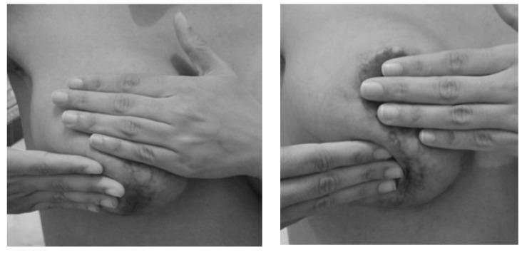
Two grayscale clinical photographs demonstrate manual therapy techniques for postoperative breast scar tissue management. In the left panel, a broad-handed compressive technique is shown, with the therapist's hand placed flat against the upper aspect of the breast and the second hand supporting the lower pole. The right panel displays a more localized manual mobilization focused on a visible, hyperpigmented, and uneven scar area near the areola. The fingertips are used to apply targeted pressure and stretching to the thickened tissue. These images illustrate physical therapy interventions following breast cancer surgery aimed at improving tissue elasticity, preventing adhesions, and normalizing the texture of hardened areas. The clinical focus is on early postoperative rehabilitation and functional recovery of the breast and chest wall.
Two grayscale clinical photographs demonstrate manual therapy techniques for postoperative breast scar tissue management. In the left panel, a broad-handed compressive technique is shown, with the therapist's hand placed flat against the upper aspect of the breast and the second hand supporting the lower pole. The right panel displays a more localized manual mobilization focused on a visible, hyperpigmented, and uneven scar area near the areola. The fingertips are used to apply targeted pressure and stretching to the thickened tissue. These images illustrate physical therapy interventions following breast cancer surgery aimed at improving tissue elasticity, preventing adhesions, and normalizing the texture of hardened areas. The clinical focus is on early postoperative rehabilitation and functional recovery of the breast and chest wall.

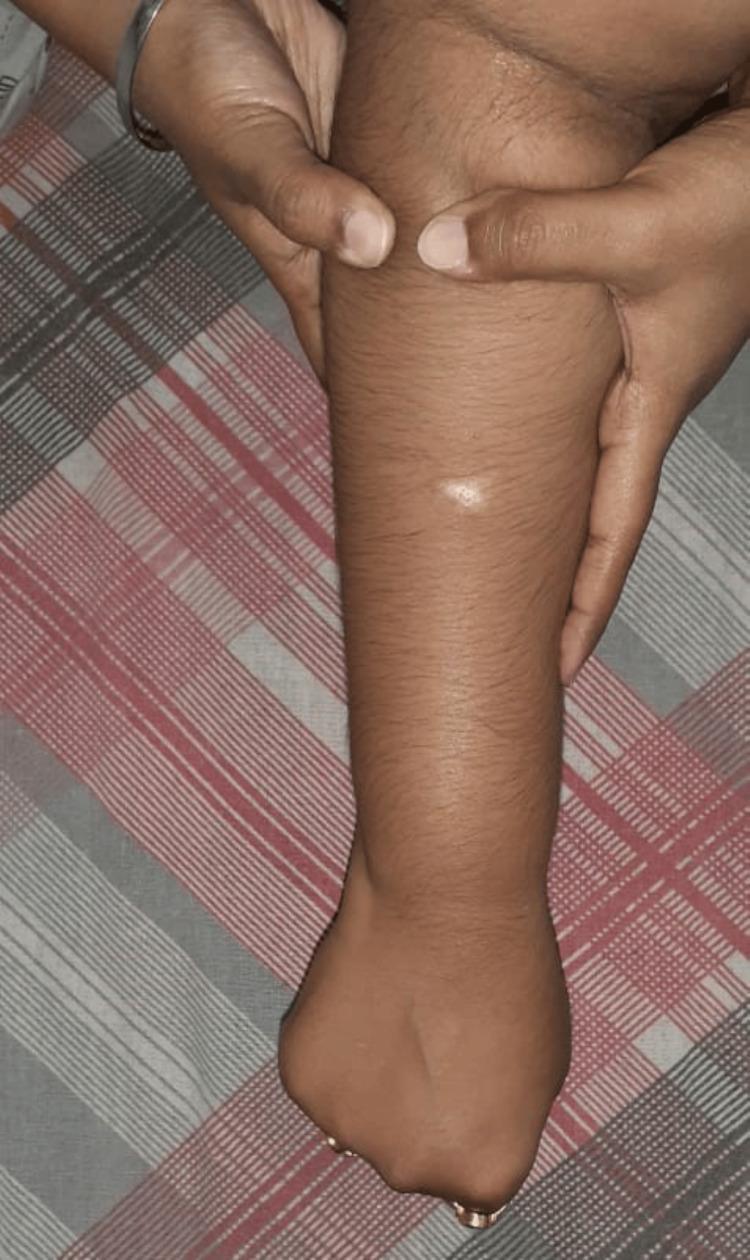
This clinical photograph demonstrates a soft tissue manual therapy intervention, specifically the Active Release Technique (ART), being applied to a patient's dorsal forearm. The image shows a practitioner's hands applying targeted manual pressure and longitudinal tension to the proximal forearm musculature. One hand is positioned near the elbow joint, while the other provides distal stabilization. A small, discrete, circular hypopigmented lesion or scar is visible on the mid-forearm's dorsal surface. The procedure is intended to address musculoskeletal dysfunction, such as adhesions or myofascial fibrosis resulting from repetitive strain or overload. The educational focus is on the clinical application of physiotherapy techniques for soft tissue mobilization and the visual presentation of skin landmarks during treatment.
This clinical photograph demonstrates a soft tissue manual therapy intervention, specifically the Active Release Technique (ART), being applied to a patient's dorsal forearm. The image shows a practitioner's hands applying targeted manual pressure and longitudinal tension to the proximal forearm musculature. One hand is positioned near the elbow joint, while the other provides distal stabilization. A small, discrete, circular hypopigmented lesion or scar is visible on the mid-forearm's dorsal surface. The procedure is intended to address musculoskeletal dysfunction, such as adhesions or myofascial fibrosis resulting from repetitive strain or overload. The educational focus is on the clinical application of physiotherapy techniques for soft tissue mobilization and the visual presentation of skin landmarks during treatment.

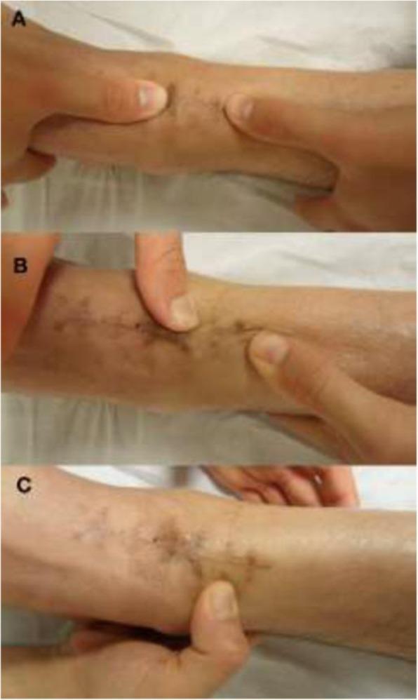
A clinical photograph series consisting of three panels (A, B, and C) demonstrating manual physical therapy techniques applied to the human forearm. The focus is on a chronic surgical scar or linear skin lesion characterized by hyperpigmented, punctate markings and slightly indurated edges. Panel A illustrates the 'push-pull' technique, where digits apply opposing traction to mobilize underlying connective tissue. Panel B shows the 'Indian burn' (torsional) maneuver, applying rotational force across the scar line to improve tissue elasticity. Panel C demonstrates a 'J stroke' technique, involving a localized, curved compression motion near the wrist. These images serve as educational material for Soft Tissue Mobilization (STM), highlighting manual intervention strategies to treat fascial restrictions and improve skin-to-subcutaneous mobility in postoperative or rehabilitative contexts. The skin surface appears treated with a topical emollient to facilitate gliding during manipulation.
A clinical photograph series consisting of three panels (A, B, and C) demonstrating manual physical therapy techniques applied to the human forearm. The focus is on a chronic surgical scar or linear skin lesion characterized by hyperpigmented, punctate markings and slightly indurated edges. Panel A illustrates the 'push-pull' technique, where digits apply opposing traction to mobilize underlying connective tissue. Panel B shows the 'Indian burn' (torsional) maneuver, applying rotational force across the scar line to improve tissue elasticity. Panel C demonstrates a 'J stroke' technique, involving a localized, curved compression motion near the wrist. These images serve as educational material for Soft Tissue Mobilization (STM), highlighting manual intervention strategies to treat fascial restrictions and improve skin-to-subcutaneous mobility in postoperative or rehabilitative contexts. The skin surface appears treated with a topical emollient to facilitate gliding during manipulation.

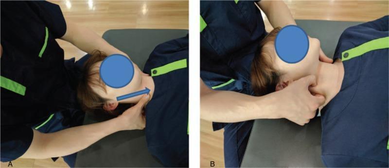
This procedural clinical photograph demonstrates two manual therapy techniques for Soft Tissue Release (SSTR) of the sternocleidomastoid (SCM) muscle. The subject is positioned supine on a treatment table with the head rotated away from the side being treated. Panel A illustrates the 'stripping technique,' where the therapist stabilizes the head with the left hand while using the thumb or finger of the right hand to apply deep longitudinal pressure along the SCM muscle belly. An overlaid blue arrow indicates a cranial-to-caudal (superior-to-inferior) stroke direction, moving from the mastoid process toward the sternum. Panel B demonstrates the 'pincer compression technique,' showing the therapist utilizing a pincer grasp with the thumb and index finger to compress and manipulate the soft tissue of the SCM muscle to address myofascial trigger points or taut bands. These physical therapy interventions are typically used to treat neck pain, reduce muscle hardness, and improve range of motion. The images emphasize proper hand placement and ergonomic positioning for manual soft tissue mobilization in clinical rehabilitation.

This procedural clinical photograph demonstrates two manual therapy techniques for Soft Tissue Release (SSTR) of the sternocleidomastoid (SCM) muscle. The subject is positioned supine on a treatment table with the head rotated away from the side being treated. Panel A illustrates the 'stripping technique,' where the therapist stabilizes the head with the left hand while using the thumb or finger of the right hand to apply deep longitudinal pressure along the SCM muscle belly. An overlaid blue arrow indicates a cranial-to-caudal (superior-to-inferior) stroke direction, moving from the mastoid process toward the sternum. Panel B demonstrates the 'pincer compression technique,' showing the therapist utilizing a pincer grasp with the thumb and index finger to compress and manipulate the soft tissue of the SCM muscle to address myofascial trigger points or taut bands. These physical therapy interventions are typically used to treat neck pain, reduce muscle hardness, and improve range of motion. The images emphasize proper hand placement and ergonomic positioning for manual soft tissue mobilization in clinical rehabilitation.

Finding Sources
Finding Sources
Reading Article

PMID: 41316557

Searching PubMed

scar mobilization physical therapy fibrosis

Reading Article

PMID: 38197283

Reading Article

PMID: 39548761

Reading Article

PMID: 35426735

Finding Sources
Reading File
Reading File
Reading File
Loading Image
I now have everything needed for a comprehensive answer. Here is the full response:

Scar Tissue Mobilisation

1. Biology of Scar Tissue (Why Mobilisation Works)
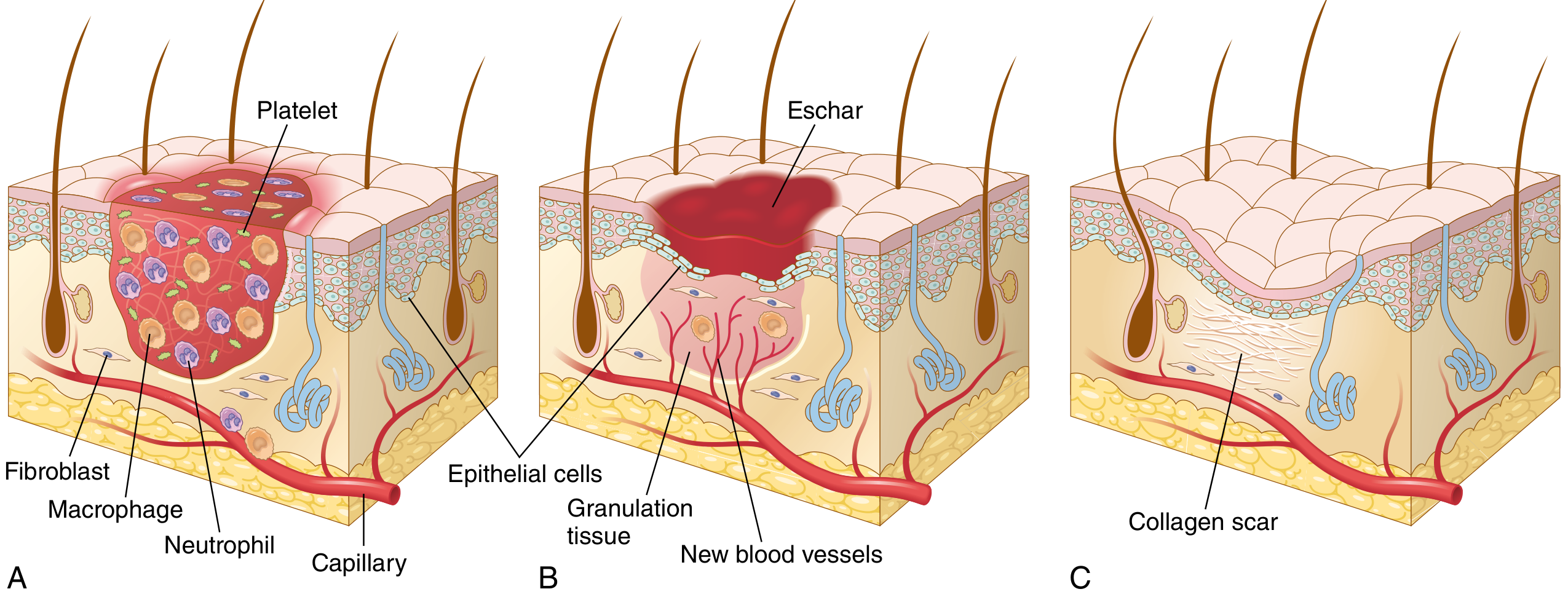
Understanding scar biology is the foundation for mobilisation rationale.
Steps in scar formation: (A) Inflammation with platelets, neutrophils, macrophages, and fibroblasts; (B) Granulation tissue with new blood vessels; (C) Final collagen scar
Wound healing progresses through three overlapping phases:
Phase 1 - Inflammation: Platelets, neutrophils, and M1 macrophages clear debris. M2 macrophages then dominate, secreting cytokines (TGF-β, PDGF) that recruit fibroblasts.
Phase 2 - Proliferation/Granulation: Fibroblasts proliferate, deposit collagen (initially type III), and new capillaries form granulation tissue.
Phase 3 - Remodelling: Granulation tissue is progressively replaced by dense collagen (type I). The matrix becomes denser, with thicker and stronger fibres over months to years. Myofibroblasts normally undergo apoptosis ~12 days after injury - in hypertrophic scars, this is delayed to 19-30 months, maintaining the hypercellular, contracted state. - Robbins, Cotran & Kumar Pathologic Basis of Disease
Scars are primarily made of disorganised type I collagen lacking the basket-weave architecture of normal dermis. They adhere to underlying structures, restricting glide between tissue layers (dermis, fascia, muscle). The goal of mobilisation is to restore this inter-tissue glide, reduce adhesion tension, and stimulate collagen reorganisation.
Key driver of hypertrophy: Persistent TGF-β signalling (and FAK-1 in burn scars) maintains myofibroblast activity and excess collagen deposition. Mechanical loading through mobilisation can influence mechanotransduction pathways and modulate this. - Mulholland & Greenfield's Surgery, 7e

2. Types of Scars Relevant to Mobilisation

Scar TypeCharacteristicsMobilisation Priority
Flat/maturePale, soft, pliableMaintenance
Adherent/tetheredBound to deep structures, restricts movementHigh
HypertrophicRaised, red, within wound marginsHigh (with care)
KeloidRaised, beyond wound margins, denseModerate (adjunct)
ContractureRestricts ROM, crosses jointsHighest

3. Manual Scar Mobilisation Techniques

3a. Basic Soft Tissue Mobilisation (STM)

The following techniques are applied directly to and around the scar, typically using a lubricant (moisturiser or oil):
Push-Pull / Transverse Friction: Digits apply opposing traction perpendicular to the scar line to mobilise underlying connective tissue from adjacent planes.
J-Stroke: A curved compression-and-glide motion applied near the scar edges, targeting fascial restrictions.
Indian Burn (Torsional technique): Rotational force applied across the scar to improve tissue elasticity along the longitudinal and transverse axes.
Circular Frictions: Small circles over and around the scar, loading the collagen fibres in multiple directions.
Three-panel series showing push-pull, Indian burn/torsional, and J-stroke techniques on a surgical forearm scar

3b. Postoperative Breast Scar Mobilisation

Two principal approaches used after breast cancer surgery:
  • Broad compressive technique: Flat hand placed against the scar area with the second hand providing counter-support - addresses large areas of thickened tissue.
  • Localised fingertip mobilisation: Targeted pressure and stretch at hyperpigmented, indurated scar zones near sensitive anatomical landmarks.
Manual therapy techniques for postoperative breast scar management - broad-hand compression (left) and localised fingertip mobilisation (right)

3c. Active Release Technique (ART)

Targeted deep pressure along the scar/muscle belly with simultaneous active or passive movement through range. Particularly used for scars associated with musculoskeletal dysfunction and myofascial fibrosis.
Active Release Technique applied to the dorsal forearm targeting adhesions and myofascial fibrosis

3d. Surgical Approaches to Scar Tension

When conservative mobilisation is insufficient, surgical options target the same principle - releasing tension:
  • Z-plasty: Lengthens scar at the expense of width; 60-degree limbs maximise tissue gain. Improves collagen remodelling after tension relief. - Mulholland & Greenfield's Surgery, 7e
  • V-Y advancement: Uses healthy adjacent tissue to advance into the contracture.
  • FTSG/STSG: Thin or full-thickness skin grafting after contracture release.

4. Timing Guidelines

PhaseTimingApproach
Acute inflammatory0-5 daysAvoid direct scar massage
Early proliferative1-3 weeksGentle peripheral mobilisation, oedema control
Late proliferative3-8 weeksBegin direct STM once wound is closed and stable
Remodelling2 months - 2 yearsProgressive loading, all STM techniques, pressure therapy
Forceful scar massage should be avoided when the scar is inflamed or breaks down early in the wound healing process. - Edger-Lacoursière et al., J Burn Care Res 2025 (PMID: 39548761)

5. Adjunct Conservative Interventions

Combined with manual mobilisation, these improve outcomes - particularly for hypertrophic/burn scars:
  • Pressure garments: >15 mmHg, >16 hours/day; started once oedema is stable and wound is adequately healed. Reduce hypertrophic scar formation by opposing collagen deposition forces.
  • Silicone gel/sheets: Extend beyond scar margins; reduce raised scar height and improve pliability. Work through occlusion and hydration.
  • Moisturisers: Applied multiple times daily; frequency more important than product type.
  • Serial casting: For contractures that interfere with function.
  • Passive stretching: Should be deprioritised relative to other modalities for pure scar management (not primary approach). - Edger-Lacoursière et al., 2025
  • Edema management: Retrograde massage, elevation, compression, and pumping exercises in the first 48-72 hours post-injury.

6. Evidence Base

Adherent post-surgical scars (extremities): A 2024 single-group study (n=19, 8 STM sessions) found a large effect on scar mobility (AS index: 0.12 → 0.41, Cohen r=0.6). Observer scar assessment (OSAS-I) also showed large effect. Clinically meaningful change exceeded MDC in 68% of subjects. - Poddighe et al., J Wound Care 2024 (PMID: 38197283)
Cesarean section scars: A 2022 clinical trial (n=32, 2 sessions) found that standardised STM improved elasticity, decreased stiffness, and improved pressure pain thresholds with small-to-moderate effects. Patients reported improved scar flexibility and surface area. - Gilbert et al., J Integr Complement Med 2022 (PMID: 35426735)
Post-abdominal surgery adhesions: A 2025 systematic review concluded that manual therapy holds potential as a non-invasive, adjunctive treatment for post-surgical adhesion symptoms, GI dysfunction, and musculoskeletal dysfunction related to adhesions. - Aziz et al., J Bodyw Mov Ther 2025 (PMID: 41316557)
Burn scars: A 2025 review recommends scar massage as part of multimodal conservative management, emphasising mechanotransduction and neurogenic inflammation as key mechanistic frameworks. Compression therapy remains the cornerstone for hypertrophic prevention. - Edger-Lacoursière et al., J Burn Care Res 2025 (PMID: 39548761)

7. Precautions and Contraindications

  • Do not mobilise over open wounds, infected tissue, or acutely inflamed/fragile scars.
  • Use reduced pressure over areas with reduced sensation (e.g. grafted/full-thickness burn sites).
  • Keloids may flare with aggressive deep friction - adjunct intralesional treatments (corticosteroid, botulinum toxin) should accompany mobilisation.
  • Pregnancy: take care with abdominal and pelvic scar work.
  • Post-oncological surgery: lymphoedema risk in axillary/groin scars - coordinate with lymphoedema team.

Summary: Scar tissue mobilisation works by mechanically loading disorganised collagen to restore inter-tissue glide, reduce adhesion tension, and stimulate remodelling. Evidence supports manual STM techniques (push-pull, J-stroke, transverse friction, ART) as effective - particularly for adherent post-surgical scars - with best results from early initiation (once wound is closed), multiple sessions, and combination with pressure therapy and silicone. Timing, scar maturity, and underlying anatomy should guide technique selection and intensity.
This is a shared conversation. Sign in to Orris to start your own chat.