
Strangulated hernia in ct
strangulated hernia CT scan findings
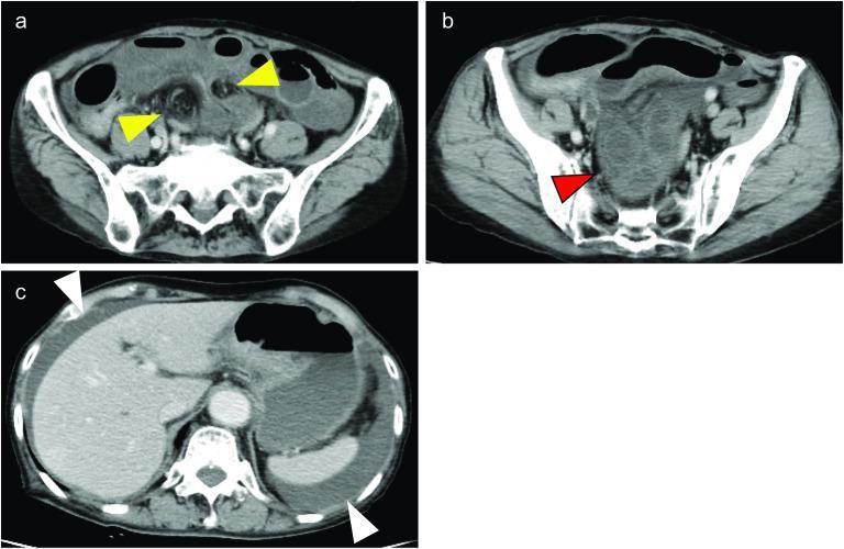
This composite diagnostic image features three axial slices (a, b, c) from a contrast-enhanced CT scan of the abdomen and pelvis, demonstrating a strangulated bowel obstruction. Panel 'a' shows the pelvic region where yellow arrows identify the hernia portal and the formation of a closed loop of the small bowel. In panel 'b', a red arrow indicates a dilated segment of the small bowel characterized by poor contrast enhancement, a visual finding highly suggestive of intestinal ischemia or necrosis due to compromised blood flow. Panel 'c' provides a superior view of the abdomen, with white arrows highlighting the presence of ascites, appearing as low-attenuation fluid collections around the liver and spleen. These findings are clinically relevant for diagnosing emergency surgical conditions such as ileo-ileal knots or internal hernias. The imaging is suitable for intermediate to advanced medical learners studying radiology, gastrointestinal emergencies, and surgical pathology.
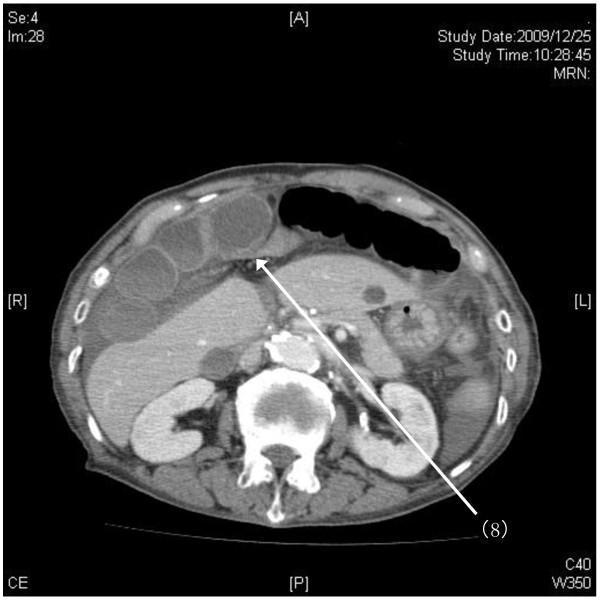
This diagnostic image is an axial (horizontal) slice from a contrast-enhanced helical CT scan of the abdomen, specifically targeting the upper abdominal region. The image demonstrates a rare case of an internal hernia. A white arrow labeled '(8)' points to a hilar defect in the falciform ligament, which is visible as a thin membranous structure in the midline. The scan reveals a strangulated distal intestinal loop herniating through this ligamentous defect. Key findings include significantly dilated, fluid-filled proximal small bowel loops positioned anteriorly and to the right, adjacent to the liver (segments S4 and S5). In contrast, distal intestinal segments appear collapsed. The liver, kidneys, and vertebral body are clearly visible, providing anatomical context. This image illustrates the pathophysiology of a closed-loop bowel obstruction caused by an internal hernia through the falciform ligament, a surgical emergency requiring prompt recognition of characteristic findings like the transition point at the ligamentous defect and associated proximal bowel dilation.
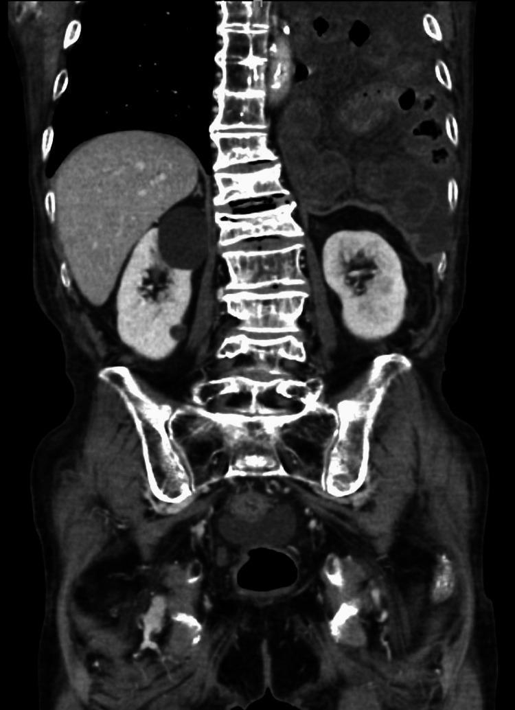
A coronal view of a contrast-enhanced computerized tomography (CT) scan of the thorax and abdomen. The diagnostic image reveals a significant diaphragmatic pathology where loops of the dilated small intestine have herniated through the esophageal hiatus into the left thoracic cavity. Key anatomical findings include a centrally positioned spine, liver in the right upper quadrant, and bilateral kidneys. Notably, the herniated segment of the small intestine demonstrates attenuated contrast enhancement of the intestinal wall, suggesting potential ischemia or vascular compromise. Associated findings include congestion of the mesentery within the thoracic segment, characteristic of strangulated intestinal obstruction. This imaging demonstrates the clinical presentation of a late-onset strangulated hiatal hernia, likely following prior gastric surgery, and highlights the radiological signs of bowel incarceration, such as wall thickening, dilatation, and poor perfusion.
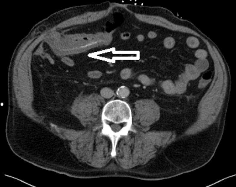
This diagnostic image is an axial computed tomography (CT) scan of the abdomen. The scan reveals a significant segment of circumferential colonic wall thickening involving the transverse colon, extending approximately 10 cm in length. A white arrow highlights a site on the abdominal wall representing a reduced Spigelian hernia, located at the lateral border of the rectus abdominis muscle. This specific segment of the transverse colon was previously herniated and now shows radiographic evidence of inflammation and edema. The surrounding mesenteric fat displays increased attenuation, suggestive of fat stranding. Other visible structures include the lumbar vertebra, abdominal aorta with calcification, and psoas muscles. In a clinical context, these findings are highly concerning for ischemic colitis following the reduction of an incarcerated or strangulated hernia. This image is used to teach medical students and residents about the complications of abdominal wall hernias and the CT appearance of secondary bowel ischemia.
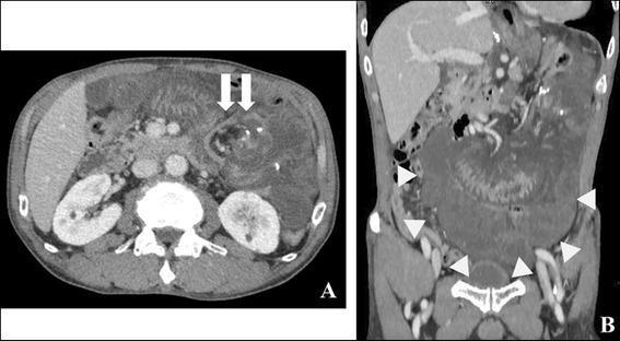
This diagnostic image consists of two panels showing a contrast-enhanced computerized tomography (CT) scan of the abdomen in axial (A) and coronal (B) planes. The scans demonstrate findings characteristic of an internal hernia and small bowel obstruction. In the axial view (A), white arrows highlight a 'whirl sign,' which is a swirling or spiral appearance of the mesenteric fat and vessels, indicating mesenteric torsion. The axial section also shows the relationship of the pathology to the liver and kidneys, with fluid (ascites) visible in the perihepatic and perisplenic spaces. In the coronal view (B), white arrowheads delineate a 'closed-loop formation' of the small intestine, characterized by a U-shaped or C-shaped cluster of dilated bowel loops. The bowel walls appear thickened, and there is evidence of surrounding free peritoneal fluid. These visual markers are critical diagnostic indicators for surgeons and radiologists to identify strangulated bowel or volvulus requiring urgent intervention.
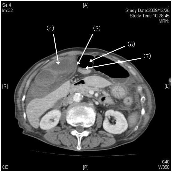
This diagnostic image is an axial (horizontal) helical computerized tomography (CT) scan of the upper abdomen, demonstrating signs of an internal hernia. The image reveals a closed loop of edematous intestine (labeled 4) positioned against the liver (segments S4 and S5). A key landmark, the falciform ligament (labeled 5), is visible as a linear membranous structure in the anterior-central abdomen. Pathological findings include significant air-filled dilation of the proximal intestines (labeled 6) on the left side of the abdomen, contrasted with the collapse of the distal intestine (labeled 7). These features are characteristic of a strangulated internal hernia through a defect in the falciform ligament, leading to mechanical bowel obstruction. The image highlights the distinctive visual differences between dilated, fluid-filled, and collapsed bowel loops in the context of emergent abdominal pathology. This clinical imaging is essential for students and residents studying surgical emergencies and radiologic interpretation of intestinal ischemia.
Strangulated Hernia on CT
Definition
CT Role
- Localization of the herniated segment and the transition point
- Characterization of completeness of obstruction
- Detection of ischemia and strangulation
- Identification of hernia type and etiology
-
90% sensitivity and >90% specificity for intestinal strangulation, with a negative predictive value of 99% - Mulholland & Greenfield's Surgery, p. 2418
Key CT Findings of Strangulation
Direct Signs of Ischemia/Strangulation
| Finding | Significance |
|---|---|
| Decreased/absent bowel wall enhancement (on IV contrast CT) | Most specific - indicates loss of bowel perfusion |
| High attenuation of bowel wall (on unenhanced CT) | Submucosal hemorrhage/ischemia |
| Bowel wall thickening >2-3 mm | Indicates impending ischemia |
| Pneumatosis intestinalis (intramural gas) | Severe ischemia/necrosis |
| Portal venous gas / mesenteric gas | Advanced ischemia, pre-gangrenous |
| Extraluminal free gas | Perforation has occurred |
Indirect/Associated Signs
| Finding | Significance |
|---|---|
| Fat stranding around herniated content | Inflammatory response to ischemia |
| Mesenteric edema and vascular engorgement | Venous congestion/obstruction |
| High-density (hemorrhagic) fluid in hernia sac | Vascular compromise |
| Free peritoneal fluid / ascites | Advanced disease |
| Closed-loop / C-shaped or U-shaped configuration | Both ends obstructed - high strangulation risk |
| Whirl sign (mesenteric twisting) | Volvulus/torsion component |
| Coffee bean sign | Closed-loop obstruction |
CT Image: Strangulated Bowel Obstruction (Closed Loop)
- (a) Yellow arrows mark the hernia portal and closed-loop small bowel formation
- (b) Red arrow shows a dilated bowel segment with poor contrast enhancement - the hallmark of intestinal ischemia
- (c) White arrows show ascites (low-attenuation free fluid) around the liver/spleen
Specific Hernia Types and CT Pearls
Inguinal Hernia (most common)
- CT shows herniated bowel in the inguinal canal - may have C-shaped configuration
- Look for bowel wall thickening, fat stranding, mesenteric engorgement, and fluid confined to the hernia sac
- Roberts & Hedges, Figure 44.7: "strangulated left inguinal hernia with C-shaped configuration... bowel wall thickening, severe fat stranding, mesenteric engorgement, extraluminal fluid confined to hernia sac"
Femoral Hernia
- Up to 40% of femoral hernias are strangulated at initial presentation, due to the narrow femoral ring
- Located below the inguinal ligament on CT
- Small ring = high strangulation risk even with small herniation
Spigelian Hernia
- Interparietal hernia at lateral rectus border
- CT shows dilated afferent loops, normal efferent loops - transition point in the hernia sac (Figure 44.6)
Obturator Hernia
- CT shows bowel herniating through the obturator foramen
- Classic: Howship-Romberg sign (pain radiating to medial thigh/knee) in >50% of strangulated cases
Internal Hernia (e.g., through falciform ligament, Peterson's space)
- CT: closed loop of edematous bowel against the liver, dilated proximal loops, collapsed distal loops
- Whirl sign at hernia portal
Hernia Angle (Prognostic CT Feature)
- Taller height + smaller (more acute) angle correlates with need for emergent repair - Fischer's Mastery of Surgery
Additional CT Images
Important Caveat
How to differentiate CT findings of strangulation from simple incarceration?
incarcerated hernia CT bowel enhancement viable vs strangulated ischemia
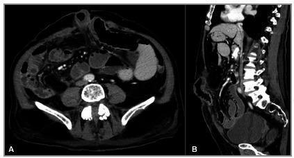
This diagnostic image consists of two panels (A: axial; B: sagittal) showing a contrast-enhanced computerized tomography (CT) scan of the abdomen and pelvis. The primary finding is a strangulated abdominal wall hernia. A small bowel loop is seen herniated through a midline abdominal wall defect. Key radiological features of strangulation and ischemia include significant parietal (wall) thickening of the incarcerated bowel loop, densification of the surrounding mesenteric fat (fat stranding), and the presence of localized fluid within the hernia sac. There is a perceptible reduction in contrast enhancement of the herniated segment compared to adjacent loops, indicative of vascular compromise. Proximal to the site of incarceration, multiple small bowel loops are dilated/distended, suggesting a secondary mechanical bowel obstruction. The images provide critical diagnostic evidence for acute surgical intervention in the context of complicated ventral hernias.

Coronal reconstruction of a contrast-enhanced abdominal computerized tomography (CT) scan. The image demonstrates a Spigelian hernia, characterized by a defect in the abdominal wall along the semilunar line. An interparietal hernia sac is visible, containing an incarcerated small bowel loop. Notably, the wall of the herniated intestinal loop shows a lack of contrast enhancement compared to surrounding loops, a diagnostic sign indicative of ischemia or necrosis. Proximal to the site of obstruction, multiple small bowel loops are markedly distended with fluid, while the distal colon appears collapsed or 'flat,' confirming a high-grade mechanical bowel obstruction. Major abdominal vascular structures are well-opacified by intravenous contrast. No significant free air or intraperitoneal effusion is visualized. This diagnostic image illustrates the radiological features of a strangulated Spigelian hernia with associated intestinal ischemia and proximal bowel obstruction.
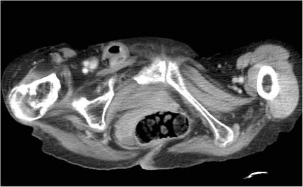
This diagnostic image is an axial contrast-enhanced Computed Tomography (CT) scan of the pelvic and upper femoral region. The primary finding is a right-sided strangulated femoral hernia, visualized as a herniated sac containing a loop of small intestine protruding through the femoral canal, medial to the femoral vessels. The incarcerated intestinal segment demonstrates wall enhancement, which is a critical finding suggesting preserved perfusion despite strangulation, though the loop appears distorted. Anatomical landmarks including the pelvic bones (pubis, ischium), femoral heads, and surrounding musculature provide orientation. The clinical significance of this imaging is to diagnose an incarcerated hernia and assess for secondary complications such as bowel ischemia or high-grade intestinal obstruction (ileus). This image is an essential educational resource for surgical residents and radiology students to identify the specific anatomical positioning of femoral versus inguinal hernias and to recognize signs of intestinal incarceration.
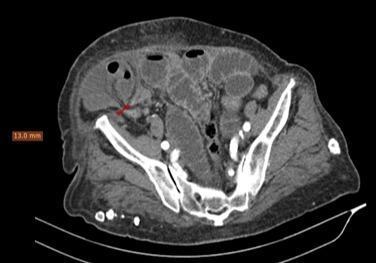
This axial contrast-enhanced CT scan of the pelvis demonstrates a Spigelian hernia on the right side. The image shows a hernia sac containing a loop of small bowel and omental fat protruding through a defect in the abdominal wall. A key finding is the 'double beak' sign at the hernial neck (indicated by a red arrow), where two transition zones of the incarcerated bowel loop converge at the narrow 13 mm fascial defect. The wall of the incarcerated bowel loop shows a lack of significant intravenous contrast enhancement compared to adjacent healthy bowel, a radiological sign highly suggestive of bowel wall ischemia and potential necrosis. Proximally, there is evidence of bowel obstruction with dilated small bowel loops, while the distal colon appears collapsed. This imaging is used in medical education to illustrate the diagnostic features of strangulated abdominal wall hernias and the importance of evaluating bowel wall enhancement in emergency surgical scenarios.
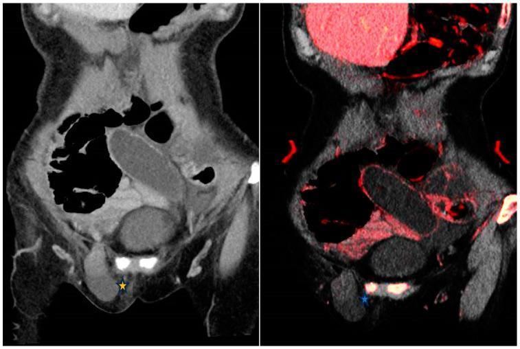
This diagnostic image displays side-by-side coronal CT views of the abdomen and pelvis, comparing a conventional 120 kVp contrast-enhanced image (left) with a dual-energy CT (DECT) iodine overlay map (IOM, right). The clinical focus is a closed-loop small bowel obstruction secondary to a right inguinal hernia, marked with an asterisk (*). On the conventional image, the incarcerated bowel loop within the inguinal region appears hypoattenuating but mural enhancement is difficult to definitively assess. The corresponding IOM image enhances clinical conspicuity by color-coding iodine distribution in red; the lack of red color in the herniated bowel wall confirms absent mural enhancement, a critical finding highly suggestive of bowel ischemia or strangulation. This comparison illustrates the utility of spectral CT in differentiating viable from non-viable bowel segments by objectively visualizing iodine uptake, which is more subtle on standard polychromatic CT. Key structures visible include the dilated proximal bowel loops and the constricted segment entering the inguinal canal.
Differentiating Strangulation vs. Simple Incarceration on CT
Core Conceptual Difference
| Incarceration | Strangulation | |
|---|---|---|
| Definition | Herniated content is irreducible but vascular supply is intact | Vascular supply is compromised - leads to ischemia and necrosis |
| Urgency | Urgent repair (risk of progressing) | Surgical emergency - immediate operation |
| Reducibility | Cannot be manually reduced | Almost always also incarcerated |
| Bowel viability | Viable | Potentially non-viable |
"A strangulated hernia is one in which the vascular supply to the herniated bowel is compromised, thus leading to ischemia. Strangulated hernias will most commonly also be incarcerated, but this is not a universal finding." - Roberts & Hedges' Clinical Procedures in Emergency Medicine
CT Differentiation: Feature by Feature
1. Bowel Wall Enhancement (Most Important Sign)
| Incarceration | Strangulation | |
|---|---|---|
| IV contrast enhancement | Normal or increased (hyperemic) | Decreased or absent - vascular compromise |
| Unenhanced CT wall attenuation | Normal (soft tissue density) | High attenuation - submucosal hemorrhage |
- Decreased/absent mural enhancement on contrast CT = loss of perfusion = strangulation
- Mulholland & Greenfield's Surgery: "Low or reduced attenuation of bowel wall on IV contrast CT scans" is a CT finding specifically suggestive of strangulation
2. Pneumatosis Intestinalis (Intramural Gas)
| Incarceration | Strangulation | |
|---|---|---|
| Intramural gas | Absent | Present (advanced ischemia/necrosis) |
3. Portal Venous / Mesenteric Gas
| Incarceration | Strangulation | |
|---|---|---|
| Portal/mesenteric gas | Absent | Present (very advanced, pre-gangrenous) |
4. Fluid in the Hernia Sac
| Incarceration | Strangulation | |
|---|---|---|
| Sac fluid character | Minimal / simple (low-density) | High-density (hemorrhagic) fluid - blood from ischemic bowel |
| Amount | Small | Often more prominent |
5. Fat Stranding and Mesenteric Changes
| Incarceration | Strangulation | |
|---|---|---|
| Fat stranding | Mild or absent | Severe fat stranding around herniated content |
| Mesenteric vessels | Normal | Vascular engorgement and mesenteric edema |
6. Bowel Wall Thickness
- Incarceration: mild edematous thickening from venous congestion
- Strangulation: moderate to severe thickening >3 mm, often with mural stratification loss
7. Closed-Loop Configuration
| Incarceration | Strangulation | |
|---|---|---|
| Loop configuration | May or may not form closed loop | C-shape / U-shape / coffee bean = closed loop, high risk |
| Whirl sign | Absent | May be present (torsion component) |
| Transition point tapering | Present | Present, often more abrupt |
8. Free Peritoneal Fluid / Ascites
| Incarceration | Strangulation | |
|---|---|---|
| Peritoneal fluid | Usually absent | Present - high-density ascites from ischemic exudate |
9. Extraluminal Gas (Perforation)
- Incarceration: absent
- Strangulation (late): extraluminal free gas = perforation has occurred
- Roberts & Hedges: "radiographic findings of extraluminal air, or poor perfusion of herniated bowel on CT with IV contrast" are hallmarks of strangulation
Summary Comparison Table
| CT Feature | Incarceration | Strangulation |
|---|---|---|
| Bowel wall enhancement (IV contrast) | Normal / hyperemic | Reduced or absent |
| Bowel wall on unenhanced CT | Normal density | High attenuation (hemorrhage) |
| Bowel wall thickness | Mild | >3 mm, moderate-severe |
| Pneumatosis intestinalis | Absent | Present (advanced) |
| Portal / mesenteric gas | Absent | Present (grave sign) |
| Sac fluid | Minimal, simple | Hemorrhagic, high-density |
| Fat stranding | Mild or absent | Severe |
| Mesenteric engorgement | Absent/mild | Present |
| Free peritoneal fluid | Absent | Present |
| Free extraluminal gas | Absent | Present (perforation) |
| Closed-loop configuration | Variable | Common (C/U shape) |
| Whirl sign | Absent | May be present |
Key CT Images