
MRD in Multiple myeloma
"minimal residual disease" AND "multiple myeloma"
MRD minimal residual disease multiple myeloma 2025 2026 guidelines clinical significance
PMID: 38768337
https://chi.scholasticahq.com/article/160933-guidelines-and-…
minimal residual disease multiple myeloma flow cytometry next generation sequencing
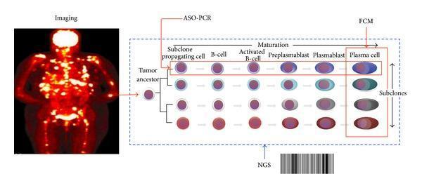
This composite educational graphic illustrates the complexity of Minimal Residual Disease (MRD) assessment in Multiple Myeloma (MM). The left panel features a full-body PET-CT maximum intensity projection (MIP) showing multifocal, hypermetabolic skeletal and extramedullary lesions (patchy involvement), emphasizing the utility of imaging for detecting non-uniform disease distribution. The right panel displays a pathophysiology diagram of clonal and cellular heterogeneity. It depicts a 'Tumor Ancestor' cell diverging into four distinct subclones. Each subclone undergoes a horizontal maturation process: subclone propagating cell, B-cell, activated B-cell, preplasmablast, plasmablast, and mature plasma cell. The diagram overlays three diagnostic detection windows: flow cytometry (FCM), which specifically targets the mature plasma cell stage; ASO-PCR, which captures the entire clonal lineage from progenitor to mature cell; and Next-Generation Sequencing (NGS), represented by a barcode, which covers the global tumor population. The figure highlights how cellular maturation and genetic subclonal variation impact the sensitivity and reach of different MRD monitoring techniques.
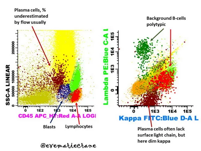
Multiparameter flow cytometry immunophenotyping of bone marrow plasma cells demonstrates clonal kappa-restricted population consistent with plasma cell myeloma. Gating on CD45, CD38, and CD138 identifies plasma cells, which reveal dim surface kappa light chain expression while cytoplasm shows bright kappa positivity; normal/reactive plasma cells display polyclonal cytoplasmic immunoglobulins and lack overt surface light chain restriction. The left panel illustrates plasma cells with a typical myeloma phenotype (CD45 dim, CD38 high, CD138 positive) against a background of polyclonal B cells and blasts; the right panel highlights the clonal kappa-restricted cluster, with dim surface κ but strong cytoplasmic κ staining, contrasting with lambda-restricted or kappa-negative populations. These features support clonality and enable minimal residual disease assessment after therapy. The assay complements morphology and serum paraprotein studies by providing quantitative, single‑cell sensitivity to detect residual malignant plasma cells below conventional thresholds. Diagnostic significance lies in confirming clonal plasma cell dyscrasia and guiding therapeutic decisions, prognosis, and MRD-directed treatment intensity. Normal variants include polyclonal plasma cells and non-neoplastic B cells, which serve as internal controls. Differential considerations include reactive plasmacytosis with polyclonal light chains and small clonal expansions. Clinically, flow cytometry MRD monitoring informs response assessment, risk stratification, and potential relapse prediction in multiple myeloma.
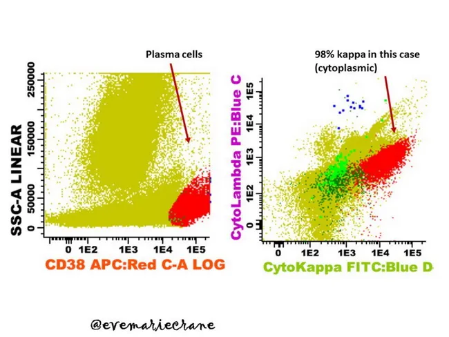
Flow cytometry immunophenotyping of a hematologic specimen reveals clonal plasma cell proliferation consistent with a plasma cell neoplasm. In the left panel, CD38 APC:Red-A log versus SSC-A linear delineates a large, bright CD38-positive plasma cell gate, marked by a dense cluster at higher CD38 intensity and substantial forward and side scatter typical of plasma cells. The right panel demonstrates intracellular light-chain analysis with CytoKappa FITC:Blue D versus Ig light chain axis, showing a dominant κ-restricted subset comprising the vast majority of κ-positive events (approximately 98%), with minimal λ-expressing cells. The data indicate strong cytoplasmic κ light-chain expression while surface immunoglobulin is absent or markedly reduced on these neoplastic cells, supporting clonality. CD138 is strongly expressed, consistent with plasmacytic differentiation. The observed immunophenotype, including CD38 bright and CD138 positivity with κ light-chain restriction, favors a malignant plasma cell process over reactive plasmacytosis. Additional markers commonly described in myeloma panels (e.g., CD19 negative/dim, CD56 variable) may be inferred but are not shown. Clinically, this pattern is critical for classification, diagnostic confirmation of plasma cell dyscrasia, and monitoring via minimal residual disease assessment, guiding staging and therapeutic decisions. This annotation is suitable for educational databases and clinical decision-support tools in hematology and research.
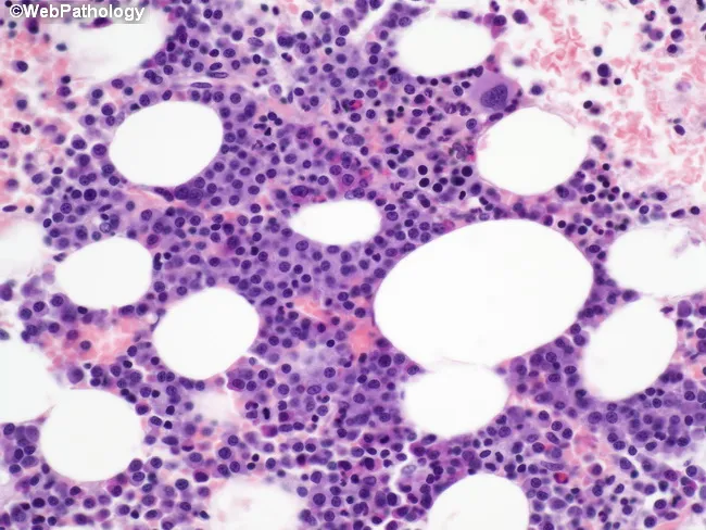
Imaging modality and technique: light microscopy of an H&E stained bone marrow trephine biopsy, demonstrating focal clusters of immature plasma cells within otherwise normal hematopoietic marrow. Primary subject: multiple myeloma plasma cell neoplasm with focal involvement in the marrow rather than uniform infiltration. Specimen: bone marrow biopsy core from iliac crest; perspective: microscopic cellular detail. Visual features: islands of small, tightly packed cells with oval to irregular nuclei, scant cytoplasm, condensed chromatin, and occasional prominent nucleoli; cytoplasm scant but adequate for recognition of plasma cell lineage. Surrounding marrow shows preserved hematopoiesis with trilineage elements; no overt osteoid or fibrotic bands described. Pathology: tumor cells exhibit high nuclear-to-cytoplasmic ratio and dysplastic, immature morphology; dispersed chromatin; some cells with conspicuous nucleoli; distribution irregular, with patches of tumor cells separated by intervening normal marrow. Diagnostic significance: histologic pattern supports early/patchy marrow involvement by plasma cell neoplasm, consistent with multiple myeloma; sampling error is possible in focal disease, emphasizing the need for regional marrow sampling and integration with ancillary studies (flow cytometry, immunoglobulin studies, cytogenetics). Clinical relevance: correlates with systemic disease burden, guides staging, prognosis, and treatment planning; spatial heterogeneity underscores importance of comprehensive marrow evaluation for monitoring response and minimal residual disease assessment.
"MRD negativity" AND "multiple myeloma" AND treatment
Minimal Residual Disease (MRD) in Multiple Myeloma
What is MRD?
Why MRD Matters: Prognostic Significance
- Goldman-Cecil Medicine notes: "Although achieving a minimal residual disease-negative state is associated with better survival, therapy usually is not changed based on the presence of minimal residual disease." (Goldman-Cecil Medicine, p. 1980) - though this caveat is being actively revisited by 2025-2026 trials.
- The landmark EVIDENCE meta-analysis (Landgren et al., Blood 2024 - PMID 38768337) included 8 newly diagnosed MM (NDMM) trials (4,907 patients) and demonstrated:
- Trial-level: MRD-negativity at 12 months correlated strongly with PFS (R² = 0.84)
- Individual-level: MRD-negativity at 12 months was associated with a global odds ratio for improved PFS of 4.02 (95% CI 2.57-5.46) in NDMM
- In relapsed/refractory MM (RRMM): individual-level OR = 7.67 (4.24-11.10)
- Conclusion: MRD-negativity is an early endpoint "reasonably likely to predict clinical benefit" and may support accelerated drug approval
- Longer progression-free survival (PFS)
- Longer overall survival (OS)
- Benefit regardless of cytogenetic risk group
- Benefit both on- and off-treatment (sustained MRD negativity > 12 months = "sustained MRD negativity")
Methods of MRD Assessment
1. Multiparameter Flow Cytometry (MFC / NGF)
- Next-Generation Flow (NGF): The EuroFlow consortium's 8-color, 2-tube panel; sensitivity up to 10^-5 to 10^-6
- Identifies aberrant plasma cell phenotype (e.g., CD38+, CD138+, CD19-, CD56+/-, CD45-/dim, CD117+/-)
- Rapid turnaround, widely available
- Limitation: requires fresh bone marrow; susceptible to sampling error with focal disease; does not detect extramedullary disease
2. Next-Generation Sequencing (NGS) - clonoSEQ / LymphoSIGHT
- Sequences the immunoglobulin heavy chain variable (IgVH) rearrangement as a clonal marker
- Sensitivity: 10^-5 to 10^-6 (FDA-cleared clonoSEQ assay)
- Advantages: objective, highly quantitative, archived samples usable
- Limitation: requires a baseline bone marrow sample for clonotype identification; ~10% of patients lack a trackable sequence
3. ASO-PCR (Allele-Specific Oligonucleotide PCR)
- Patient-specific primers designed for the clonal Ig rearrangement
- Sensitivity: up to 10^-5 to 10^-6
- Labor-intensive, used mainly in research settings
4. Imaging-based MRD
- PET-CT (FDG-PET): Detects extramedullary or focal lesions not sampled by bone marrow biopsy; "MRD-negative by bone marrow + PET-negative" = deepest sustained remission
- Whole-body MRI (WB-MRI): Alternative imaging for focal lesions, avoids radiation
- IMWG criteria require imaging to be negative for complete MRD-negative state to be "imaging-validated"
5. Emerging: Liquid Biopsy
- Cell-free DNA (cfDNA) / circulating tumor DNA (ctDNA) from peripheral blood
- Mass spectrometry (MASS-FIX) for detecting monoclonal immunoglobulin at high sensitivity
- Avoids bone marrow biopsy; can monitor disease continuously
- Not yet standardized for routine MRD assessment
IMWG Response Criteria and MRD Categories
| Category | Definition |
|---|---|
| CR | No M-protein by SPEP/UPEP; normal FLC ratio; <5% plasma cells in BM |
| Stringent CR (sCR) | CR + normal FLC ratio + absence of clonal PCs by IHC or 2-color flow |
| MRD-negative (10^-5) | sCR + no clonal PCs by NGF or NGS at ≥10^-5 sensitivity |
| MRD-negative (10^-6) | As above, at ≥10^-6 sensitivity |
| Imaging-validated MRD-negative | MRD-negative by NGF/NGS + negative PET-CT |
| Sustained MRD-negative | MRD-negative maintained for ≥12 months |
MRD in Clinical Practice: When to Test
- After induction/post-transplant: Test at end of each treatment phase in all patients achieving CR
- During maintenance: MRD-positive patients: every 6 months; MRD-negative patients: every 12 months
- On clinical suspicion of relapse: Test at any point
- Combine bone marrow MRD with imaging (PET-CT or WB-MRI) for a comprehensive assessment, especially in high-risk patients
MRD and Key Clinical Trials (2024-2025)
| Trial | Regimen | Key MRD Finding |
|---|---|---|
| CASSIOPEIA (Moreau et al., Lancet Oncol 2024 - PMID 38889735) | Dara-VTd vs VTd (transplant-eligible NDMM) | Dara-VTd: 64% MRD-neg vs 44% with VTd; sustained MRD-neg correlated with OS benefit |
| MAIA (Facon et al., Leukemia 2025 - PMID 40016302) | Dara-Rd vs Rd (transplant-ineligible NDMM) | MRD-neg rate 26% with Dara-Rd vs 7% with Rd; MRD-neg patients had markedly improved long-term OS |
| CEPHEUS (Usmani et al., Nat Med 2025 - PMID 39910273) | Dara-VRd vs VRd (transplant-ineligible/deferred NDMM) | Superior MRD-neg rate with Dara-VRd; MRD-negativity key secondary endpoint |
| BENEFIT (Leleu et al., Nat Med 2024 - PMID 38830994) | Isa-VRd (transplant-ineligible NDMM) | High MRD-neg rates with quadruplet; CD38 antibody intensification drives deeper responses |
MRD-Adapted Treatment: The Frontier
- MRD-driven treatment cessation: If sustained MRD-negativity, could maintenance be safely stopped? (MASTER, GEM-CESAR, AURIGA trials exploring this)
- MRD-driven intensification: MRD-positive patients at end of induction may benefit from treatment escalation
- The Pan-Pacific 2026 consensus confirms: CD38-based maintenance is recommended regardless of MRD status, with potentially greater benefit in MRD-positive or high-risk patients
Regulatory Landscape: FDA 2026 Draft Guidance
- MRD = absence of detectable cancer cells in bone marrow in patients who have already achieved CR
- Measured by flow cytometry or NGS at ≥10^-5 sensitivity
- Sustained MRD-negativity (≥12 months) preferred endpoint
Limitations and Challenges
| Challenge | Detail |
|---|---|
| Sampling heterogeneity | Focal/patchy marrow involvement means single biopsy may miss disease (need imaging as complement) |
| Assay standardization | NGF vs clonoSEQ - results not always interchangeable; sensitivity thresholds vary |
| Baseline sample needed for NGS | ~10% of patients lack a trackable Ig sequence |
| Extramedullary disease | Bone marrow MRD-negative but PET-positive = not true MRD-negative |
| No universal clinical action threshold | MRD positivity alone currently does not trigger treatment change outside trials |
| Cost and access | NGS-based MRD (clonoSEQ) is expensive; NGF requires specialized labs |
Summary
- MRD-negativity is the deepest and most prognostically meaningful response measure in MM, superior to CR or sCR alone
- Two main validated methods: NGF (EuroFlow) and NGS (clonoSEQ) - both FDA-cleared at 10^-5 sensitivity
- Clinical trials consistently show MRD-negativity confers superior PFS and OS regardless of treatment era or line
- The EVIDENCE meta-analysis formally validated MRD-negativity as a surrogate endpoint for PFS (R² = 0.84)
- FDA Jan 2026 draft guidance formalizes MRD + CR as endpoints for accelerated approval, accelerating drug development
- MRD-adapted therapy (stopping, reducing, or intensifying based on MRD) remains investigational but is the clear direction of the field
- Imaging (PET-CT) must complement bone marrow MRD to exclude extramedullary disease
- Goldman-Cecil Medicine International Edition, p. 1980
- EVIDENCE meta-analysis: Landgren et al., Blood 2024
- Pan-Pacific MM Working Group MRD Consensus, 2026