
pelvimetry and pelvic assesment in obstetrics- iam a pg resident - act like professor -explain to me as a professor in ms obs & gyn
Reading File
Reading File
Reading File
Searching the Web
WHO intrapartum care routine clinical pelvimetry not recommended guideline
Searching PubMed
obstetric pelvimetry
Finding Sources
Finding Sources
Finding Sources
Searching Images
obstetric pelvic inlet diameters landmarks diagram obstetric conjugate diagonal conjugate
Finding Sources
Finding Sources
Finding Sources
Reading File
Reading File
Finding Sources
Finding Sources
Reading Web Page
https://iris.who.int/server/api/core/bitstreams/7e9a5c8e-4bc…
Pelvimetry and pelvic assessment in obstetrics
Professor's opening point: do not confuse pelvimetry with pelvic assessment. Pelvimetry is measurement of bony pelvic dimensions. Pelvic assessment is the broader obstetric judgement of whether the passage, passenger, and powers are compatible in this particular labour.
A pelvis does not deliver a baby in isolation. A woman with “borderline” measurements may deliver a 3.0 kg well-flexed occipito-anterior fetus normally, while a woman with apparently normal measurements may develop dystocia with deflexion, persistent occipito-posterior position, macrosomia, or poor uterine action.
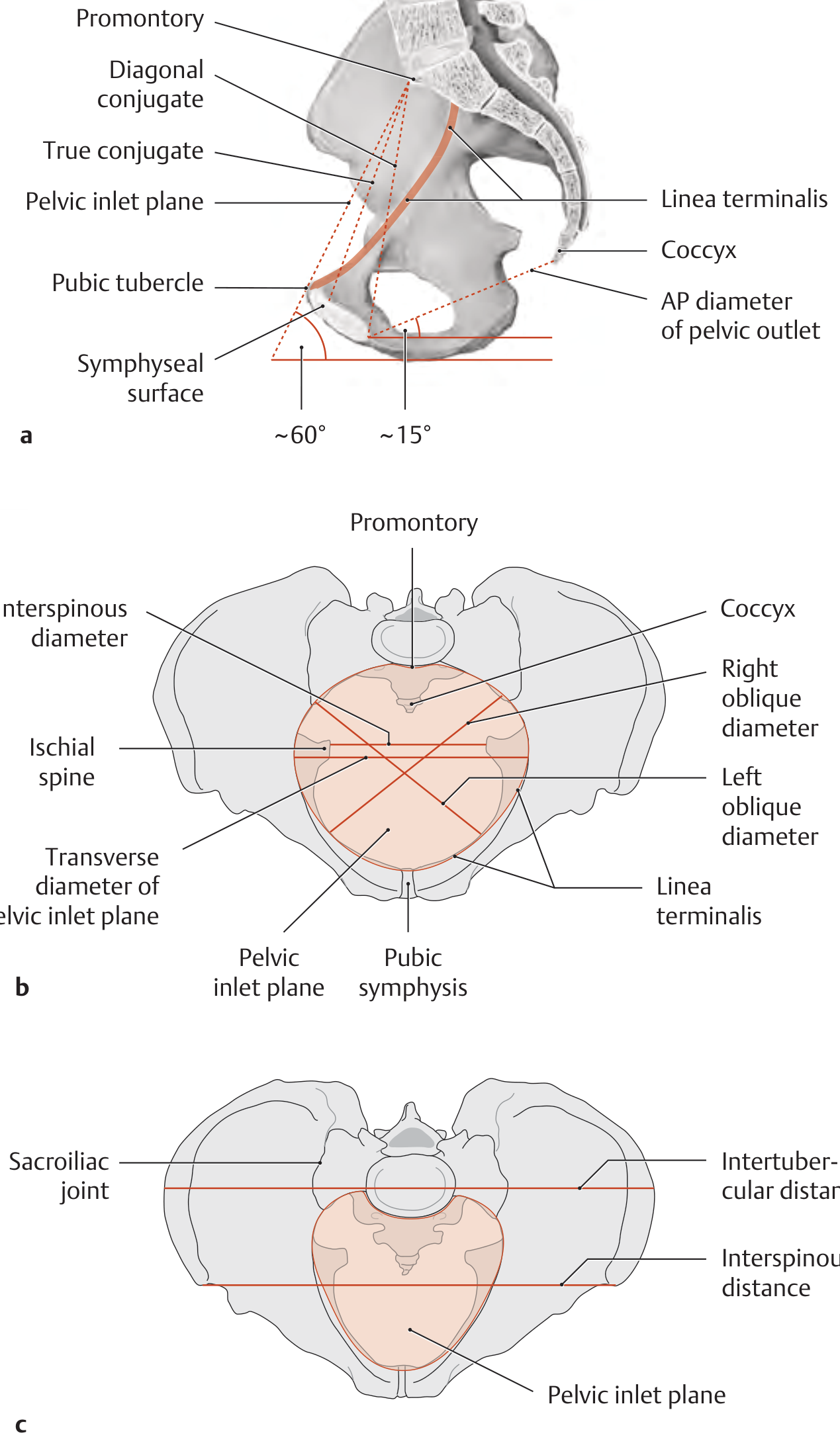
The bony pelvis is a curved canal, not a rigid straight tube. The inlet, midpelvis, and outlet must each be considered. - General Anatomy and Musculoskeletal System (THIEME Atlas), p. 161
1. Definitions
Pelvimetry
Measurement of the dimensions and shape of the maternal bony pelvis, by:
- Clinical pelvimetry: digital vaginal examination.
- Radiological pelvimetry: X-ray, CT, or MRI.
- Ultrasound-based assessment: limited role for bony dimensions, but very useful for fetal head position, attitude, and descent.
Pelvic assessment
A clinical assessment that includes:
- Pelvic bony architecture and capacity
- Soft-tissue obstruction
- Fetal size, lie, presentation, position, attitude, and station
- Progress of labour
- Uterine contractions
- Evidence of fetopelvic disproportion or obstruction
The meaningful diagnosis is therefore usually cephalopelvic disproportion (CPD) or, more accurately, fetopelvic disproportion, and it is often a diagnosis made dynamically in labour, not from an isolated antenatal measurement.
2. Functional obstetric anatomy of the pelvis
Think of the pelvis in three levels.
| Level | Important landmarks | Obstetric importance |
|---|---|---|
| Pelvic inlet | Sacral promontory, linea terminalis, upper border of symphysis pubis | Engagement of the fetal head |
| Midpelvis / pelvic cavity | Ischial spines, sacrum, side walls | Usually the narrowest and most clinically important plane |
| Pelvic outlet | Lower border of symphysis, ischial tuberosities, sacrococcygeal region, pubic arch | Internal rotation, extension, and delivery of the head |
A. Pelvic inlet
Important diameters
| Diameter | Definition | Approximate value | Clinical relevance |
|---|---|---|---|
| Obstetric conjugate | Sacral promontory to nearest point on posterior surface of symphysis pubis | About 10 to 10.5 cm | Narrowest AP diameter of inlet, cannot be directly measured vaginally |
| Anatomical or true conjugate | Promontory to upper border of symphysis | About 11 cm | Anatomical measurement |
| Diagonal conjugate | Promontory to lower border of symphysis | About 12.5 to 13 cm | The clinically measurable conjugate |
| Transverse diameter | Widest distance across pelvic brim | About 13 cm | Important in inlet capacity |
| Oblique diameter | Sacroiliac joint to contralateral iliopectineal eminence | About 12 cm | Relevant to engagement in oblique diameter |
The diagonal conjugate is approximately 12.5 to 13 cm; the true conjugate is about 11 cm. - General Anatomy and Musculoskeletal System (THIEME Atlas), p. 161
Estimating the obstetric conjugate
[
\textbf{Obstetric conjugate} \approx \textbf{Diagonal conjugate} - 1.5\text{ to }2 \text{ cm}
]
Example:
- Diagonal conjugate = 12.5 cm
- Estimated obstetric conjugate = 10.5 to 11 cm
Clinical teaching: if you can easily reach the promontory, the AP diameter may be reduced. But do not make a diagnosis of contracted inlet only from that finding. Finger length, technique, and maternal position matter.
B. Midpelvis
This is the level residents must not neglect.
The interspinous diameter
- Distance between the tips of the ischial spines
- Approximately 10 to 10.5 cm, with some texts citing about 11 cm
- It is commonly the narrowest transverse diameter of the pelvis
The ischial spines define the plane of station 0.
During examination, assess:
- Are the spines blunt or prominent?
- Are they projecting medially?
- Is the sacrum well curved and hollow, or flat?
- Are the lateral pelvic walls parallel, convergent, or divergent?
Red-flag configuration:
- Prominent ischial spines
- Convergent side walls
- Flat sacrum
- Poorly mobile coccyx
This combination suggests a contracted midpelvis and raises concern for deep transverse arrest or persistent occipito-posterior arrest.
C. Pelvic outlet
Important dimensions
| Diameter | Approximate value | How to assess |
|---|---|---|
| AP outlet diameter | About 11 cm, increased by backward movement of coccyx | Mobility of coccyx, posterior sagittal space |
| Intertuberous or bituberous diameter | About 10 to 11 cm | Knuckle or fist test, with caution |
| Subpubic angle | Usually >85-90° in a typical gynecoid pelvis | Width of pubic arch |
An AP outlet measurement may be described as about 9 cm plus the mobility-related increase from posterior displacement of the coccyx. - General Anatomy and Musculoskeletal System (THIEME Atlas), p. 161
The pubic arch
- A broad arch is favorable for delivery of the fetal head.
- A narrow, acute arch may be associated with anterior asynclitism, delayed descent, or difficulty in outlet delivery.
- Anatomically, the female subpubic angle is generally about 80 to 85°, wider than in males. - Gray's Anatomy for Students, pelvic anatomy section
3. Pelvic types: useful description, not a delivery decision
The classic Caldwell-Moloy classification is historical and descriptive.
| Pelvic type | Features | Obstetric association |
|---|---|---|
| Gynecoid | Round or slightly transverse inlet, wide subpubic angle, non-prominent spines, curved sacrum | Traditionally most favorable |
| Android | Heart-shaped inlet, narrow anterior segment, prominent spines, narrow pubic arch | Persistent OP, deep transverse arrest |
| Anthropoid | Oval inlet with long AP diameter, relatively narrow transverse diameter | OP engagement and persistent OP may occur, but vaginal birth can be normal |
| Platypelloid | Flattened pelvis, wide transverse but short AP inlet | Difficulty with engagement, transverse lie of head at brim |
Do not write “android pelvis = cesarean.” This is incorrect. Pelvic type neither reliably predicts the outcome nor substitutes for intrapartum observation.
4. Clinical pelvimetry: examination technique
When to do it
Clinical pelvic examination may be appropriate when there is a reason to assess the pelvis, for example:
- Previous obstructed labour or prior cesarean for proven CPD
- Past pelvic fracture, pelvic surgery, or known skeletal deformity
- Marked short stature with other risk factors
- Suspicion of pelvic mass or lower genital tract obstruction
- Malpresentation, especially selected vaginal breech cases under a protocol
- Labour abnormality where the vaginal examination may clarify the level or nature of obstruction
It should be done gently, with consent, privacy, a chaperone where appropriate, and strict attention to infection prevention.
How to perform it
Step 1: Prepare
- Explain the purpose and obtain consent.
- Ask the patient to empty her bladder.
- Dorsal lithotomy or modified dorsal position, with adequate exposure and privacy.
- Use a gentle, systematic bimanual vaginal examination.
Step 2: Assess the inlet
- Reach for the sacral promontory with the middle finger.
- If reached, mark the point at the inferior margin of the symphysis with your examining hand.
- Measure the distance after withdrawal: this is the diagonal conjugate.
- Estimate obstetric conjugate by subtracting 1.5 to 2 cm.
Also assess:
- Linea terminalis: can it be followed easily?
- Sacral promontory: prominent or not?
- Is there an obstructing mass?
Step 3: Assess the midpelvis
Palpate:
- Ischial spines: blunt, average, or prominent
- Sacrum: broad concavity or flat
- Side walls: parallel, converging, or diverging
- Sacrosciatic notch: wide or narrow
Step 4: Assess the outlet
Assess:
- Intertuberous width
- Subpubic angle
- Mobility of coccyx
- Posterior sagittal space
- Any vaginal, cervical, vulval, rectal, or pelvic mass
A practical examination statement
Instead of writing only “pelvis adequate,” document the actual findings:
“Sacral promontory not reached. Ischial spines blunt and non-prominent; side walls parallel; sacrum well curved; subpubic arch wide; coccyx mobile. No clinically evident bony pelvic contraction or soft tissue obstruction.”
Or, if concerning:
“Sacral promontory easily reached, prominent ischial spines with convergent side walls, flat sacrum, and narrow subpubic angle. Findings suggest reduced midpelvic capacity. Correlate with fetal size, position, and labour progress.”
5. What does a “contracted pelvis” mean?
It is not one single entity.
| Type | Main feature | Likely labour problem |
|---|---|---|
| Inlet contraction | Reduced obstetric conjugate or transverse inlet | Delayed or absent engagement |
| Midpelvic contraction | Prominent spines, convergent walls, flat sacrum | Arrest of descent, deep transverse arrest |
| Outlet contraction | Narrow intertuberous diameter or narrow pubic arch | Difficulty at outlet and delivery of head |
| Generally contracted pelvis | Multiple dimensions reduced | Recurrent labour obstruction possible |
| Asymmetrical pelvis | One-sided distortion, previous fracture, scoliosis | Asynclitism, malrotation, obstruction |
Important caveat
“Contracted pelvis” based on a single measurement is a poor clinical conclusion. The relevant question is:
Is there clinically significant fetopelvic disproportion in this labour?
6. Antenatal pelvic assessment
History is highly informative
Ask specifically about:
- Previous spontaneous vaginal delivery, particularly birth weight and labour duration
- Previous operative vaginal delivery and indication
- Previous cesarean, especially whether it was for true arrest disorder after adequate labour
- Previous obstructed labour, symphysiotomy, destructive operation, or stillbirth related to labour
- Pelvic trauma or fracture
- Skeletal disease, rickets, spinal deformity, hip disease
- Symptoms suggesting pelvic tumour or genital tract obstruction
Professor's pearl
A previous uncomplicated vaginal delivery of an average or large baby is a much stronger reassuring datum than a “normal pelvimetry” report.
But it does not guarantee success in a new pregnancy with macrosomia, diabetes, malpresentation, or malposition.
7. Intrapartum pelvic assessment: where real obstetrics happens
Pelvic adequacy is assessed by progress of labour with a well-assessed fetus, not by a one-time pelvic examination.
At each vaginal examination, correlate:
- Cervical dilatation and effacement
- Membrane status and liquor
- Presenting part
- Position and attitude
- Station and descent
- Caput and moulding
- Pelvic landmarks
- Contraction frequency, duration, and strength
- Fetal well-being
Findings that suggest possible disproportion or obstruction
- High, unengaged head at term in a primigravida, particularly with other risk factors
- No descent despite adequate contractions and full dilatation
- Increasing caput and severe moulding
- Persistent transverse position or persistent OP with poor descent
- Head not applying to cervix, with cervical oedema
- Bandl ring, rising maternal pulse, dehydration, ketonuria, haematuria, or fetal compromise in advanced cases
- Failure of descent despite correction of ineffective uterine action and malposition
Never diagnose CPD before excluding:
- Inadequate uterine activity
- Malposition
- Deflexion
- Asynclitism
- Full bladder
- Inappropriate timing or inaccurate assessment of active labour
- Incorrect estimation of fetal position or station
8. Pelvimetry in modern practice
Routine clinical pelvimetry
Routine pelvimetry on admission in labour is not recommended for healthy women in spontaneous labour. The WHO intrapartum guideline specifically advises against routine clinical pelvimetry on labour admission in this group.
Why?
- Poor predictive value for vaginal birth
- Poor reproducibility between examiners
- It does not account for fetal size, head attitude, moulding, position, or uterine contractions
- It can lead to unnecessary intervention and cesarean birth
- A normal pelvis does not exclude labour dystocia, and an apparently small pelvis does not prove obstruction
Radiological pelvimetry
X-ray pelvimetry
- Historical method
- Rarely justified now because of radiation exposure and limited predictive value
CT pelvimetry
- More accurate bony measurement than plain radiography
- Still involves ionizing radiation
- Not a routine test in pregnancy
MRI pelvimetry
- No ionizing radiation
- Can provide detailed bony and soft-tissue anatomy
- May be selectively used in certain protocols, including selected vaginal breech planning, pelvic deformity, prior trauma, or suspected pelvic mass
- It should not replace senior clinical judgement or individualized counselling
Maternal-fetal medicine texts describe CT and MRI pelvimetry in selected breech contexts, but this is not an endorsement of routine pelvimetry for cephalic labour. - Creasy & Resnik’s Maternal-Fetal Medicine, Breech Vaginal Delivery section
9. Pelvic assessment in breech presentation
For a planned vaginal breech birth, many institutional protocols require or strongly consider assessment of:
- Clinically adequate pelvis
- Estimated fetal weight within protocol limits
- Frank or complete breech
- Flexed fetal head
- No hyperextension of fetal neck
- No major fetal anomaly
- Immediate availability of cesarean delivery and a skilled breech team
Here pelvimetry, particularly MRI pelvimetry in some centers, is a selective adjunct. It is never the sole criterion for allowing or refusing vaginal breech birth.
10. Viva questions and model answers
“Can you assess the obstetric conjugate directly?”
No. It cannot normally be directly measured clinically. We measure the diagonal conjugate and estimate the obstetric conjugate by subtracting approximately 1.5 to 2 cm.
“What is the narrowest plane of the pelvis?”
The midpelvis at the level of the ischial spines, represented by the interspinous diameter, is usually considered the narrowest transverse plane.
“What is the most useful pelvimetry?”
The most useful assessment is functional pelvimetry during labour: serial assessment of descent, rotation, cervical dilatation, caput, moulding, contractions, and fetal well-being.
“Does a prominent sacral promontory mean cesarean delivery?”
No. It suggests possible reduction in AP inlet diameter. The final decision depends on the overall pelvis, fetus, labour progress, and maternal-fetal condition.
“What is the importance of the ischial spines?”
They are the landmark for station 0 and indicate midpelvic capacity. Prominent spines may contribute to midpelvic contraction and arrest of rotation or descent.
“What is the best evidence of adequate pelvis?”
A prior uncomplicated vaginal birth is reassuring. In a nulliparous woman, the best practical test is normal progress of a properly monitored labour with an appropriately sized, well-positioned fetus.
11. Take-home messages for a PG resident
- Do not label a pelvis simply adequate or inadequate. Describe inlet, midpelvis, outlet, and soft tissues.
- The midpelvis matters most, especially ischial spines, sacral curve, and side walls.
- Clinical pelvimetry is subjective and should not determine mode of delivery by itself.
- Routine pelvimetry in healthy labour is not recommended.
- CPD is usually a dynamic intrapartum diagnosis.
- Evaluate the three Ps together: passenger, passage, and powers.
- In suspected dystocia, first exclude poor contractions, malposition, deflexion, asynclitism, and bladder distension.
- Document positive and negative findings clearly, not merely “pelvis adequate.”
This is a shared conversation. Sign in to Orris to start your own chat.