
Can you explain me the difference between, uh, pluripotent cells, multipotent cells, uh, totipotent cells, and unipotent cells?
These four terms describe the developmental potential of stem cells - how many different cell types they can give rise to. Here's a quick rundown before I pull from the medical library:
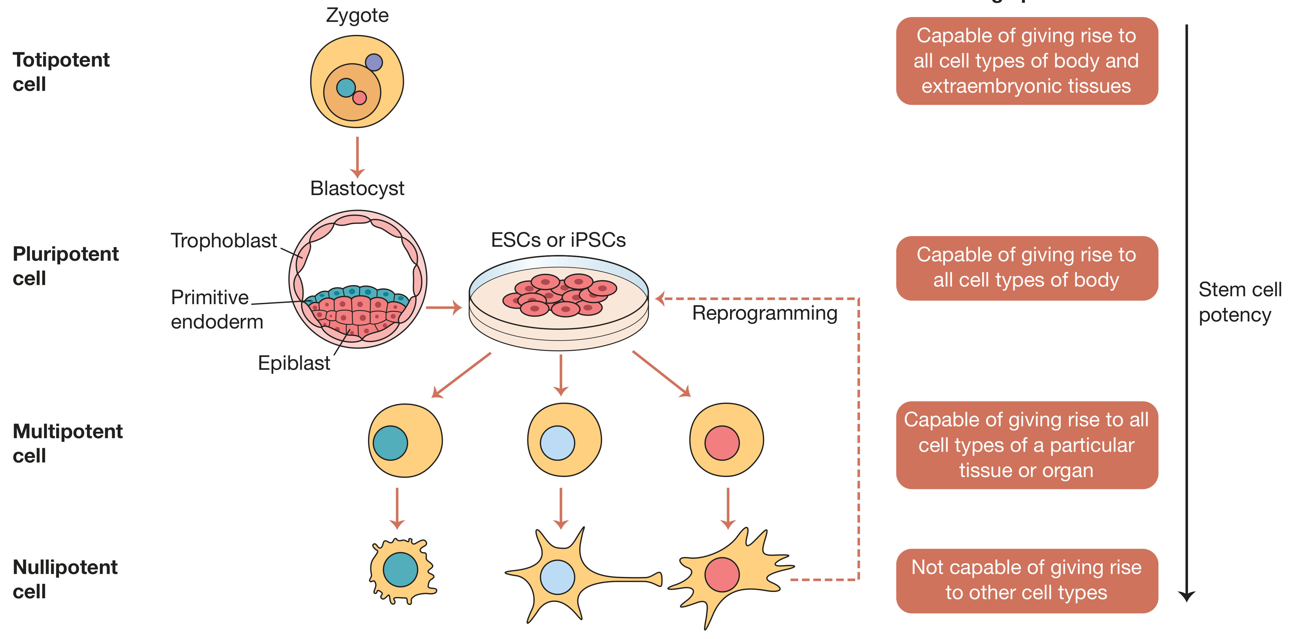
- Totipotent: can become any cell, including placenta/extraembryonic tissue
- Pluripotent: can become almost any cell except extraembryonic tissue
- Multipotent: limited to cell types within a specific tissue lineage
- Unipotent: can only produce one cell type
Searching sources...
Reading File
Finding Sources
Finding Sources
Reading File
Reading File
Reading File
Loading Image
Loading Image
Here is a full breakdown, drawn from multiple medical textbooks:
Stem Cell Potency: The Four Types
All stem cells share two defining properties: self-renewal (they can keep dividing to produce more of themselves) and potency (they can differentiate into specialized cell types). The four categories below describe how broad that potency is, from greatest to least.
1. Totipotent ("all powerful")
A totipotent cell can give rise to every single cell type in the organism - both the cells of the body itself (embryonic tissue) and the extraembryonic structures like the placenta and yolk sac.
- Example: The fertilized egg (zygote) and the first few cells produced after it divides (roughly up to the 4-8 cell morula stage).
- This is the broadest possible potential. No other cell type can match it.
- As Medical Physiology puts it: "the zygote is the ultimate stem cell because through its divisions, it gives rise to every cell lineage present in the complete organism."
2. Pluripotent ("many powerful")
A pluripotent cell can differentiate into cells from all three embryonic germ layers (ectoderm, mesoderm, endoderm) - meaning virtually any cell type in the human body - but it cannot form extraembryonic tissues like the placenta.
- Examples: Embryonic stem cells (ESCs) derived from the inner cell mass of the blastocyst; induced pluripotent stem cells (iPSCs), which are adult cells reprogrammed back to a pluripotent state.
- This is the type most used in regenerative medicine research.
Key distinction from totipotent: pluripotent cells are "inside the blastocyst" cells - they can build a whole person, but not the placenta needed to sustain the pregnancy.
3. Multipotent ("several powerful")
A multipotent cell can differentiate into multiple cell types, but only within a specific tissue family or lineage. Their range is much narrower than pluripotent cells.
- Examples:
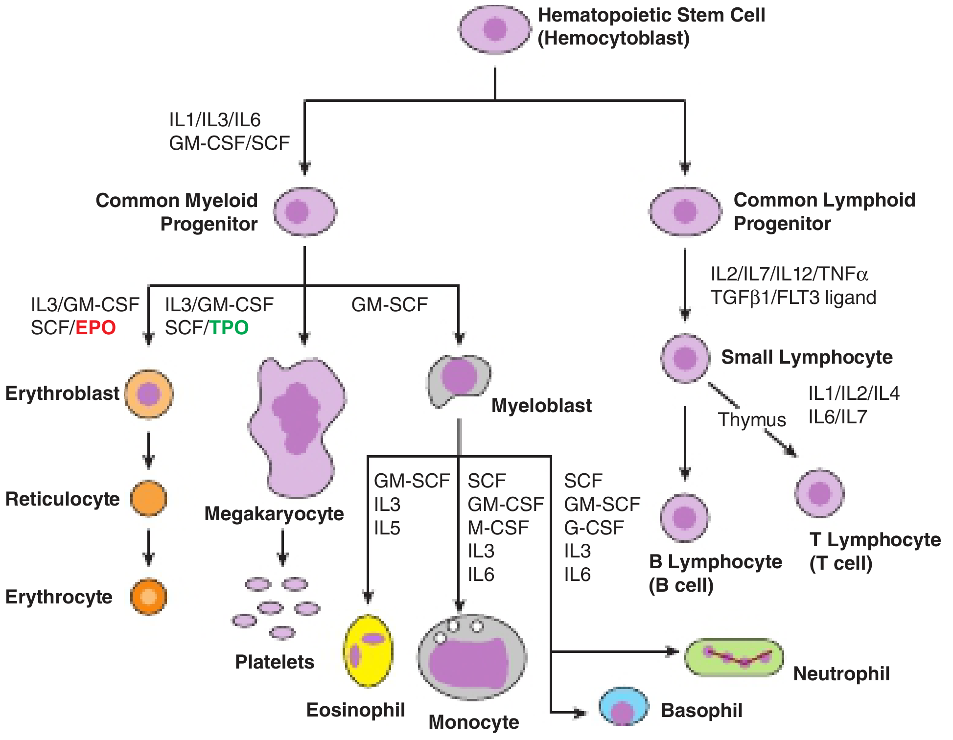
- Hematopoietic stem cells (HSCs) in bone marrow - can produce all blood cells (red cells, white cells, platelets) but cannot become neurons or muscle.
- Mesenchymal stem cells (MSCs) - can become bone, cartilage, or fat cells.
- Neural stem cells - can produce neurons, astrocytes, and oligodendrocytes.
- These are the stem cells resident in adult tissues, responsible for routine cell replacement and repair.
4. Unipotent ("one powerful")
A unipotent cell can produce only one type of differentiated cell. They still have self-renewal capacity (which distinguishes them from non-stem progenitor cells), but their fate is essentially fixed.
- Examples:
- Spermatogonial stem cells (give rise only to sperm)
- Muscle satellite cells (give rise only to skeletal muscle fibers)
- Some skin stem cells that only replenish a single layer of the epidermis
Visual Summary
Here is the hierarchy from a surgical textbook (Bailey & Love's), showing the progression from totipotent to nullipotent (fully differentiated):
And here is a classic example of multipotent stem cell differentiation - the hematopoietic stem cell giving rise to all blood cell lineages:
Quick Comparison Table
| Term | Differentiation Range | Key Example |
|---|---|---|
| Totipotent | All body cells + placenta/extraembryonic tissue | Zygote, early morula cells |
| Pluripotent | All 3 germ layers (body only) | ESCs, iPSCs |
| Multipotent | Multiple types within one lineage | Hematopoietic, mesenchymal stem cells |
| Unipotent | One cell type only | Spermatogonial stem cells, satellite cells |
The key conceptual ladder is: totipotent > pluripotent > multipotent > unipotent, with each step representing a narrowing of developmental options. This narrowing is generally irreversible in nature, though iPSC technology has shown it can be artificially reversed by reprogramming adult cells back to pluripotency.
Sources: Bailey and Love's Short Practice of Surgery 28th Ed, p. 62; Harper's Illustrated Biochemistry 32nd Ed, p. 665; Medical Physiology (Boron & Boulpaep)
What are M-O-R-P-H-O-G-E-N-S?
Morphogens are signaling molecules that spread through developing tissue and instruct cells to adopt different fates depending on their concentration. A cell close to the source gets a high dose and becomes one cell type; a cell far away gets a low dose and becomes another. Classic examples include Sonic Hedgehog (SHH), Wnt proteins, and BMPs.
Searching sources...
Finding Sources
Reading File
Reading File
Reading File
Reading File
Loading Image
Morphogens
A morphogen is a substance produced by cells in one region of a developing embryo that diffuses outward to form a concentration gradient across surrounding tissue. Cells read that gradient and adopt different identities depending on how much morphogen they are exposed to - high concentration near the source, low concentration far away.
The formal definition from Thompson & Thompson Genetics and Genomics in Medicine:
"A substance produced by cells in a particular region of an embryo that diffuses from its point of origin through the tissues of the embryo to form a concentration gradient. Cells undergo specification and then determination to different fates, depending on the concentration of morphogen they experience."
How They Work
The core logic is straightforward:
- A small group of cells acts as the source, secreting the morphogen
- The molecule diffuses through neighboring tissue, forming a gradient (highest near the source, falling off with distance)
- Cells at different positions along that gradient express different threshold-activated transcription factors, committing them to different fates
- This creates spatial patterning - different cell types in defined, reproducible positions
This is called positional information: a cell's fate depends on where it sits relative to the morphogen source.
Key Examples
1. Sonic Hedgehog (SHH) - the most studied morphogen
SHH is secreted by the notochord (a midline structure beneath the developing spinal cord). It diffuses upward through the ventral neural tube, creating a concentration gradient:
- Highest concentration (directly above the notochord) → induces the floor plate
- Intermediate concentration → induces motor neurons
- Lowest concentration (more lateral/dorsal) → other neuronal subtypes
SHH also patterns the limb bud: produced by the "zone of polarizing activity" at the posterior side of the limb, its gradient instructs cells to become digit 5 (pinky, near the source) vs. digit 1 (thumb, far from the source).
This diagram shows both roles of SHH as a morphogen - neural tube patterning (top) and limb bud digit specification (bottom):
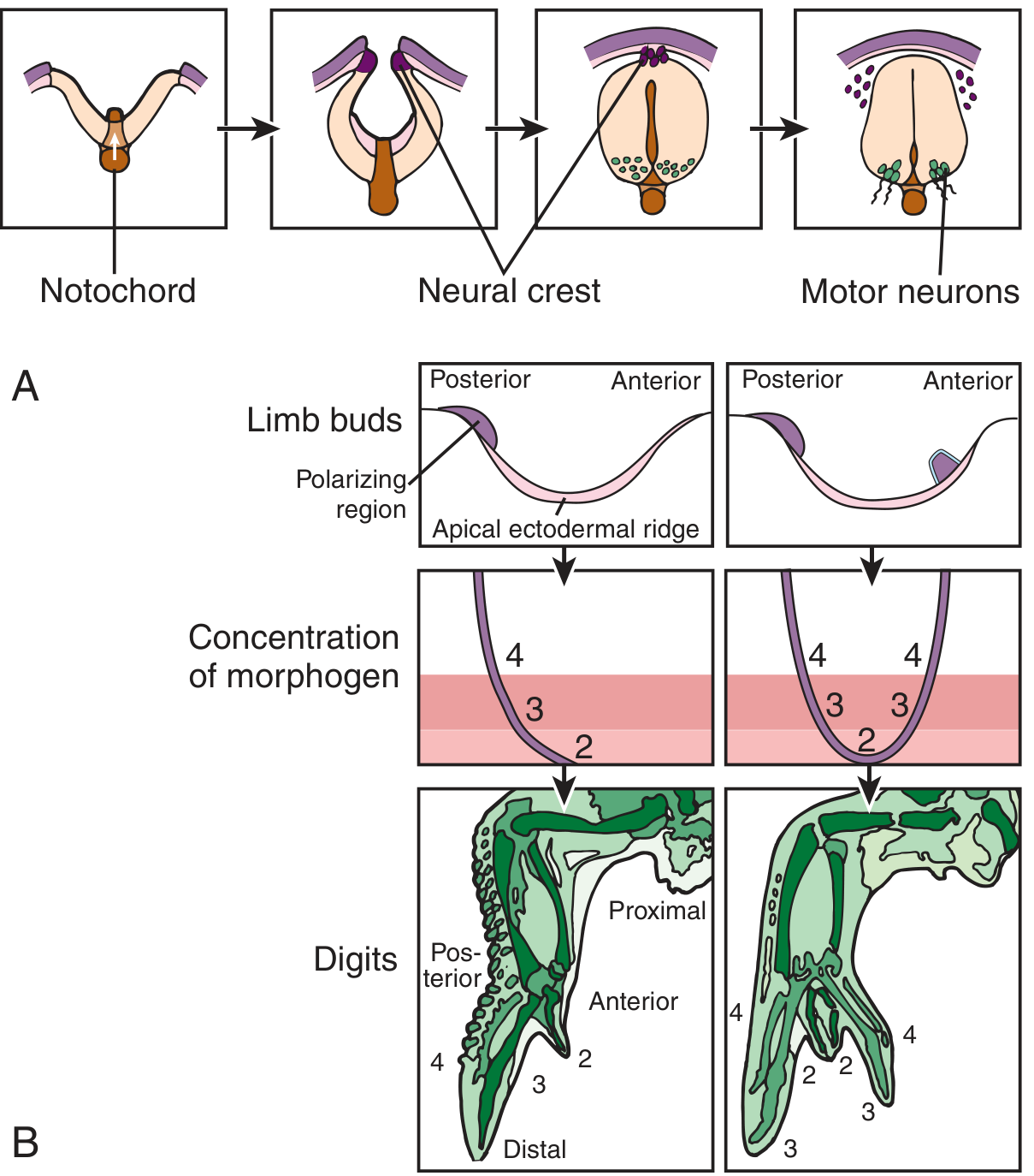
Note the right panel: when an ectopic (extra) polarizing region is placed on the anterior side of the limb bud, it creates a mirror-image gradient - and you get a duplicated set of posterior digits. This is one of the classic experiments proving morphogen logic.
2. Bone Morphogenetic Proteins (BMPs)
BMPs work from the dorsal side of the neural tube (secreted from the ectoderm and then the roof plate), countering SHH from below. They induce:
- Neural crest cells at the dorsal margin
- Sensory relay neurons in the dorsal spinal cord
So the neural tube is patterned by two opposing morphogen gradients: SHH ventrally and BMPs dorsally.
3. Wnt Proteins
Wnts are secreted by the roof plate after neural tube closure. They primarily promote proliferation of dorsal neural tube progenitors, and also play major roles in anterior-posterior body axis specification and gut/limb development.
4. Fibroblast Growth Factors (FGFs)
23 members in humans. They act as ligands for tyrosine kinase receptors and are critical morphogens in craniofacial and limb development. Mutations in FGF receptors cause achondroplasia (the most common form of dwarfism) and craniosynostosis syndromes (premature skull suture fusion).
Why Morphogens Matter Clinically
Mutations that disrupt morphogen gradients cause predictable birth defects:
| Morphogen | Mutation Effect |
|---|---|
| SHH loss | Holoprosencephaly (forebrain fails to divide), cleft lip/palate, hypotelorism (close-set eyes), single central incisor |
| BMP pathway | Bone/cartilage malformations |
| FGF receptors | Achondroplasia, craniosynostosis |
| Wnt pathway | Wide range - cancers, neural defects |
Even a 50% reduction in SHH (heterozygous loss) is enough to alter the gradient magnitude and produce an abnormal phenotype, which is why many morphogen gene mutations act as autosomal dominant conditions.
Summary
Morphogens are the embryo's coordinate system - they convert a featureless field of cells into organized tissue by providing position-dependent chemical instructions. The same principle (gradient → threshold → transcription factor → fate) is reused across many different tissues and organisms, from fly wings to human brains.
Sources: Thompson & Thompson Genetics and Genomics in Medicine, 9th Ed, pp. 343, 353-354; Eric Kandel Principles of Neural Science, 6th Ed, pp. 1165-1166
Definition of morphogens
From the textbook definition I retrieved in our previous conversation (Thompson & Thompson Genetics and Genomics in Medicine, 9th Ed):
Morphogen: a substance produced by cells in a particular region of an embryo that diffuses from its point of origin through the tissues of the embryo to form a concentration gradient. Cells undergo specification and then determination to different fates, depending on the concentration of morphogen they experience.
In plain terms: a morphogen is a signaling molecule that spreads through developing tissue as a gradient, and cells decide what to become based on how much of it they detect.
This is a shared conversation. Sign in to Orris to start your own chat.