
Case where ipt is negative but pregnant
A negative IPT (immunochromatographic pregnancy test) but clinically pregnant scenario is an important obstetric/diagnostic concept. Let me look this up properly.
Searching sources...
Reading File
Now let me search the medical library and PubMed in parallel for this topic.
Finding Sources
Finding Sources
Searching PubMed
false negative urine pregnancy test hCG hook effect
Now let me read the key textbook sections in detail:
Reading File
Reading File
Reading File
Excellent - I now have comprehensive textbook data. Let me compile the full answer.
False-Negative Pregnancy Test (Negative IPT) in a Pregnant Woman
A negative immunochromatographic pregnancy test (IPT/urine hCG test) in a truly pregnant woman is a clinically important pitfall. Here are all the recognized causes, grouped by mechanism:
1. Hook Effect (Most Clinically Important)
This is the classic exam and clinical scenario to know.
- When hCG levels are extremely high (typically >1,000,000 mIU/mL), the assay is overwhelmed
- Excess hCG saturates both the fixed solid-phase antibody and the labeled soluble antibody simultaneously, preventing sandwich complex formation - so no colored line appears
- Classically seen in:
- Gestational trophoblastic disease (hydatidiform mole, choriocarcinoma) - this is the high-yield scenario
- Multiple gestation pregnancy (twins/triplets can raise hCG enough to trigger hook effect)
- Fix: Dilute the urine sample 1:10 or 1:50 and repeat the test - a positive result will appear
A 2024 case report (Phillipo et al., PMID 38292164) documented a false-negative qualitative hCG test with classic ultrasound findings of complete molar pregnancy due to the hook effect. A 2022 case report (Reitz et al., PMID 36447678) documented the same phenomenon in a multiple gestation pregnancy.
- Tintinalli's Emergency Medicine, p. 736-739
- Tietz Textbook of Laboratory Medicine 7th Ed, p. 445-446
2. hCG Beta Core Fragment Interference
- After the 5th week of pregnancy, the predominant hCG variant in urine is the beta core fragment (hCGβcf), not intact hCG
- hCGβcf can bind one of the two anti-hCG antibodies in the sandwich assay, but because it lacks the other epitope, it blocks the intact hCG from forming a complete sandwich
- This leads to a false-negative result even at moderate-to-high concentrations
- This is more device-specific - affects some POC devices more than others
- Tietz Textbook of Laboratory Medicine 7th Ed, p. 445-446
3. Too Early in Pregnancy (Timing Error)
- Most IPT devices have a detection threshold of ~20-25 mIU/mL
- In the first days after implantation, hCG may be <20 mIU/mL
- At 1 week post-conception: only 5-50 mIU/mL
- Home tests are only 50-97% sensitive on the first day of missed period, rising to ~100% at 11 days after missed period
- Fix: Repeat the test in 48-72 hours
- Tintinalli's Emergency Medicine, p. 737
- Textbook of Family Medicine 9e, p. 814
4. Dilute Urine
- Particularly problematic when hCG is <50 mIU/mL (very early pregnancy)
- Low urine specific gravity (e.g., after high fluid intake) can bring hCG below the detection threshold
- Fix: Use first-morning urine (most concentrated), or send serum quantitative hCG
- Tintinalli's Emergency Medicine, p. 739
5. Drug Interference
- Phenothiazines (chlorpromazine, perphenazine, promethazine) can cause both false-positive and false-negative pregnancy test results
- Mechanism involves interference with the immunologic reagents
- Kaplan & Sadock's Comprehensive Textbook of Psychiatry
Summary Table
| Cause | Mechanism | Clinical Setting | Fix |
|---|---|---|---|
| Hook effect | Antibody saturation from extremely high hCG | Molar pregnancy, choriocarcinoma, multiple gestation | Dilute urine, repeat |
| Beta core fragment | Competitive inhibition of sandwich assay | After 5th week, device-dependent | Serum quantitative hCG |
| Too early | hCG below detection threshold | <1 week post-conception | Repeat in 48-72h |
| Dilute urine | Low hCG concentration in sample | High fluid intake, early pregnancy | First-morning urine or serum hCG |
| Drug interference | Phenothiazine assay interference | Psychiatric patients | Serum quantitative hCG |
Key Management Point
When clinical suspicion for pregnancy is high (symptoms, missed period, ultrasound findings) and the urine IPT is negative:
- Send quantitative serum beta-hCG - nearly 100% sensitive when threshold is ≥5 mIU/mL
- Dilute the urine sample and repeat if hook effect is suspected (e.g., very high clinical suspicion + gestational trophoblastic disease signs)
- Do a pelvic ultrasound - particularly important to rule out ectopic pregnancy even when the test is negative but clinical suspicion persists
Negative upt
The previous answer covered IPT (immunochromatographic pregnancy test) - a UPT (urine pregnancy test) is essentially the same thing. The causes of a false-negative UPT in a pregnant woman are identical to what was just covered.
Here is a focused summary specific to UPT:
False-Negative UPT in a Pregnant Woman
Causes
1. Hook Effect (High-Dose Hook Effect)
- hCG extremely elevated (>1,000,000 mIU/mL) saturates both antibodies in the lateral flow assay - no sandwich complex forms, no line appears
- Classic settings: hydatidiform mole, choriocarcinoma, multiple gestation
- Fix: Dilute urine 1:10 to 1:50 and repeat
2. Too Early in Pregnancy
- hCG <20-25 mIU/mL (below detection threshold of most UPT strips)
- At <1 week post-conception, hCG is only 5-50 mIU/mL
- Home UPTs are only 50-97% sensitive on day 1 of missed period
- Fix: Repeat in 48-72 hours using first-morning urine
3. Dilute Urine
- High fluid intake lowers urine hCG concentration below the detection threshold
- More significant when hCG is already low (very early pregnancy)
- Fix: Use first-morning urine (most concentrated)
4. hCG Beta Core Fragment Interference
- After 5th week, beta core fragment dominates urine hCG - it blocks one antibody arm of the assay, preventing intact hCG from forming a positive signal
- Device-specific; affects some POC/strip UPTs more than others
- Fix: Serum quantitative beta-hCG
5. Drug Interference
- Phenothiazines (promethazine, chlorpromazine, perphenazine) can cause false-negative results
- Fix: Serum quantitative beta-hCG
What To Do When UPT is Negative But Pregnancy Suspected
| Step | Action |
|---|---|
| 1 | Repeat UPT with first-morning urine |
| 2 | Send serum quantitative beta-hCG (nearly 100% sensitive at ≥5 mIU/mL threshold) |
| 3 | If hook effect suspected - dilute urine 1:10 and repeat UPT |
| 4 | Pelvic ultrasound - especially to exclude ectopic pregnancy |
Bottom line: A negative UPT does NOT rule out pregnancy. Serum beta-hCG is the gold standard when clinical suspicion remains. The hook effect in gestational trophoblastic disease is the highest-yield false-negative scenario in exams and clinical practice.
Prozone phenomenon
The prozone phenomenon is a well-known immunology/lab medicine concept - antibody excess causing a false-negative precipitin/agglutination reaction. Let me pull the authoritative textbook content.
Searching sources...
Finding Sources
Finding Sources
Good results. Let me now read the key sections in detail:
Reading File
Reading File
Reading File
Now let me also check for the broader immunology/general context of prozone and where else it applies (IgG4, cryptococcal antigen):
Finding Sources
Finding Sources
Reading File
Excellent. Let me fetch the precipitin curve diagram - it's perfect for explaining this concept visually:
Loading Image
Loading Image
Perfect - I have everything needed for a comprehensive answer with textbook diagrams.
Prozone Phenomenon
Definition
The prozone phenomenon is a false-negative result in a precipitation or agglutination-based immunoassay caused by antibody excess relative to antigen. Despite high antibody concentrations (and true positivity), no visible precipitate or agglutination forms - the test appears negative.
Immunological Basis - The Precipitin Curve
The precipitin curve explains it best:
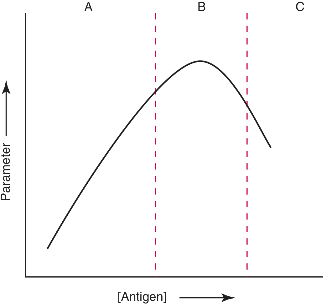
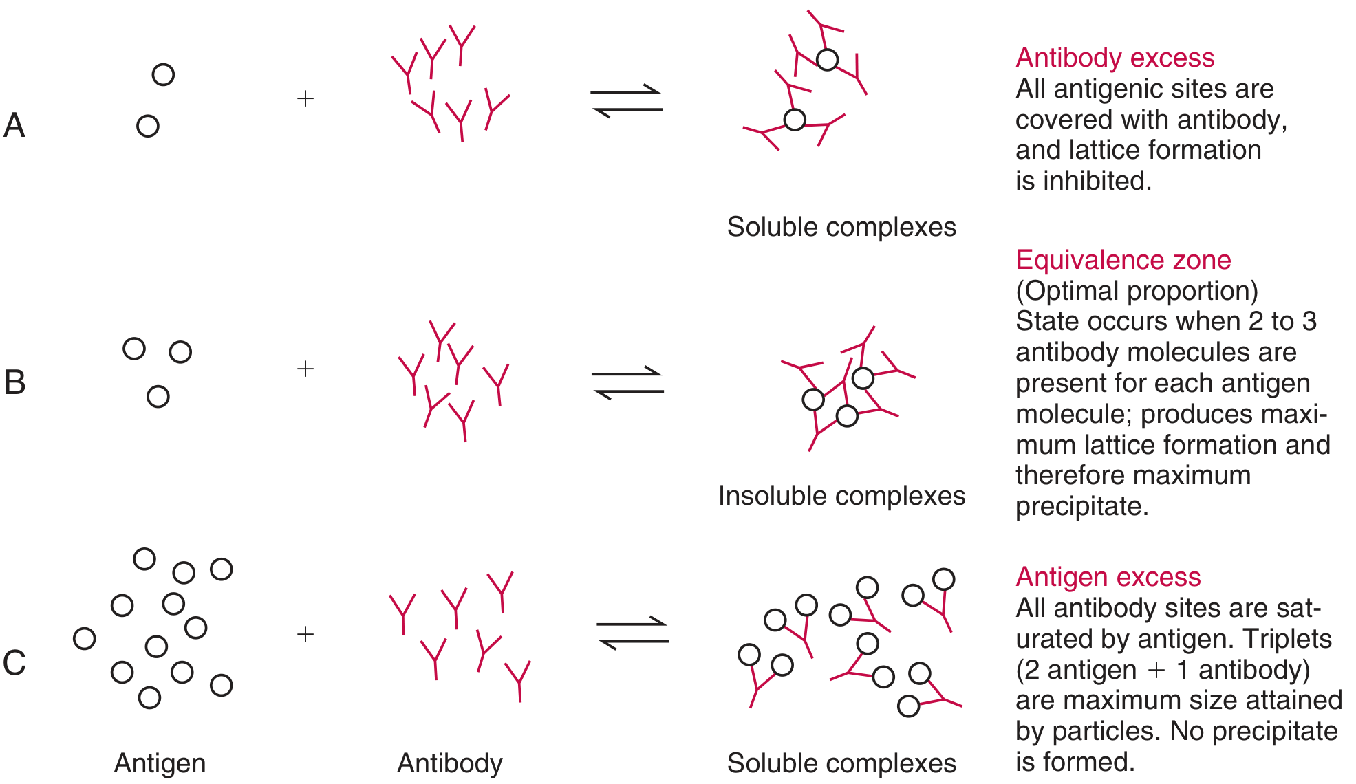
There are three zones on the precipitin curve:
| Zone | Condition | Result | Name |
|---|---|---|---|
| A | Antibody excess | Soluble complexes - no precipitate | Prozone |
| B | Equivalence (2-3 Ab per Ag) | Maximum insoluble lattice - maximum precipitate | Equivalence zone |
| C | Antigen excess | Soluble complexes - no precipitate | Postzone |
Why no precipitate in antibody excess?
When antibodies are in massive excess, every antigenic epitope gets covered by its own antibody molecule. This prevents cross-linking between antigen molecules - no large insoluble lattice can form. The complexes remain small and soluble, so the test reads negative.
- Tietz Textbook of Laboratory Medicine 7th Ed, p. 3513-3520
Classical Clinical Example: Syphilis Serology
The prozone phenomenon is most classically described with non-treponemal syphilis tests (VDRL and RPR):
- In secondary syphilis, antibody titers can become so high that the test is paradoxically negative
- This is why secondary syphilis is sometimes called "the great imitator" - the rash appears, but the VDRL/RPR is negative
- The prozone phenomenon occurs more frequently in HIV co-infected patients (immune dysregulation causes massively elevated antibody levels)
Fix: Dilute the serum (e.g., 1:2, 1:4, etc.) and repeat - a positive result will appear at the correct dilution (equivalence zone)
Many laboratories do NOT routinely check for the prozone phenomenon, so clinicians must specifically request it in the appropriate setting (e.g., a patient with a suspicious rash and a negative nontreponemal test).
- Fitzpatrick's Dermatology, p. 1979; Red Book 2021, p. 131
Other Clinical Settings Where Prozone Occurs
| Condition | Test Affected | Notes |
|---|---|---|
| Secondary syphilis (classic) | VDRL, RPR | Especially in HIV co-infection |
| Hydatidiform mole / choriocarcinoma | urine hCG / UPT | This is the "hook effect" - same mechanism |
| Multiple myeloma / IgG4-RD | Serum IgG4 quantitation | Spuriously low IgG4 due to prozone |
| Cryptococcal meningitis | Serum/CSF cryptococcal lateral flow antigen assay | High fungal burden → antibody excess |
Prozone vs. Hook Effect
These are essentially the same phenomenon with different names depending on context:
| Term | Used In | Excess Molecule |
|---|---|---|
| Prozone | Precipitation/agglutination assays (VDRL, RPR) | Antibody excess |
| Hook effect | Sandwich immunoassay (hCG, tumor markers) | Antigen excess (overwhelms both antibody arms) |
Note: In the hook effect (hCG/UPT), it is technically antigen excess (the postzone equivalent) in a sandwich assay - but the result is the same: false negative due to failure of lattice/signal formation.
Key Points for Exams
- Prozone = antibody excess = false negative in precipitation assays
- Classic setting: secondary syphilis with high VDRL/RPR titers - especially with HIV co-infection
- Fix = dilute the serum and repeat
- Equivalent in sandwich immunoassays = hook effect (antigen excess)
- Clinicians must specifically request dilution studies - labs do not routinely perform them
Mirena
Mirena is a levonorgestrel-releasing intrauterine system (LNG-IUS). Let me pull comprehensive textbook content on it.
Searching sources...
Finding Sources
Finding Sources
Reading File
Finding Sources
Finding Sources
Finding Sources
Finding Sources
Finding Sources
Reading File
Reading File
Loading Image
I now have comprehensive textbook data. Here is the full answer:
Mirena (Levonorgestrel Intrauterine System / LNG-IUS)
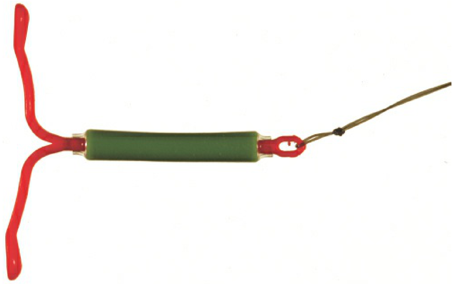
Basic Profile
| Feature | Detail |
|---|---|
| Type | T-shaped levonorgestrel-releasing intrauterine system (LNG-IUS) |
| Size | 32 × 32 mm |
| Hormone load | 52 mg levonorgestrel |
| Release rate | Initial 20 µg/day, declining to ~10 µg/day by 5 years |
| Serum LNG levels | 130-200 pg/mL (low - much less than oral progestin-only pills) |
| Duration of use | Up to 5 years (some evidence supports longer) |
- Berek & Novak's Gynecology, p. 203; Textbook of Family Medicine 9e, p. 691
Mechanism of Action (Triple Action)
- Thickened, scant cervical mucus - impedes sperm penetration
- Endometrial atrophy - profound progestogenic effect makes endometrium hostile to implantation
- Local intrauterine inflammatory response - impairs sperm motility and function
About 85% of cycles remain ovulatory - the primary mechanism is NOT anovulation but local uterine/cervical effects. Systemic hormone levels are too low to reliably suppress ovulation.
The IUD is NOT an abortifacient - studies of serum beta-hCG and tubal washings confirm that fertilization is prevented, not implantation.
- Berek & Novak's Gynecology, p. 202-203
Contraceptive Efficacy
- Failure rate: <0.2 per 100 woman-years (among the most effective methods available)
- Total pregnancies over 7 years: only 1.1 per 100 women
- Comparable to tubal sterilization but fully reversible
Non-Contraceptive / Therapeutic Uses
| Indication | Notes |
|---|---|
| Heavy menstrual bleeding (menorrhagia) | Reduces bleeding by up to 90%; used as alternative to hysterectomy in Europe and UK |
| Uterine fibroids | Reduces menorrhagia from fibroids (may be less effective with distorting submucosal fibroids) |
| Endometriosis | Reduces pain and lesion activity |
| Adenomyosis | Significant symptom improvement |
| Endometrial protection in HRT | Delivers progestin locally in postmenopausal women on estrogen therapy |
| Endometrial cancer risk reduction | Protective effect via endometrial atrophy |
- Berek & Novak's Gynecology, p. 222-226
Contraindications
- Current or suspected pregnancy
- Abnormal uterine cavity (distorting fibroids, congenital anomaly)
- Active or recurrent PID / endometritis
- Known or suspected endometrial/cervical malignancy
- Unexplained genital bleeding
- Allergy to IUD components
Not contraindicated (unlike combined OCP) in:
- Breastfeeding/lactation
- History of VTE
- Smokers
- Women at increased cardiovascular risk
Risks and Complications
1. Infection (PID)
- Risk is elevated only in the first 20 days after insertion (insertion-related)
- After that, rate returns to baseline (~1.6 per 1,000 women/year) - the same as the general population
- The major risk factor is STI exposure, not the IUD itself
- Actinomycosis is the one pelvic infection unequivocally linked to IUD use
- If PID develops: treat with antibiotics; remove only if no improvement after 72 hours
2. Ectopic Pregnancy
- IUD prevents most pregnancies, so overall ectopic rate is very low (0.02 per 100 woman-years)
- BUT: if pregnancy occurs despite an IUD, the proportion that are ectopic is higher
- Always rule out ectopic if a Mirena user presents with pelvic pain + positive beta-hCG
3. Expulsion
- Occurs in ~2-10% of users, more common in the first year
- Higher risk in women with heavy periods or younger age
4. Perforation
- Rate: 0.6-1.6 per 1,000 insertions
- Higher risk in the 6-8 weeks postpartum (so postpartum placement is delayed)
5. Menstrual Changes
- Irregular spotting common in the first 3-6 months
- Amenorrhea or oligomenorrhea develops in 20-50% by 1 year (from endometrial atrophy) - this is expected and benign, not a sign of pregnancy
Fertility After Removal
- Contraceptive effects reverse promptly after removal
- 1-year pregnancy rate after removal: 89 per 100 women under age 30 - equivalent to women who used no contraception
- No evidence of tubal factor infertility from LNG-IUD itself (only from STI exposure)
- Berek & Novak's Gynecology, p. 252
Key Summary Points
- Mirena is one of the most effective reversible contraceptives available (failure rate <0.2%)
- Works mainly via local cervical/endometrial effects - not primarily by blocking ovulation
- Major therapeutic value for heavy menstrual bleeding, endometriosis, adenomyosis, and endometrial protection
- PID risk is insertion-related only (first 20 days); thereafter no increased risk
- Amenorrhea with Mirena = expected effect, not a pathological finding
- If pregnancy occurs while on Mirena - always exclude ectopic
Medical management of fibroid
Searching sources...
Finding Sources
Finding Sources
Finding Sources
Finding Sources
Finding Sources
Finding Sources
Finding Sources
Reading File
Finding Sources
Finding Sources
Found the key sections. Let me read the Treatment section and surrounding content:
Reading File
Reading File
Reading File
I have rich content on medical management. Let me compile the comprehensive answer now.
Medical Management of Uterine Fibroids (Leiomyomas)
Fibroids are estrogen- and progesterone-dependent benign smooth muscle tumors. Medical therapy targets this hormonal dependence to reduce symptoms (heavy bleeding, bulk, pain) or shrink fibroids pre-operatively. Medical treatment does not permanently cure fibroids - they typically regrow after stopping treatment.
When to Treat (Indications)
Most fibroids are asymptomatic and managed with watchful waiting. Medical treatment is indicated when:
- Heavy menstrual bleeding (HMB) / menorrhagia
- Pelvic pressure/bulk symptoms
- Pre-operative downsizing before myomectomy or hysterectomy
- Treating anaemia before surgery
- Women approaching menopause (bridging therapy - fibroids regress after menopause)
- Berek & Novak's Gynecology, p. 4695-4697
Medical Therapy Options
1. GnRH Agonists (e.g. Leuprolide, Goserelin, Nafarelin)
Mechanism: Downregulate pituitary GnRH receptors → suppress LH/FSH → profound hypo-oestrogenic state → fibroid shrinkage
| Feature | Detail |
|---|---|
| Fibroid volume reduction | 35-65% within 3-6 months |
| Bleeding effect | Amenorrhoea in most patients |
| Pre-op benefit | Corrects anaemia, reduces uterine size, may convert laparotomy to laparoscopy |
| Duration | Maximum 3-6 months (bone loss limits long-term use) |
| Add-back therapy | Low-dose oestrogen/progestin added to prevent bone loss and vasomotor symptoms |
| Major limitation | Fibroids regrow to pre-treatment size within 3-6 months of stopping |
GnRH agonists are the most widely studied pre-operative medical therapy for fibroids.
2. GnRH Antagonists (e.g. Elagolix, Relugolix, Linzagolix)
Mechanism: Direct competitive blockade of GnRH receptors → rapid onset of hypo-oestrogenism (no initial flare, unlike agonists)
- Faster onset of action than agonists (no initial stimulatory "flare" phase)
- Elagolix (Oriahnn) - FDA approved specifically for heavy menstrual bleeding due to fibroids
- Relugolix combination tablet - approved for fibroids with add-back oestrogen/progestin
- Dose-dependent amenorrhoea and fibroid volume reduction
- Used with add-back therapy for longer-term use
3. Progestin-Releasing IUD (LNG-IUS / Mirena)
Mechanism: Local endometrial atrophy → dramatic reduction in menstrual blood loss
- Reduces HMB by up to 90%
- Does not shrink fibroids or reduce bulk
- Most effective for controlling bleeding when cavity is not significantly distorted
- May be less effective with submucosal or distorting fibroids
- Also treats dysmenorrhoea and offers contraception
4. Selective Progesterone Receptor Modulators (SPRMs)
Mifepristone and Ulipristal acetate (UPA)
Mechanism: Bind progesterone receptors → antagonise progesterone action → endometrial atrophy + direct antiproliferative effect on fibroid cells
| Drug | Effect |
|---|---|
| Ulipristal acetate | 20-40% volume reduction; amenorrhoea in >70%; intermittent courses used |
| Mifepristone | Reduces fibroid volume and HMB; less studied than UPA |
- UPA (Esmya) was approved in Europe for intermittent long-term use but faced regulatory restrictions due to rare hepatotoxicity cases (European Medicines Agency review 2020)
- Unlike GnRH agonists, SPRMs cause PAEC (progesterone receptor modulator-associated endometrial changes) - a benign, reversible histological change that must not be confused with endometrial hyperplasia
5. Combined Oral Contraceptives (COCPs)
- Control HMB and dysmenorrhoea effectively
- Do not shrink fibroids and do not consistently reduce fibroid growth
- No definitive relationship between OCP use and fibroid development or growth
- Useful for symptom control in women who also need contraception
- Avoid if large fibroids with pressure symptoms
6. Progestin-Only Pills / Depot Medroxyprogesterone Acetate (DMPA)
- Reduce HMB by inducing endometrial atrophy
- Do not reliably shrink fibroids
- DMPA (Depo-Provera) causes amenorrhoea in many users
7. Tranexamic Acid
- Antifibrinolytic - inhibits plasminogen activation in the endometrium
- Reduces HMB by ~40-50% per cycle
- Taken only during menstruation (not continuous)
- No effect on fibroid size
- Non-hormonal option - suitable for women wanting to conceive
8. NSAIDs (e.g. Mefenamic Acid, Ibuprofen)
- Reduce HMB by ~20-35% via prostaglandin inhibition
- Good for dysmenorrhoea associated with fibroids
- Weakest effect on bleeding among the medical options
- Non-hormonal; safe and well tolerated
9. Iron Supplementation
- Not directly treating the fibroid but essential for managing iron-deficiency anaemia from chronic HMB
- Often used in combination with any of the above before surgery
Summary Comparison Table
| Drug/Class | Reduces HMB | Shrinks Fibroid | Duration | Key Limitation |
|---|---|---|---|---|
| GnRH agonist | Yes (amenorrhoea) | Yes (35-65%) | Max 6 months | Regrowth on stopping; bone loss |
| GnRH antagonist | Yes | Yes | Longer with add-back | Cost; menopausal SE |
| LNG-IUS (Mirena) | Yes (up to 90%) | No | 5 years | Ineffective if cavity distorted |
| SPRM (UPA/mifepristone) | Yes | Yes (20-40%) | Intermittent courses | Hepatotoxicity (UPA); PAEC |
| COCP | Yes | No | Long-term | Does not reduce size |
| Tranexamic acid | Yes (~40-50%) | No | During menses only | Symptomatic only |
| NSAIDs | Modest (~20-35%) | No | During menses only | Weakest effect |
Key Points
- GnRH agonists are the most potent pre-operative medical therapy - used to shrink fibroids, correct anaemia, and reduce surgical complexity; not a long-term solution
- GnRH antagonists (elagolix, relugolix) are the newer oral agents approved specifically for fibroid-related HMB with add-back therapy
- LNG-IUS is best for controlling HMB without shrinking fibroids - highly effective when cavity is not distorted
- SPRMs offer fibroid volume reduction with amenorrhoea but are limited by hepatotoxicity concerns
- None of the medical treatments permanently cure fibroids - all are either temporizing or symptom-controlling measures
- Watchful waiting is appropriate for mildly symptomatic women, especially those approaching menopause
- Berek & Novak's Gynecology, pp. 4632-4634, 4695-4715
Adenomyosis
Searching sources...
Finding Sources
Finding Sources
Good - found dedicated sections across multiple textbooks. Let me read them all in parallel:
Reading File
Reading File
Reading File
Reading File
Reading File
Reading File
Reading File
Let me also fetch the MRI image of adenomyosis and check the Robbins pathology section:
Loading Image
Reading File
I now have comprehensive content from multiple textbooks. Here is the full answer:
Adenomyosis
Definition
Adenomyosis is defined as the presence of endometrial glands and stroma within the myometrium, accompanied by reactive smooth muscle hypertrophy and hyperplasia of the surrounding myometrium. The ectopic endometrial tissue remains in continuity with the endometrium, consistent with downward invasion rather than implantation.
- Can be diffuse (throughout the myometrium) or focal (adenomyoma - a discrete mass)
- Often coexists with endometriosis and uterine fibroids
- Robbins & Kumar Basic Pathology, p. 548; Sabiston Textbook of Surgery, p. 2982
Epidemiology
- Prevalence: Extremely variable (1-70% depending on diagnostic criteria used) - historically underdiagnosed because it required histology at hysterectomy
- Found in ~30% of women of reproductive age; increases with age toward menopause
- More common in multiparous women than nulliparous
- Predominantly affects women in their 4th and 5th decades
Pathogenesis
Not fully understood. Leading theories:
- Invagination/downward invasion - endometrial epithelium invades the endomyometrial junction and penetrates into myometrium
- Molecular interaction between displaced endometrial cells and adjacent myometrial cells causes hypertrophy and inflammation
- Shares pathophysiology with endometriosis
- The junctional zone (JZ) is the target tissue - disruption of the JZ by uterine instrumentation, surgery, or repeated trauma may facilitate invasion
Risk Factors
| Category | Factors |
|---|---|
| Hormonal | Early menarche (≤10 yrs), short cycles (≤24 days), obesity, tamoxifen use, increasing age, OCP use |
| Obstetric/surgical | Multiparity, spontaneous miscarriage, caesarean section, curettage, surgical termination of pregnancy |
| Coexistent pathology | Endometriosis, fibroids breaching the endometrial-myometrial interface, endometrial hyperplasia |
| Other | Smoking |
- Bailey & Love's Short Practice of Surgery 28th Ed, p. 2624-2635
Clinical Features
Symptoms (the classic triad)
- Menorrhagia / heavy menstrual bleeding (AUB) - most common
- Dysmenorrhoea - secondary, colicky, worsening premenstrually
- Chronic pelvic pain / dyspareunia
Also: subfertility, irregular bleeding, noncyclic pelvic pain
33% are asymptomatic - found incidentally on imaging or at hysterectomy
Signs
- Enlarged, globular, tender uterus - symmetrically enlarged (unlike fibroids which cause irregular enlargement)
- Uterine tenderness on bimanual examination, especially premenstrually
- Abnormalities at hysteroscopy: irregular endometrium, cystic haemorrhagic lesions, altered vascularisation
Diagnosis
1. Transvaginal Ultrasound (TVUS)
- Accuracy: 68-86% for diffuse adenomyosis
- Typical features (MUSA criteria - ≥3 features usually required):
- Enlarged globular uterus with asymmetric wall thickening
- Myometrial cysts (2-6 mm subendometrial cysts) - present in 50%
- Echogenic subendometrial linear striations
- Hyperechogenic islands within myometrium
- Fan-shaped shadowing
- Poor definition of the endomyometrial interface
- "Rain shower" appearance - multiple fine areas of attenuation
- Speckled pattern on colour Doppler
Focal adenomyosis is harder to distinguish from fibroids on USS.
2. MRI (Most Accurate Non-Invasive Tool)
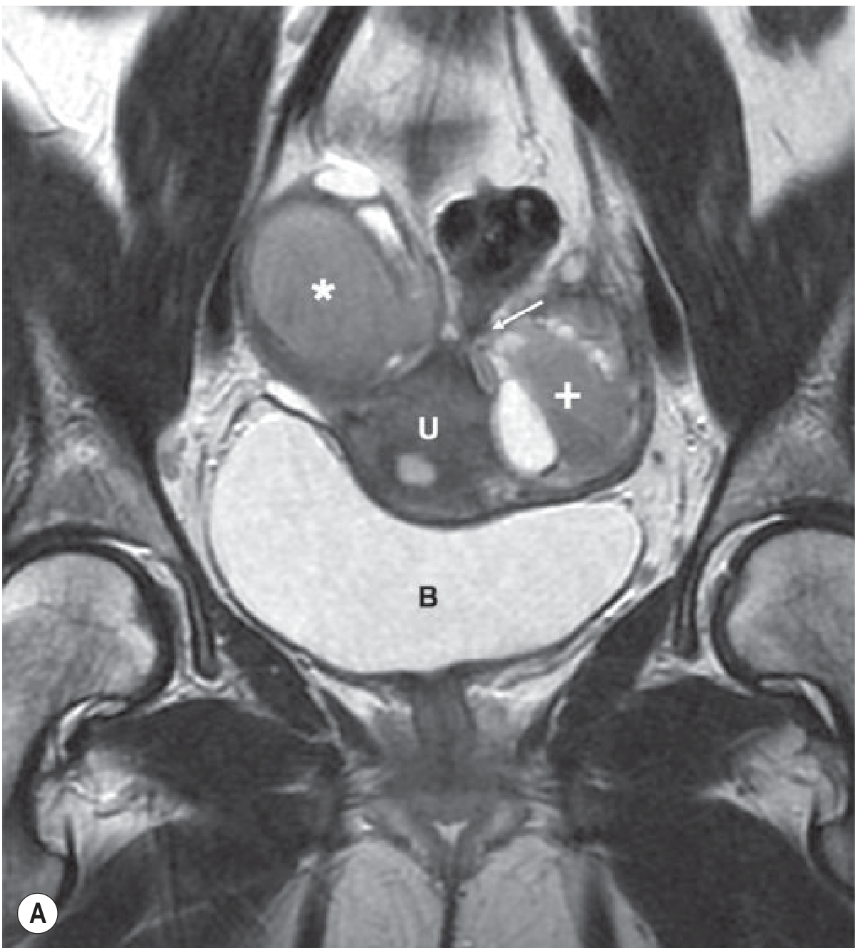
Key MRI findings on T2-weighted images:
- Junctional zone (JZ) thickening - the key diagnostic criterion:
- JZ ≥12 mm → high accuracy for adenomyosis
- JZ ≤8 mm → excludes adenomyosis
- JZ 8-12 mm → indeterminate (need ancillary criteria)
- Ill-defined low-signal intensity areas within myometrium (smooth muscle hyperplasia)
- High T2 signal foci within low-signal myometrium = ectopic endometrial islands
- High T1 signal foci (punctate haemorrhage within ectopic tissue) - 95% PPV for adenomyosis
- High T2 linear striations (finger-like projections from endometrium into myometrium)
- Rare: cystic adenomyosis (extensive haemorrhage)
MRI best differentiates adenomyoma from fibroid (important for surgical planning).
3. Histology (Gold Standard)
- Nests of endometrial glands and stroma deep in the myometrium, interposed between muscle bundles
- Traditionally required hysterectomy specimen - limits pre-operative diagnosis
- Not feasible in women wishing to preserve fertility
Adenomyosis vs. Fibroids - Key Differences
| Feature | Adenomyosis | Fibroid |
|---|---|---|
| Uterine shape | Globular, symmetrical enlargement | Irregular, asymmetric |
| Margins | Ill-defined | Well-defined pseudocapsule |
| MRI | Low JZ signal, ill-defined | Well-defined, variable signal, displaces JZ |
| Tenderness | Often tender | Usually not tender |
| Effect on fertility | Reduces implantation | Depends on location |
| Response to GnRH | Partial shrinkage | 35-65% shrinkage |
| After menopause | Regresses | Regresses |
Management
Medical (First-Line for Symptom Control)
| Drug | Effect |
|---|---|
| NSAIDs (e.g. ibuprofen, mefenamic acid) | Reduce dysmenorrhoea and HMB via prostaglandin inhibition |
| LNG-IUS (Mirena) | Reduces HMB by up to 90%; improves dysmenorrhoea; local endometrial atrophy; first-choice medical therapy |
| Combined OCP | Controls HMB and dysmenorrhoea |
| Progestogens (e.g. dienogest) | Good evidence for adenomyosis pain; continuous use preferred |
| GnRH agonists (leuprolide, goserelin) | Induce temporary amenorrhoea; reduce uterine volume; used pre-operatively or as bridge to menopause; max 6 months |
| GnRH antagonists (elagolix) | Rapid effect; used with add-back therapy |
| Danazol | Androgenic, induces amenorrhoea; rarely used due to side effects |
| Aromatase inhibitors (letrozole) | Block local oestrogen production; used in refractory cases |
| SPRMs (ulipristal acetate) | Reduces HMB and uterine volume; regulatory restrictions now apply |
Radiological / Minimally Invasive
- Uterine Artery Embolisation (UAE) - reduces blood supply to adenomyotic tissue; symptoms recur in some; post-embolisation syndrome (fever, pain, nausea) is common SE; concerns about ovarian reserve
- High-Intensity Focused Ultrasound (HIFU) / MR-guided Focused Ultrasound (MRgFUS) - non-invasive thermal ablation of adenomyotic tissue; SE include abdominal pain, skin burns, rare sciatic nerve injury
Surgical (Uterus-Preserving)
For women desiring fertility:
- Non-excisional: thermal coagulation of diseased myometrium
- Partial reductive surgery: wedge resections, H-incisions (for diffuse disease)
- Complete adenomyotic excision: double/triple-flap method, asymmetric dissection (for focal adenomyoma)
- Endometrial ablation/resection - for HMB without fertility wish; works best for superficial disease
- Risk: uterine rupture ~6% after adenomyomectomy (cf. 0.26% after myomectomy)
Surgical (Definitive)
- Total hysterectomy - only definitive cure
- Preferred over subtotal as adenomyosis can recur in the cervical stump and rectovaginal septum
- Reserved for women who have failed medical management and do not desire future fertility
Key Points
- Adenomyosis = endometrial tissue within the myometrium (vs. endometriosis = outside the uterus)
- Classic presentation: globular tender uterus + menorrhagia + secondary dysmenorrhoea in a multiparous woman in her 40s
- MRI is the best imaging - JZ ≥12 mm is diagnostic; T1 bright foci (haemorrhage) have 95% PPV
- LNG-IUS (Mirena) is the first-choice medical treatment - up to 90% reduction in HMB
- Hysterectomy is the only definitive cure
- Commonly coexists with endometriosis and fibroids - always look for both
- Regresses after menopause (oestrogen-dependent)
- Bailey & Love's 28th Ed, pp. 2587-2706; Robbins Basic Pathology p. 548-550; Grainger & Allison's Radiology pp. 987-1013; Sabiston Surgery p. 2982-2984
This is a shared conversation. Sign in to Orris to start your own chat.