
Make comprehensive and detailed notes on Myasthenia Gravis presentation for my MD medicine PGship, starting the notes/ presentation from a clinical case with a mcqs regarding the differentials about differentials . then differential diagnosis and how each differential is different from myasthenia , then starting from main topic form it's etiology , incidence , pathophysiology , clinical presentations , types of myasthenia, different clinical signs , different associations of the disease with other diseases , investigation and tests for diagnsosis , treatment and management., prognosis ...add pictures diagrams where necessary, .also add medically relevant and easily recallable mnemonics ( related to disease) for the topic ..
myasthenia gravis ptosis neuromuscular junction
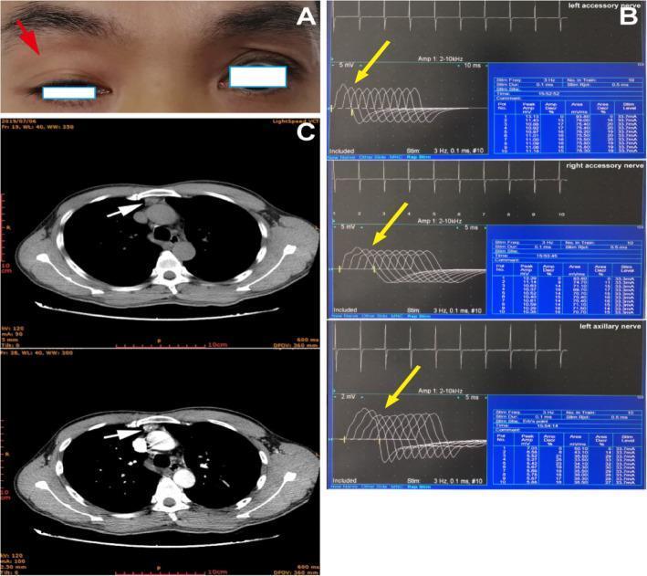
A composite medical image illustrating diagnostic manifestations of myasthenia gravis across three modalities. Panel A is a clinical photograph of the periorbital region showing asymmetrical ophthalmoparesis; a red arrow indicates significant ptosis of the right upper eyelid, which obscures the upper half of the pupil compared to the normal left eyelid position. Panel B displays results from a repetitive nerve stimulation (RNS) test of the bilateral accessory and left axillary nerves; yellow arrows highlight a decremental response in compound muscle action potential (CMAP) amplitudes, characteristic of neuromuscular junction dysfunction. Panel C contains two axial computed tomography (CT) images of the chest; white arrows point to an enlarged soft tissue mass in the anterior mediastinum, consistent with thymic hyperplasia. Together, these panels demonstrate the classic clinical triad of ocular symptoms (ptosis), electrophysiological evidence of fatigue, and associated anatomical abnormalities (thymus) often found in autoimmune myasthenia gravis.
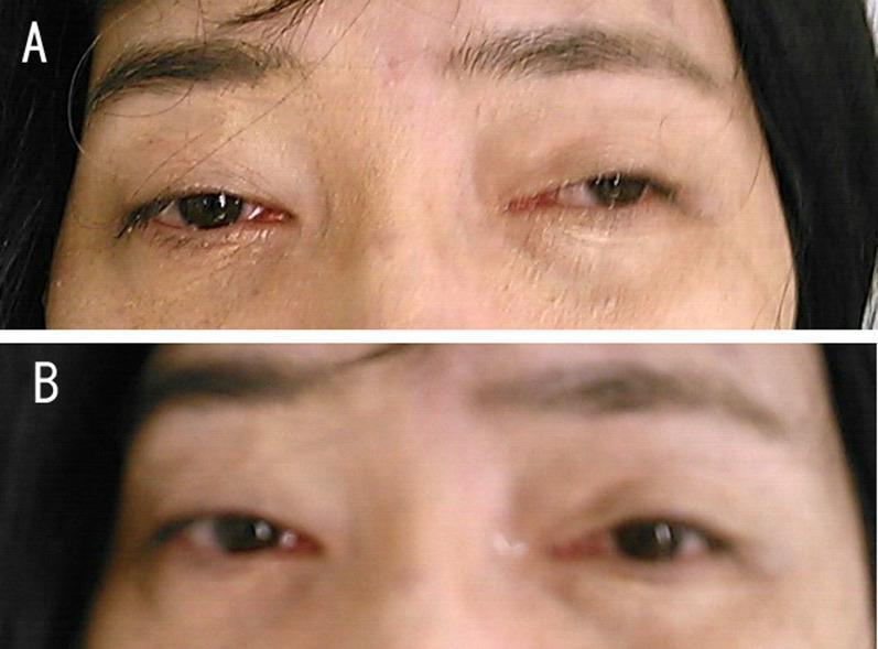
This clinical photograph is a side-by-side comparison illustrating a positive Tensilon (edrophonium) test in a patient with Myasthenia Gravis (MG). Panel A shows the patient's orbital region at baseline, demonstrating significant bilateral asymmetrical ptosis (drooping of the upper eyelids). The left eyelid is more severely affected, covering a substantial portion of the visual axis. Panel B shows the same patient after the administration of edrophonium, an acetylcholinesterase inhibitor. There is a marked improvement in the bilateral ptosis, characterized by an increased palpebral fissure width and elevation of the upper lid margins, exposing more of the iris and pupil. This visual change is a classic diagnostic indicator for disorders of the neuromuscular junction, specifically MG associated with anti-acetylcholine receptor (AchR) antibodies. The image serves as a clinical teaching tool for neurology and ophthalmology to demonstrate the transient reversal of fatiguable muscle weakness through pharmacological intervention.
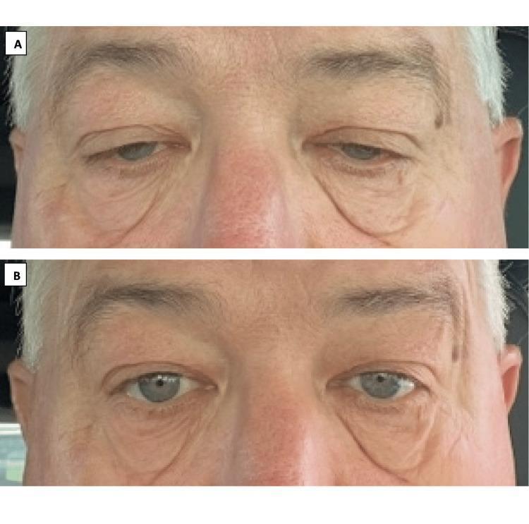
This clinical photograph consists of a side-by-side comparison (labeled A and B) demonstrating the pharmacological management of bilateral eyelid ptosis in a patient with Myasthenia Gravis (MG). Panel A (Baseline): Displays severe bilateral upper eyelid drooping, which obscures approximately 30-40% of the superior iris and limits the palpebral fissure width. The periorbital skin shows significant laxity and skin folds characteristic of chronic ptosis and aged skin. Panel B (Post-Treatment): Shows the same patient 30 minutes after the administration of oxymetazoline hydrochloride 0.1% ophthalmic solution. There is a notable elevation of both upper eyelids, resulting in increased exposure of the iris and sclera, with only 5-10% of the iris remaining covered. Educational Focus: The images illustrate the clinical efficacy of alpha-adrenergic agonists in stimulating Müller’s muscle (sympathetic smooth muscle) to improve eyelid elevation. This comparison highlights a non-surgical management option for symptomatic ptosis in patients with neuromuscular junction disorders or refractory weakness.
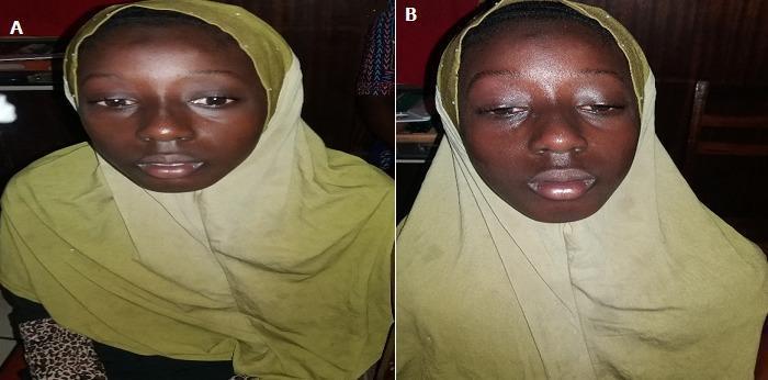
Two side-by-side clinical photographs (labeled A and B) documenting ocular symptoms in a pediatric patient. Image A demonstrates moderate bilateral ptosis, where the upper eyelids are abnormally low, partially obscuring the superior portion of the iris and pupil. Image B shows the same patient exhibiting significantly worsened or 'majoré' bilateral ptosis, with the eyelids nearly fully occluding the pupils. This visual progression illustrates muscular fatigability, a classic clinical sign of juvenile ocular myasthenia gravis. The comparison demonstrates how sustained upward gaze or prolonged activity can exacerbate levator palpebrae superioris weakness. The images are categorized under ophthalmology and clinical neurology, serving as an educational resource for identifying ptosis severity and the diagnostic hallmark of fatigability in neuromuscular junction disorders.
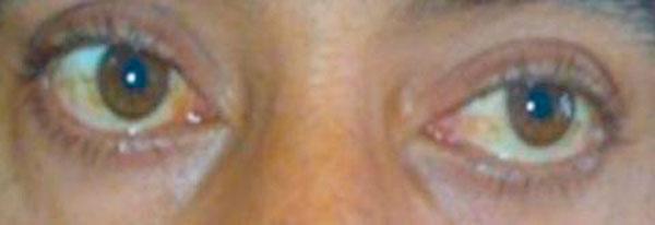
This clinical photograph shows a close-up view of a patient's eyes demonstrating bilateral asymmetric ptosis (drooping of the upper eyelids). The right upper eyelid (left side of the image) exhibits a more severe degree of ptosis compared to the left, resulting in a narrower palpebral fissure and significant coverage of the superior iris. Conversely, the left upper eyelid margin is positioned higher, revealing more of the ocular surface. The image captures the clinical response during a neostigmine test, a diagnostic investigation used in the evaluation of Myasthenia Gravis to assess for the temporary reversal of muscle weakness. Mild conjunctival injection is visible, particularly in the nasal region of the right eye. This visual is significant for medical education regarding the diagnostic workup of neuromuscular junction disorders and the clinical manifestation of ocular myasthenia.
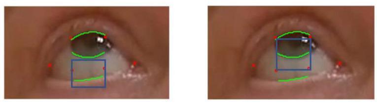
This clinical image consists of two side-by-side close-up photographs of a human eye, demonstrating computer vision and machine learning techniques for ophthalmologic assessment. The images utilize annotated overlays to track key anatomical landmarks related to Myasthenia Gravis (MG) evaluation, specifically for ptosis (eyelid droop). Red dots represent machine learning-derived landmarks at the eye corners and lid margins. Green curved lines delineate the upper eyelid contour, the lower eyelid margin, and the limbus (the interface between the iris and sclera). Blue bounding boxes define 'Regions of Interest' (ROI); in the left frame, the box targets the lower eyelid-sclera junction, while in the right frame, it frames the inferior iris-sclera boundary. This methodology is designed for automated, quantitative monitoring of eyelid distance and eye area during sustained upward gaze, providing objective data for telemedicine-based clinical examinations of neuromuscular junction disorders.
neuromuscular junction acetylcholine receptor antibody diagram
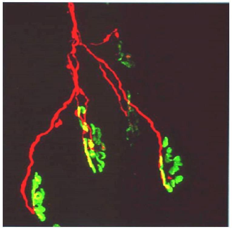
This diagnostic fluorescence microscopy image illustrates the neuromuscular junction (NMJ) architecture. The image features a single motor axon labeled with red fluorescence (TRITC-conjugated anti-neurofilament antibody), showing a characteristic distal branching pattern. These red axonal branches terminate at multiple motor end-plates, which are visualized in green (FITC-conjugated α-bungarotoxin) to reveal the high density of postsynaptic acetylcholine receptors (AChRs). The spatial relationship demonstrates a single axon innervating a cluster of six individual end-plates. Areas of colocalization between the presynaptic nerve terminal and the postsynaptic membrane appear yellow, indicating functional synaptic contact. This visual serves as an educational model for neuroanatomy and toxicology, particularly in the study of neurotoxic snake venoms (like those from the elapid family) that target presynaptic proteins or postsynaptic receptors, leading to neuromuscular paralysis and axonal degeneration.
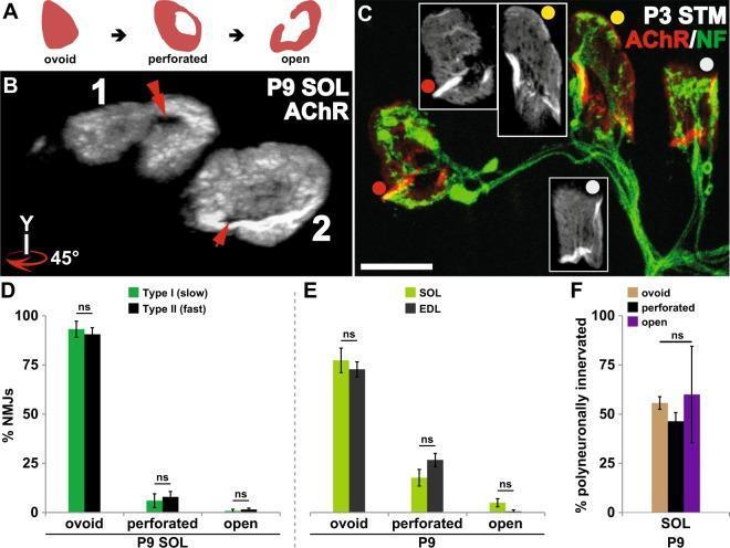
This composite educational image illustrates the maturation of postsynaptic acetylcholine receptor (AChR) aggregates at the neuromuscular junction (NMJ) and its relationship to synapse elimination. Panel A provides a schematic of morphological progression from ovoid plaque to perforated plaque to an open configuration. Panel B displays grayscale confocal images of P9 mouse soleus AChR aggregates, rotated 45 degrees to highlight central perforations (red arrows). Panel C shows a fluorescence micrograph of P3 sternomastoid NMJs labeled for AChR (red) and Neurofilament (green), demonstrating varied maturation stages: open (red dot), perforated (yellow dot), and ovoid (grey dot) within polyneuronally innervated synapses. Panels D and E are bar graphs comparing AChR maturation across muscle fiber types (Type I vs. Type II) and muscles (Soleus vs. EDL), showing no significant difference ('ns'). Panel F quantifies that the percentage of polyneuronally innervated NMJs does not significantly differ based on postsynaptic morphology. The data indicates that postsynaptic AChR maturation occurs independently of the timing of axonal input removal during development.
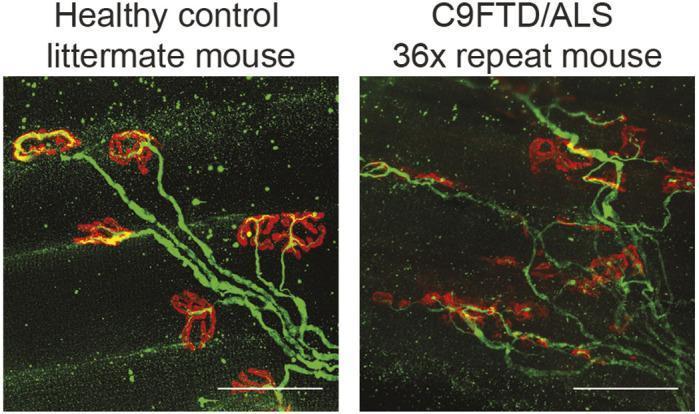
A side-by-side comparison of immunofluorescence diagnostic images showing neuromuscular junction (NMJ) pathology in a murine model of C9orf72 amyotrophic lateral sclerosis and frontotemporal dementia (ALS/FTD). The microscopy displays whole-mount staining of the extensor digitorum longus (EDL) muscle. Post-synaptic acetylcholine receptors are labeled with red α-bungarotoxin, while pre-synaptic axons and motor nerve terminals are labeled with green neurofilament antibody. The 'Healthy control' panel shows highly organized, linear axonal projections terminating in distinct, continuous, and robust 'pretzel-like' motor endplates. In contrast, the 'C9FTD/ALS 36x repeat' panel demonstrates significant neurodegeneration characterized by disorganized, fragmented axonal branching and 'dissolving' boutons with reduced or sparse red fluorescence. This visual evidence highlights the pathophysiology of denervation and synaptic instability in motor neuron disease. Scale bars represent 50 µm.
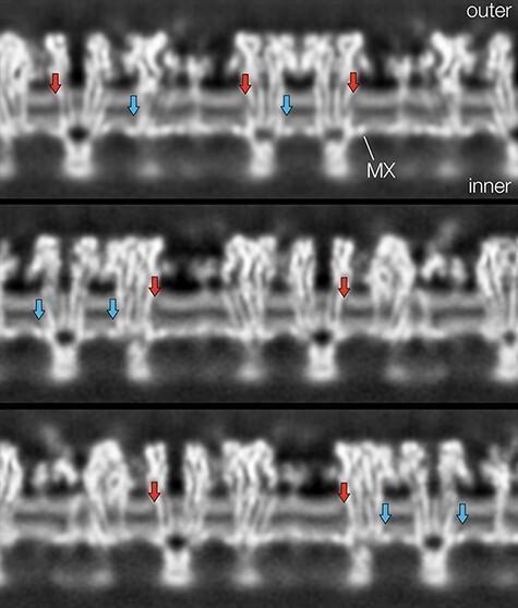
This diagnostic visualization presents cross-sections through a helical reconstruction of a cholinergic postsynaptic membrane, likely derived from cryo-electron microscopy. The image displays the profile of a lipid bilayer at the neuromuscular junction, characterized by two parallel tracks of high-intensity light grey densities representing phospholipid headgroups. The total thickness between these headgroup peaks is approximately 30 Å. Interspersed within this lipid matrix are irregular blocks of density representing nicotinic acetylcholine receptor proteins, including transmembrane (TM) helices and cytoplasmic MX helices. Red arrows highlight localized gaps or weakening of density in the outer leaflet adjacent to protein surfaces, while blue arrows indicate similar regions of weakened density in the inner leaflet above the MX helices. These specific visual gaps are attributed to high concentrations of cholesterol, which lacks a large electron-dense headgroup compared to phospholipids. The arrangement illustrates the non-uniform distribution of cholesterol and its role in forming protein-stabilized microdomains within the postsynaptic membrane.
myasthenia gravis thymus thymoma CT scan anterior mediastinum
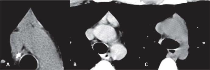
This diagnostic image contains three axial CT scan slices (labeled A, B, and C) demonstrating different morphological configurations of the thymus in the anterior mediastinum. (A) Displays an 'arrowhead' configuration where the two thymic lobes are confluent, forming a single triangular mass with soft-tissue attenuation similar to skeletal muscle. (B) Shows a 'bilobed' thymus presenting as two distinct, separate soft-tissue masses, appearing diffusely enlarged with density exceeding chest wall musculature. (C) Illustrates a single visualized lobe appearing as an asymmetrical soft-tissue mass. These imaging variants are critical in evaluating patients with myasthenia gravis, where thymic hyperplasia or thymoma may be present. The scans highlight essential anatomical relationships between the thymus and adjacent cardiovascular structures in the superior mediastinum, useful for differentiating normal thymic variants from pathology in adult patients.
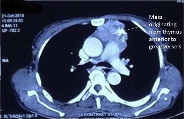
This diagnostic image is an axial contrast-enhanced computed tomography (CT) scan of the thorax at the level of the great vessels. The scan reveals a large, heterogeneously enhancing mass located in the anterior mediastinum. An annotation on the image indicates that the lesion originates from the thymus and is situated immediately anterior to the great vessels, including the ascending aorta and pulmonary trunk. The mass exhibits irregular borders and varying densities, which are characteristic findings for thymic neoplasms such as thymoma. The surrounding anatomical landmarks, including the vertebral body, descending aorta, and sternum, are clearly visible. This clinical image illustrates a common presentation of an anterior mediastinal tumor, often associated with paraneoplastic syndromes like myasthenia gravis. It serves as an educational example for radiological identification of mediastinal compartment pathologies and the evaluation of tumor relationship to vascular structures.
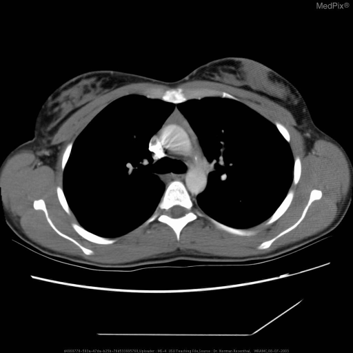
**Imaging Modality:** Computed Tomography (CT) scan of the chest. **Anatomical Region:** Axial section of the superior mediastinum at the level of the aortic arch. **Observed Pathology:** The image demonstrates a soft-tissue mass located in the anterior mediastinal compartment, specifically within the prevascular space. The morphology and location are consistent with thymic tissue, which in the context of myasthenia gravis, may represent thymic hyperplasia or a thymic neoplasm such as a thymoma. **Characteristic Visual Features:** * **Location:** Anterior to the ascending aorta and the superior vena cava, posterior to the sternum. * **Density:** The lesion exhibits soft-tissue attenuation, appearing denser than the surrounding mediastinal fat. * **Margins:** The mass appears relatively well-defined but lacks clear fatty cleavage planes from adjacent vascular structures, a common finding in thymic pathology. * **Surrounding Structures:** The lungs appear clear and the bony structures of the thoracic cage are intact. **Clinical Context:** Evaluation of the thymus is a critical diagnostic step for patients with myasthenia gravis to identify thymic abnormalities that may necessitate surgical intervention (thymectomy). **Diagnostic Significance:** This imaging is essential for differentiating normal age-related thymic involution (fatty replacement) from pathological thymic persistence or enlargement.
repetitive nerve stimulation decremental response EMG myasthenia gravis
A composite medical image illustrating diagnostic manifestations of myasthenia gravis across three modalities. Panel A is a clinical photograph of the periorbital region showing asymmetrical ophthalmoparesis; a red arrow indicates significant ptosis of the right upper eyelid, which obscures the upper half of the pupil compared to the normal left eyelid position. Panel B displays results from a repetitive nerve stimulation (RNS) test of the bilateral accessory and left axillary nerves; yellow arrows highlight a decremental response in compound muscle action potential (CMAP) amplitudes, characteristic of neuromuscular junction dysfunction. Panel C contains two axial computed tomography (CT) images of the chest; white arrows point to an enlarged soft tissue mass in the anterior mediastinum, consistent with thymic hyperplasia. Together, these panels demonstrate the classic clinical triad of ocular symptoms (ptosis), electrophysiological evidence of fatigue, and associated anatomical abnormalities (thymus) often found in autoimmune myasthenia gravis.
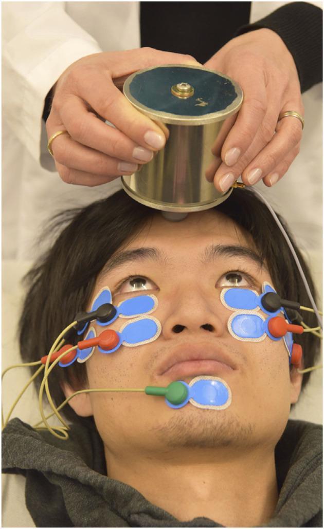
This clinical photograph demonstrates the experimental setup for recording repetitive ocular vestibular evoked myogenic potentials (roVEMP). The procedure involves a patient in a supine position with maximal upward gaze to facilitate recording from the inferior oblique extraocular muscles. A handheld cylindrical metallic mini-shaker is applied to the midline of the forehead at the hairline (standard AFz point) to deliver bone-conducted vibration stimuli. Recording is achieved via a multi-electrode montage on the face: active electrodes (black leads) are placed at the infraorbital margins, reference electrodes (red leads) are positioned directly below them, and a grounding electrode (green lead) is attached to the chin. All electrodes are secured with light-blue adhesive pads. This neurophysiological setup is used to assess vestibular function and neuromuscular transmission, specifically in the diagnosis and monitoring of conditions such as ocular myasthenia gravis, where fatigue or decrement in muscle response to repetitive stimulation is clinically significant.
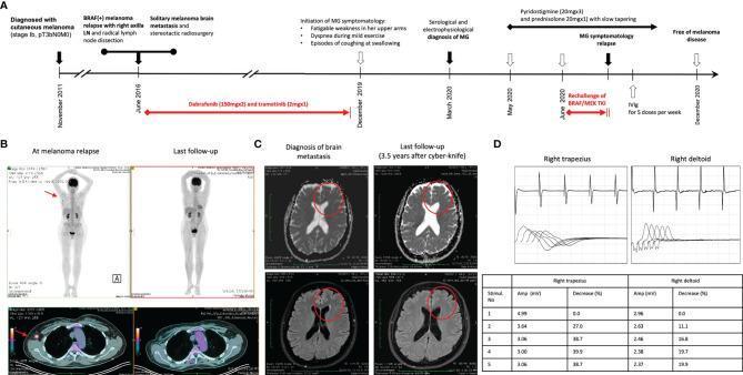
A multi-panel medical figure documenting a clinical case of metastatic melanoma and secondary myasthenia gravis (MG). Panel A presents a longitudinal timeline (2011–2020) tracking cutaneous melanoma diagnosis, BRAF(+) relapse, brain metastasis, and the subsequent onset of MG during targeted therapy with BRAF/MEK inhibitors (dabrafenib/trametinib). Panel B contains whole-body PET/CT scans and axial CT slices; the 'At melanoma relapse' images show a hypermetabolic focus in the right axilla (indicated by red arrows), which resolves at 'Last follow-up'. Panel C displays axial brain MRI slices (T1 and FLAIR) showing a solitary left frontal lobe metastasis at diagnosis, with subsequent volume reduction and scarring observed 3.5 years post-stereotactic radiosurgery (Cyber-knife). Panel D illustrates neurophysiological diagnostics for MG, including repetitive nerve stimulation (RNS) waveforms from the right trapezius and deltoid muscles. The accompanying table quantifies a pathological decrement in compound muscle action potential (CMAP) amplitude (up to 39.9%), diagnostic of a neuromuscular junction disorder.
🧠 MYASTHENIA GRAVIS - Comprehensive MD Medicine Notes
PART 1: CLINICAL CASE (Opening the Topic)
📋 CASE VIGNETTE
Mr. Raj, 28F, presents to Medicine OPD with a 4-month history of bilateral drooping of eyelids, worse in the evening and after prolonged reading. She also complains of double vision, difficulty chewing food by the end of meals, and nasal voice when talking for long periods. She denies any sensory symptoms, muscle pain, or recent infection. Examination reveals fatigable ptosis (worsens on sustained upward gaze), ophthalmoplegia, and proximal limb weakness that worsens after repetitive movements. Deep tendon reflexes are normal. Pupils are normal and reactive.
❓ MCQ BANK - DIFFERENTIALS (Attempt Before Reading On!)
- A. Lambert-Eaton Myasthenic Syndrome
- B. Myasthenia Gravis ✅
- C. Guillain-Barre Syndrome
- D. Botulism
Rationale: Fatigable, worsening-with-activity weakness + ptosis + normal reflexes + no sensory involvement = MG. LEMS has improving strength with activity; GBS has ascending weakness + areflexia; Botulism has descending weakness + autonomic signs.
- A. Myasthenia Gravis
- B. Botulism
- C. Lambert-Eaton Myasthenic Syndrome ✅
- D. Multiple Sclerosis
Rationale: LEMS = presynaptic VGCC antibodies. Classically associated with small cell lung cancer. Weakness IMPROVES briefly with repetition (post-tetanic potentiation). Autonomic features (dry mouth, impotence) are hallmark. Reflexes are ABSENT but reappear after exercise.
- A. Myasthenia Gravis
- B. GBS
- C. Botulism ✅
- D. LEMS
Rationale: Botulism = food-borne, descending, dilated fixed pupils, no fever, NO sensory loss, presynaptic ACh release block.
- A. Incremental response at 50Hz
- B. Decremental response at 2-3 Hz ✅
- C. Absent F-waves
- D. Fibrillation potentials
- A. IgG1 activating complement
- B. Anti-AChR IgG1
- C. IgG4 non-complement fixing ✅
- D. Anti-VGCC IgG2
PART 2: DIFFERENTIAL DIAGNOSIS - MG vs. Its Mimics
🔄 COMPARISON TABLE: MG vs. All Differentials
| Feature | MG | LEMS | Botulism | GBS | Eaton-Lambert Overlap | Mitochondrial Myopathy |
|---|---|---|---|---|---|---|
| Mechanism | Post-synaptic AChR (anti-AChR Ab) | Pre-synaptic VGCC (P/Q type Ca²⁺ channels) | Pre-synaptic (Clostridium toxin blocks ACh vesicle release) | Peripheral nerve (anti-ganglioside Ab) | Both | Mitochondrial DNA mutations |
| Onset | Subacute, fluctuating | Subacute | Acute (hours-days) | Acute ascending | Variable | Chronic |
| Weakness pattern | Ocular first (ptosis, diplopia) > bulbar > limb | Proximal LEGS first → arms → bulbar | Descending: cranial → bulbar → limb | Ascending: limb → respiratory | Mixed | Proximal, chronic |
| Weakness with activity | WORSENS (fatigue) | Initially WORSENS, then IMPROVES briefly | Constant | Constant | Variable | Constant |
| Ptosis | Bilateral, fatigable | Less common | Present | Absent | Present | Present (bilateral, symmetric) |
| Pupils | Normal | Normal | Dilated, fixed (mydriasis) | Normal | Normal | Normal or mild abnormality |
| Deep Tendon Reflexes | Normal | Absent (improve post-exercise) | Normal or absent | Absent (areflexia) | Variable | Normal |
| Sensory involvement | Absent | Absent | Absent | Present (glove-stocking) | Absent | Absent |
| Autonomic | Absent | Present (dry mouth, impotence, orthostasis) | Present (constipation, urinary retention, dry mouth) | Present (BP fluctuations, arrhythmia) | Variable | Absent |
| CSF | Normal | Normal | Normal | Cytoalbuminous dissociation | Normal | Normal |
| EMG (RNS at 2-3Hz) | Decremental >10% | Incremental >200% at 50Hz | Incremental at high-frequency | Normal or prolonged latencies | Both | Myopathic |
| Key antibody | Anti-AChR (85%), Anti-MuSK (10%) | Anti-P/Q VGCC | None | Anti-GQ1b (Miller Fisher), Anti-GM1 | Both | None |
| Association | Thymoma, autoimmune diseases | Small cell lung cancer | Contaminated canned food, honey (infants) | Campylobacter, CMV, EBV | Thymoma + SCLC | Family history |
| Treatment | AChE inhibitors, immunosuppression, thymectomy | 3,4-DAP, IVIG, treat underlying cancer | Antitoxin, supportive | IVIG, plasmapheresis, supportive | Both approaches | Supportive, supplements |
🔑 KEY DIFFERENTIATING MNEMONICS
- Myasthenia Gravis = Weakness Worsens with activity
- LEMS = Limbering up Lessens weakness (briefly improves)
- Botulism = Descending paralysis (cranial → trunk → limbs)
- GBS = Ascending paralysis (feet → legs → arms → face)
PART 3: MYASTHENIA GRAVIS - THE MAIN TOPIC
🔵 DEFINITION
"Myasthenia Gravis" = Greek/Latin for "grave muscle weakness"
📊 EPIDEMIOLOGY / INCIDENCE
| Parameter | Data |
|---|---|
| Prevalence | ~20/100,000 (US); approximately 60,000 patients |
| Trend | Increasing prevalence over past 50 years (better ascertainment, ageing population) |
| Gender - Age <40 | Women 3x more commonly affected than men |
| Gender - Age >50 | Males more commonly affected than females |
| Gender - Puberty | Roughly equal |
| Peak age (female) | 2nd-3rd decade |
| Peak age (male) | 6th-7th decade |
| Current US trend | Majority of patients now >50 years old; more men than women overall |
Mnemonic - "Young Women, Old Men": Early-onset MG = young women (thymic hyperplasia). Late-onset MG = older men (thymic atrophy/thymoma).
⚙️ ETIOLOGY
- ~10% of MG patients have a thymoma (thymic epithelial neoplasm)
- ~30% (especially young patients) have thymic hyperplasia with B-cell follicles
- Thymoma MG: usually in men, age >40, AChR+ antibodies with anti-titin and anti-ryanodine antibodies
- Genetic predisposition (HLA haplotypes)
- Other autoimmune diseases (thyroid disease, SLE, RA)
- Drugs: D-penicillamine, checkpoint inhibitors (pembrolizumab), interferon-alpha
- Pregnancy (can precipitate MG or neonatal MG)
🔬 PATHOPHYSIOLOGY
Normal Neuromuscular Junction:
- Motor nerve action potential arrives at the NMJ
- Voltage-gated Ca²⁺ channels (VGCCs) open at presynaptic terminal
- Ca²⁺ influx triggers ACh vesicle exocytosis into the synaptic cleft
- ACh binds to nicotinic AChR on the postsynaptic membrane (junctional folds)
- Na⁺ influx → endplate potential → muscle action potential → contraction
In Myasthenia Gravis:
- T-helper cells in thymus activate B-cells
- B-cells produce anti-AChR IgG1 antibodies (in 80-85% of cases)
- Antibodies directed against the alpha subunit of the nicotinic AChR
| Mechanism | Description |
|---|---|
| 1. Complement-mediated destruction | Anti-AChR IgG1 activates complement → C5-C9 membrane attack complex → destruction of postsynaptic junctional folds |
| 2. Antigenic modulation | Antibody cross-links adjacent AChRs → accelerated internalization and degradation (reduces receptor number) |
| 3. Functional blockade | Antibody directly blocks ACh binding site on AChR (less common) |
- AChR number reduced dramatically
- Postsynaptic membrane simplified (loss of junctional folds)
- Safety factor for neuromuscular transmission reduced
- At high rates of stimulation (exercise), depleting ACh causes transmission failure
- Result = fatigable, worsening-with-use weakness
Why Fatigable?
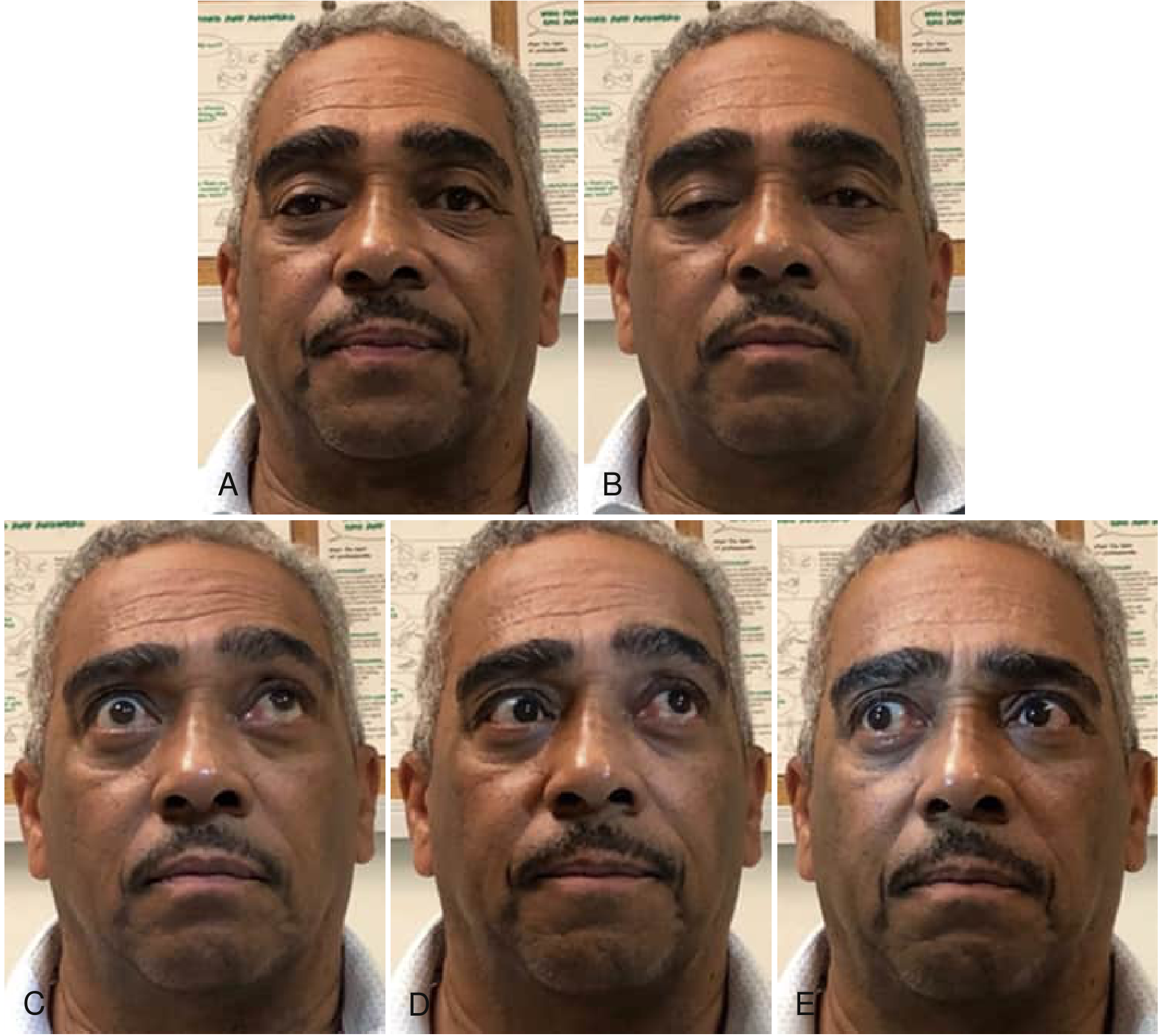
🖼️ Ocular Motility Abnormalities in MG
🩺 CLINICAL PRESENTATION
🔑 Mnemonic: "PODBC" (Ptosis Opens the Door Before Crisis)
- Ptosis + diplopia - first in 2/3 of patients
- Oropharyngeal/bulbar weakness (chewing, swallowing, speech)
- Dyspnea (respiratory involvement in crisis)
- Body/limb weakness (proximal > distal)
- Confirmation: fatigability, diurnal variation (worse evening)
Ocular Symptoms (Onset in ~67% of patients)
- Ptosis - bilateral and asymmetric; worsens with sustained upward gaze ("fatigue ptosis")
- Diplopia - from ophthalmoplegia
- Virtually all patients develop BOTH within 2 years of onset
- Cogan's Lid Twitch sign: Brief upward overshoot of lid when moving eyes from downward to primary gaze
Bulbar Symptoms (Onset in ~15%)
- Dysarthria - nasal or dysarthric speech (cranial nerve X, XII weakness)
- Dysphagia - chewing difficulty worsened by end of meals ("jaw fatigue")
- Facial weakness - inability to smile ("snarling smile"), difficulty closing eyelids (lagophthalmos)
- "Myasthenic Snarl": Weakness of facial muscles + attempt to smile = snarl-like appearance
Limb/Axial Symptoms (Onset in ~10%)
- Neck weakness: Neck flexors > extensors ("dropped head syndrome")
- Proximal > distal limb weakness
- Deltoids, triceps, wrist extensors, ankle dorsiflexors commonly affected
- No sensory symptoms
- Normal reflexes (IMPORTANT distinguishing feature!)
Respiratory
- Respiratory muscle weakness can lead to Myasthenic Crisis (20% of patients)
- Diaphragm and intercostal muscles affected
Diurnal Pattern
- Least in the morning, worst in evening
- Worsens with heat, exercise, illness, stress
🔄 CLINICAL SIGNS
| Sign | Description |
|---|---|
| Fatigable ptosis | Ptosis worsens on sustained upward gaze for 30-60 seconds |
| Cogan's lid twitch | Brief upward twitch of lid when gaze returns from downgaze to primary position |
| Simpson's test | Sustained upward gaze >30 seconds → increasing ptosis |
| Peek sign | Patient asked to close eyes tightly → gradually opens slightly (orbicularis weakness) |
| Nasal speech | Soft palate weakness |
| Neck drop sign | Chin drops to chest on neck flexor testing |
| Ice pack test | Ice on ptotic lid for 2 min → ptosis improves (cooling slows AChE activity → more ACh available) |
| Snarl sign | Attempt to smile produces a snarl-like expression (facial weakness) |
📐 TYPES / CLASSIFICATION
A. By Osserman Classification (Classic):
| Grade | Description |
|---|---|
| Grade I | Ocular MG only |
| Grade IIA | Mild generalized MG, no respiratory |
| Grade IIB | Moderate generalized MG, no respiratory |
| Grade III | Severe generalized MG (acute onset) |
| Grade IV | Severe generalized MG (late severe) |
| Grade V | Myasthenic Crisis (respiratory failure) |
B. By MGFA (Myasthenia Gravis Foundation of America) Classification:
| Class | Description |
|---|---|
| Class I | Any ocular muscle weakness only |
| Class II | Mild weakness other than ocular |
| IIa | Predominantly limb/axial |
| IIb | Predominantly bulbar/respiratory |
| Class III | Moderate weakness |
| IIIa / IIIb | Limb/axial or Bulbar |
| Class IV | Severe weakness |
| Class V | Intubation needed (crisis) |
C. By Serological Subtypes (MOST IMPORTANT FOR EXAMS):
| Subtype | Antibody | Thymus | Age | Sex | Features |
|---|---|---|---|---|---|
| Early-onset generalized | AChR | Hyperplasia | <50 years | F>M (1:3) | Classic presentation |
| Late-onset generalized | AChR + Titin/Ryanodine | Normal/atrophic | >50 years | M>F | Thymectomy less beneficial |
| Thymoma-associated | AChR + Titin + Ryanodine | Thymoma | Any (usually >40) | Equal | Anti-titin ab = severe disease |
| MuSK MG | Anti-MuSK (IgG4) | Normal | <40 years | F >> M | Prominent facial/bulbar/respiratory; POOR response to AChEI; atrophy |
| LRP4 MG | Anti-LRP4 | Unknown | 30-50 years | F predominant | Oropharyngeal + respiratory selective weakness |
| Seronegative | None detectable (some: anti-clustered AChR, agrin, cortactin) | Hyperplasia in some | Variable | Variable | Treatment response similar to AChR MG |
| Ocular MG | AChR 50% | Unknown | Adults (West); Children (Asia) | Variable | Eye only; 50% generalize within 2 years |
| Neonatal MG | Maternal AChR ab (transient) | Normal | Neonates | Equal | Transient (2-8 weeks); hypotonia, poor feeding |
Mnemonic - MuSK MG "FARMS":
- Facial/bulbar prominence
- Atrophophy of affected muscles
- Refractory to AChE inhibitors
- More common in young Females
- Sensitive to Steroids and Rituximab
🔗 ASSOCIATIONS WITH OTHER DISEASES
Mnemonic: "MG MATES with Thymes"
- Myositis
- Graves' disease / Thyroid disease (most common - 10-15%)
- MG-Autoimmune overlap
- Thymoma (10%) and Thymic hyperplasia (30%)
- Epithelial autoimmune diseases (SLE, RA, Sjogren's)
- SLE, Sarcoidosis
Full List of Associations:
| Disease | Notes |
|---|---|
| Thyroid disease | Most common (10-15%): Graves' disease, Hashimoto's thyroiditis |
| Thymoma | 10% of MG have thymoma; 40% of thymoma patients have MG |
| Thymic hyperplasia | 30% of MG (especially early-onset) |
| Rheumatoid Arthritis | Autoimmune overlap |
| SLE | Autoimmune overlap |
| Pernicious anemia | Autoimmune anti-parietal cell Ab |
| Type 1 Diabetes | Autoimmune |
| Pemphigus vulgaris | Anti-desmoglein Ab |
| Sarcoidosis | Rare |
| Lambert-Eaton Overlap | Rare; both VGCC and AChR Ab |
| Immune checkpoint inhibitor therapy | Pembrolizumab, nivolumab can CAUSE MG |
🧪 INVESTIGATIONS & DIAGNOSIS
🔑 Mnemonic: "ACE IT"
- Antibodies (anti-AChR, anti-MuSK, anti-LRP4)
- Chest CT (thymoma/thymic hyperplasia)
- Electrophysiology (RNS, SFEMG)
- Ice pack test / edrophonium test
- Thyroid function tests (associated thyroid disease)
1. Antibody Tests
| Test | Sensitivity | Specificity | Notes |
|---|---|---|---|
| Anti-AChR Ab (RIPA) | 85% (generalized MG); 50% (ocular MG) | >99% | First-line test; IgG1 |
| Anti-MuSK Ab | Up to 50% of AChR-seronegative GMG | High | IgG4; not complement-fixing; poor AChEI response |
| Anti-LRP4 Ab | ~3-5% of seronegative | High | IgG1 |
| Anti-Titin Ab | ~30% of generalized | High | Marker for thymoma MG; severe disease |
| Anti-Ryanodine Ab | Variable | Moderate | Associated with thymoma and severe disease |
| Low-affinity anti-AChR | ~65% of doubly seronegative | Moderate | Cell-based assay (clustered AChR) |
2. Pharmacological Tests
- Edrophonium = ultra-short-acting AChE inhibitor (onset 30-60 sec, duration 5-10 min)
- Technique: IV edrophonium 2mg test dose → then 8mg if no adverse effect
- Positive test: Rapid, dramatic improvement in ptosis/diplopia/weakness
- Sensitivity ~70-90% for ocular MG
- Atropine must be at bedside (muscarinic side effects: bradycardia, bronchospasm)
- Contraindications: Severe COPD, cardiac arrhythmias
- Alternative where edrophonium unavailable
- 1.5 mg IM; peak effect at 30-45 minutes
- Give atropine 0.6mg beforehand
- Non-pharmacological; cold slows AChE activity → more ACh at NMJ
- Ice pack on ptotic lid for 2 minutes
- Positive: Ptosis improves by ≥2mm
- Sensitivity ~77-96%, Specificity ~98%
- Cannot be done in all patients (iris damage risk if corneal disease)
3. Electrophysiology
- Stimulate nerve at 2-3 Hz; decremental response >10% in 4th-5th potential = positive
- Sensitivity: 53-89% in generalized MG; 48-67% in ocular MG
- More sensitive in proximal/facial muscles
- Post-exercise exhaustion test: Exercise the muscle for 30-60 seconds → RNS again → increased decrement
- Most sensitive test for NMJ dysfunction (close to 100%)
- Measures jitter (variability of inter-potential interval between 2 muscle fibers of same motor unit)
- Increased jitter = abnormal NMT
- Normal jitter in a weak muscle EXCLUDES MG
- Not specific for MG (abnormal in other NMJ disorders too)
| Test | Sensitivity (Gen MG) | Sensitivity (Ocular MG) | Notes |
|---|---|---|---|
| Anti-AChR Ab | 85% | 50% | Highly specific |
| RNS | 53-89% | 48-67% | Low sensitivity in mild/ocular disease |
| SFEMG | ~99% | ~90% | Most sensitive; not specific |
| Ice pack test | 77-96% (for ptosis) | High | Non-specific for NMJ |
| Edrophonium | 70-90% | 70-90% | False positives possible |
4. Imaging
- Mandatory in ALL MG patients to look for thymoma or thymic hyperplasia
- MRI superior for soft tissue characterization
- PET-CT if malignant thymoma suspected
5. Other Tests
- TFTs + anti-thyroid antibodies (Graves/Hashimoto)
- ANA, RF (screen for associated autoimmune diseases)
- CBC, LFTs, RFTs (baseline before immunosuppression)
- Pulmonary function tests (FVC, NIF - monitor respiratory reserve in crisis)
💊 TREATMENT AND MANAGEMENT
Overview - The "Staircase" Approach
    Myasthenic Crisis → URGENT: ICU + Mechanical Ventilation + IVIG / Plasmapheresis
         ↑
    Immunosuppression (Azathioprine / Mycophenolate / Cyclosporine / Methotrexate)
         ↑
    Corticosteroids (Prednisolone - start low, escalate)
         ↑
    Thymectomy (if thymoma or thymic hyperplasia, age 18-65)
         ↑
    Symptomatic: AChE Inhibitors (Pyridostigmine)
1. Symptomatic Treatment - Acetylcholinesterase (AChE) Inhibitors
| Drug | Dose | Duration | Notes |
|---|---|---|---|
| Pyridostigmine (Mestinon) | 30-60mg every 4-6h | 3-6 hours | First-line symptomatic; titrate to symptoms |
| Neostigmine | 15-30mg oral 4x/day | 2-4 hours | Less used for chronic MG |
- Managed with propantheline or glycopyrrolate
2. Thymectomy
| Indication | Recommendation |
|---|---|
| Thymoma | MANDATORY - regardless of MG severity |
| Early-onset MG (AChR+, age 18-50, thymic hyperplasia) | Recommended - improves long-term outcome |
| Late-onset MG (>50 years, atrophic thymus) | Less clear benefit |
| MuSK MG | Generally NOT recommended |
3. Corticosteroids
- Prednisolone - first-line immunosuppressant for MG
- Start LOW (15-20mg alternate days) and escalate slowly
- WARNING: Transient worsening in first 1-2 weeks (initial exacerbation is common - warn patient/hospitalize if severe MG)
- Taper slowly once stable
- Long-term: Use lowest effective dose; alternate-day dosing reduces side effects
4. Steroid-Sparing Immunosuppressants
| Drug | Dose | Onset of Action | Notes |
|---|---|---|---|
| Azathioprine | 2-3 mg/kg/day | 6-18 months | Check TPMT enzyme before starting; hepatotoxic |
| Mycophenolate mofetil | 1-1.5g twice daily | 3-6 months | Good tolerability |
| Cyclosporine | 3-5 mg/kg/day | 1-6 months | Monitor BP and renal function |
| Tacrolimus | 0.1 mg/kg/day | 1-3 months | Useful in refractory MG |
| Methotrexate | 10-20 mg/week | 6-12 months | Alternative steroid-sparer |
| Rituximab | 375 mg/m²/week × 4 | 3-6 months | Excellent for MuSK MG; anti-CD20 B-cell depletion |
5. Acute Immunotherapy (Short-term, Fast-acting)
| Treatment | Mechanism | Onset | Duration of Effect | Use |
|---|---|---|---|---|
| Plasmapheresis (PLEX) | Removes circulating AChR abs | Days | 4-8 weeks | Crisis, pre-operative, refractory |
| IVIG | Modulates immune response, Fc receptor blockade | Days | 4-8 weeks | Crisis, pre-operative; equivalent to PLEX |
6. Novel / Targeted Therapies
| Drug | Target | Class | Notes |
|---|---|---|---|
| Eculizumab (Soliris) | C5 complement | Anti-C5 monoclonal antibody | FDA-approved for refractory AChR+ generalized MG; blocks complement-mediated AChR destruction |
| Ravulizumab | C5 complement | Long-acting anti-C5 | Every 8-week dosing |
| Efgartigimod (Vyvgart) | FcRn receptor | Anti-FcRn antibody | Reduces IgG (including AChR abs) recycling; FDA-approved 2021 |
| Rozanolixizumab | FcRn | Anti-FcRn | Subcutaneous; reduces pathogenic IgG |
| Zilucoplan | C5 | Anti-C5 | Subcutaneous self-injection |
| Rituximab | CD20 B-cells | Anti-CD20 | Especially effective in MuSK MG |
⚠️ DRUGS TO AVOID IN MG (Precipitate Weakness!)
Mnemonic: "ABCDE + FAMS"
- Aminoglycosides (gentamicin, tobramycin)
- Beta-blockers
- Chloroquine/Hydroxychloroquine
- D-penicillamine (can CAUSE MG!)
- Erythromycin/macrolides, Echelating agents (desferrioxamine)
- Fluoroquinolones (ciprofloxacin, levofloxacin)
- Anti-arrhythmics: Procainamide, quinidine
- Magnesium (IV - for eclampsia)
- Statins (can worsen/precipitate MG)
🚨 MYASTHENIC CRISIS vs. CHOLINERGIC CRISIS
CRITICAL FOR EXAMS!
| Feature | Myasthenic Crisis | Cholinergic Crisis |
|---|---|---|
| Cause | Undertreatment, disease exacerbation, infection, surgery | Overdose of AChE inhibitors |
| Mechanism | Too LITTLE ACh activity at NMJ | Too MUCH ACh activity (muscarinic + nicotinic) |
| Pupils | Dilated (mydriatic) | Constricted (miotic) |
| Secretions | Minimal | Excessive (SLUDGE) |
| Heart rate | Normal/elevated | Bradycardia |
| GI | Normal | Abdominal cramps, diarrhea |
| Fasciculations | Absent | Present |
| Tensilon test | Improves weakness | Worsens weakness (or no change) |
| Treatment | IVIG / Plasmapheresis + Increase AChEI | STOP AChEI + Atropine |
Mnemonic for Cholinergic Crisis - "SLUDGE + BAM":
- Salivation, Lacrimation, Urination, Defecation, GI distress, Emesis
- Bradycardia, Abdominal cramps, Miosis
📋 MANAGEMENT ALGORITHM
Confirmed MG Diagnosis
        |
        ├── Myasthenic Crisis?
        │      YES → ICU, intubation if FVC < 15ml/kg or NIF < -20 cmH₂O
        │             Plasma exchange OR IVIG (equivalent efficacy)
        │             Temporary hold AChEI if cholinergic crisis suspected
        │
        ├── Thymoma present?
        │      YES → Thymectomy (after medical stabilization)
        │
        ├── ALL patients: AChE inhibitor (Pyridostigmine)
        │
        ├── Insufficient control?
        │      → Prednisolone (start low, increase slowly)
        │      → Add Azathioprine (or MMF) as steroid-sparer
        │
        ├── Refractory generalized AChR+ MG?
        │      → Eculizumab or Efgartigimod
        │
        └── MuSK MG?
               → Rituximab (highly effective)
               → Avoid or minimize AChEI
               → Immunosuppression (steroids + MMF)
🔮 PROGNOSIS
| Parameter | Data |
|---|---|
| Overall prognosis | Good with treatment; most patients achieve "minimal manifestations" |
| Spontaneous remission | ~10-20% in early-onset MG |
| Complete stable remission | 50% with thymectomy (early-onset AChR+ MG) |
| Ocular → Generalized | 50% of ocular MG generalizes within 2 years; after 2 years of ocular only, generalization is unlikely |
| Mortality | <5% with modern management (mainly from myasthenic crisis complications) |
| Crisis mortality | ~3-4% with mechanical ventilation and ICU care |
| Thymoma MG | Less likely to remit; removal of thymoma does NOT cure MG |
| Predictors of poor prognosis | Older age, late onset, thymoma, MuSK antibodies, anti-titin/ryanodine antibodies |
| MuSK MG | More severe, atrophy; less responsive to AChEI; but responds well to rituximab |
- ~1/3 remain ocular only
- ~1/3 have mild generalized disease
- ~1/3 have severe or life-threatening disease
PART 4: COMPLETE MNEMONIC SUMMARY SHEET
🧠 MASTER MNEMONICS FOR MG
- Ptosis
- Ophthalmoplegia (diplopia)
- Dysphagia/Dysarthria (bulbar)
- Body (limb) weakness proximal > distal
- Characteristic: fatigability, worse in evening
- AChR antibody (80-85%) - IgG1, complement fixing
- MuSK antibody (10% of seronegative) - IgG4, NO complement
- LRP4 antibody (rare)
- Complement-mediated destruction of junctional folds
- Cross-linking and accelerated endocytosis of AChR (antigenic modulation)
- Confiscation of ACh binding site (functional blockade)
- Antibodies (AChR, MuSK, LRP4)
- Chest CT (thymoma/hyperplasia)
- Electrophysiology (RNS decrement, SFEMG jitter)
- Ice pack + edrophonium test
- Thyroid function (associated disease)
- Aminoglycosides
- Beta-blockers
- Chloroquine
- D-penicillamine
- Erythromycin/macrolides
- Fluoroquinolones
- Anti-arrhythmics (procainamide)
- Magnesium IV
- Statins
- Myasthenic = Under-treated = Dilated pupils
- Cholinergic = SLUDGE + Bradycardia + Miosis
- Facial/bulbar prominent
- Atrophy of muscles
- Refractory to pyridostigmine
- More female
- Sensitive to steroids and rituximab
- Pyridostigmine (symptomatic)
- Azathioprine (steroid-sparer)
- Thymectomy (if indicated)
- IVIG / Plasmapheresis (crisis / pre-op)
- Steroids (prednisolone - backbone immunotherapy)
- Rituximab (MuSK MG)
- Eculizumab (refractory AChR+ GMG)
- Crisis = ICU + Ventilator
- Myositis
- Graves' + thyroid disease (most common)
- Malignancy (thymoma)
- Autoimmune: RA, SLE, Sjogren's
- Thymoma 10% / Thymic hyperplasia 30%
- Endocrine: DM type 1, pernicious anemia
- Sarcoidosis
- LEMS: incremental (UP) response at high-frequency RNS
- MG: decremental (DOWN) response at low-frequency RNS (2-3 Hz)
PART 5: QUICK REVISION - HIGH-YIELD EXAM POINTS
| # | High-Yield Point |
|---|---|
| 1 | MG = MOST COMMON primary disorder of neuromuscular transmission |
| 2 | Anti-AChR ab present in 85% generalized, 50% ocular MG |
| 3 | Ptosis + diplopia = presenting feature in 2/3 of patients |
| 4 | Weakness WORSENS with activity, IMPROVES with rest |
| 5 | Deep tendon reflexes NORMAL (key differentiator from GBS/LEMS) |
| 6 | Ice pack test: improves ptosis in MG; sensitivity ~77-96% |
| 7 | Tensilon test: edrophonium IV → rapid improvement confirms MG |
| 8 | RNS decremental >10% at 2-3 Hz = positive for MG |
| 9 | SFEMG (jitter) = most sensitive test (~99%) |
| 10 | Anti-MuSK ab = IgG4, no complement, poor AChEI response, treat with rituximab |
| 11 | Thymoma in 10% MG; 40% thymoma patients have MG |
| 12 | All MG patients need CT chest at diagnosis |
| 13 | Myasthenic crisis: FVC <15 ml/kg → intubate; treat with IVIG or PLEX |
| 14 | Myasthenic (dilated pupils) vs Cholinergic crisis (miotic, SLUDGE) |
| 15 | Steroids can cause initial WORSENING in first 2 weeks |
| 16 | D-penicillamine can CAUSE drug-induced MG |
| 17 | Ocular MG: 50% generalize within 2 years of onset |
| 18 | Neonatal MG: transient (2-8 weeks); from passive transfer of maternal AChR abs |
| 19 | Eculizumab: anti-C5, FDA-approved for refractory AChR+ generalized MG |
| 20 | Efgartigimod: anti-FcRn, FDA-approved 2021; reduces total IgG including AChR abs |
Sources: Bradley & Daroff's Neurology in Clinical Practice, Harrison's Principles of Internal Medicine 22e (2025), Goldman-Cecil Medicine, Washington Manual of Therapeutics, Rosen's Emergency Medicine, Adams & Victor's Principles of Neurology, Robbins & Kumar Pathologic Basis of Disease, Medical Physiology.