
Femoral triangle
Reading File
The femoral triangle is a clinically important fascial compartment in the upper anterior thigh containing the femoral nerve, artery, and vein. Let me search the medical library for comprehensive content.
Searching sources...
Finding Sources
Finding Sources
Reading File
Reading File
Reading File
Reading File
Reading File
Loading Image
Loading Image
Loading Image
Here is a comprehensive overview of the femoral triangle, sourced from Gray's Anatomy for Students, the THIEME Atlas of Anatomy, and Fischer's Mastery of Surgery.
Femoral Triangle
The femoral triangle is a wedge-shaped (pyramid-shaped) depression formed by muscles in the upper anterior thigh, at the junction between the anterior abdominal wall and the lower limb. It serves as a major passageway for neurovascular structures entering the lower extremity.
Boundaries
| Structure | Role |
|---|---|
| Inguinal ligament | Superior border (base of triangle) |
| Medial margin of sartorius | Lateral border |
| Medial margin of adductor longus | Medial border |
The apex points inferiorly and is continuous with the adductor canal (Hunter's canal), which descends medially down the thigh and passes through the adductor hiatus in adductor magnus to open into the popliteal fossa behind the knee.
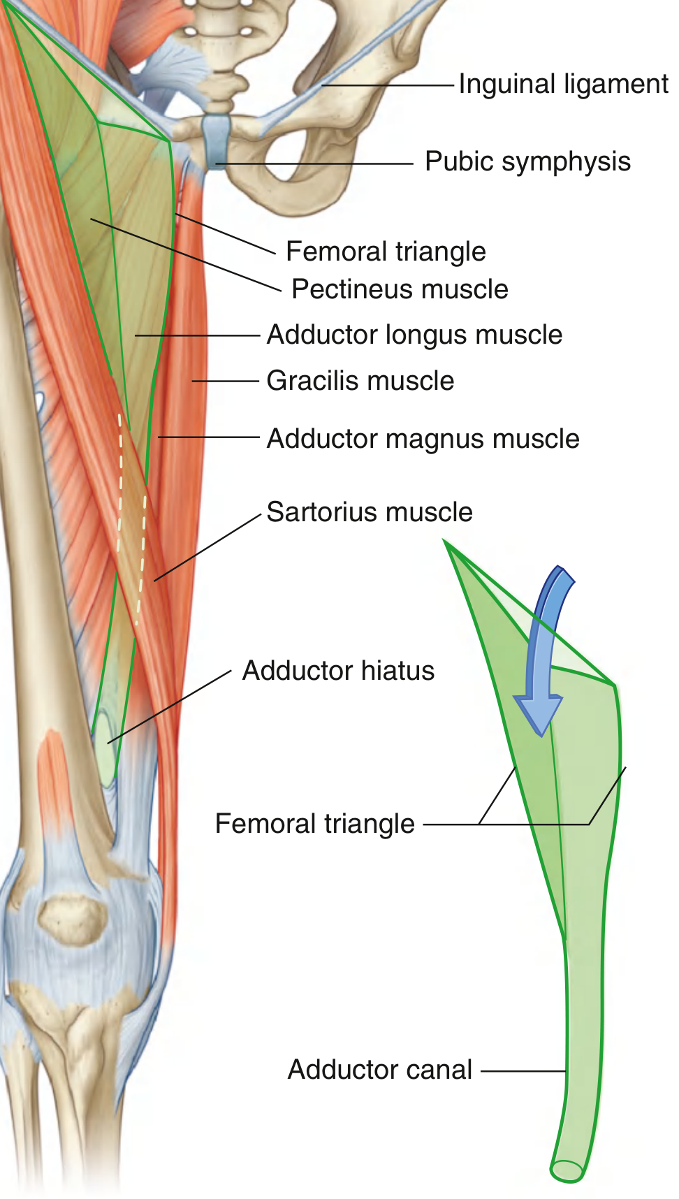
Floor
The floor is formed by two muscles, from lateral to medial:
- Iliopsoas (laterally) - descending from the abdomen
- Pectineus (medially)
- Adductor longus completes the medial floor
Roof
The roof is formed by the fascia lata (deep fascia of the thigh) and skin, pierced by the great saphenous vein as it passes through the saphenous opening (fossa ovalis) to drain into the femoral vein.
Contents (lateral to medial)
The mnemonic NAVY (from lateral to medial) is commonly used:
| Structure | Position | Notes |
|---|---|---|
| Femoral Nerve | Most lateral | Lies outside the femoral sheath |
| Femoral Artery | Middle | Continuation of external iliac artery under inguinal ligament |
| Femoral Vein | Medial to artery | Common femoral vein; receives great saphenous vein |
| Lymphatics / Femoral Canal | Most medial | Deep inguinal lymph nodes; medial compartment of femoral sheath |
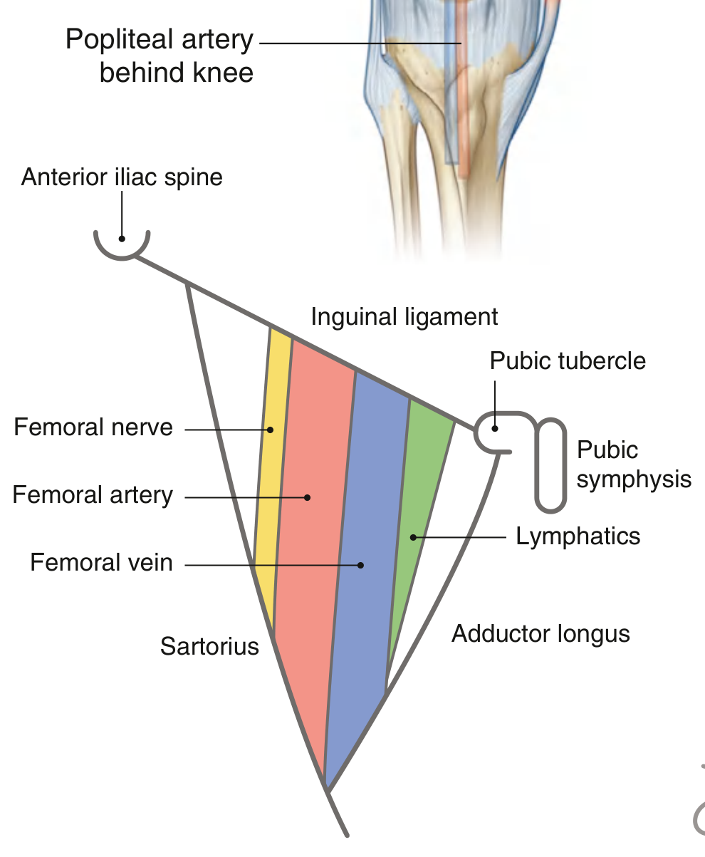
Femoral Sheath
The femoral artery, vein, and lymphatics (but NOT the femoral nerve) are enclosed within the femoral sheath - a funnel-shaped fascial sleeve:
- Continuous superiorly with transversalis fascia (anterior) and iliac fascia (posterior) of the abdomen
- Divided into 3 compartments:
- Lateral - femoral artery
- Intermediate - femoral vein
- Medial (femoral canal) - lymphatics + Cloquet's (Rosenmüller) node
The femoral canal is the most medial compartment. Its superior opening (the femoral ring) is a potential weak point and is the site of femoral hernias. The femoral ring is bounded by:
- Anteriorly: inguinal ligament
- Posteriorly: pectineus muscle/pectineal ligament
- Medially: lacunar ligament
- Laterally: femoral vein
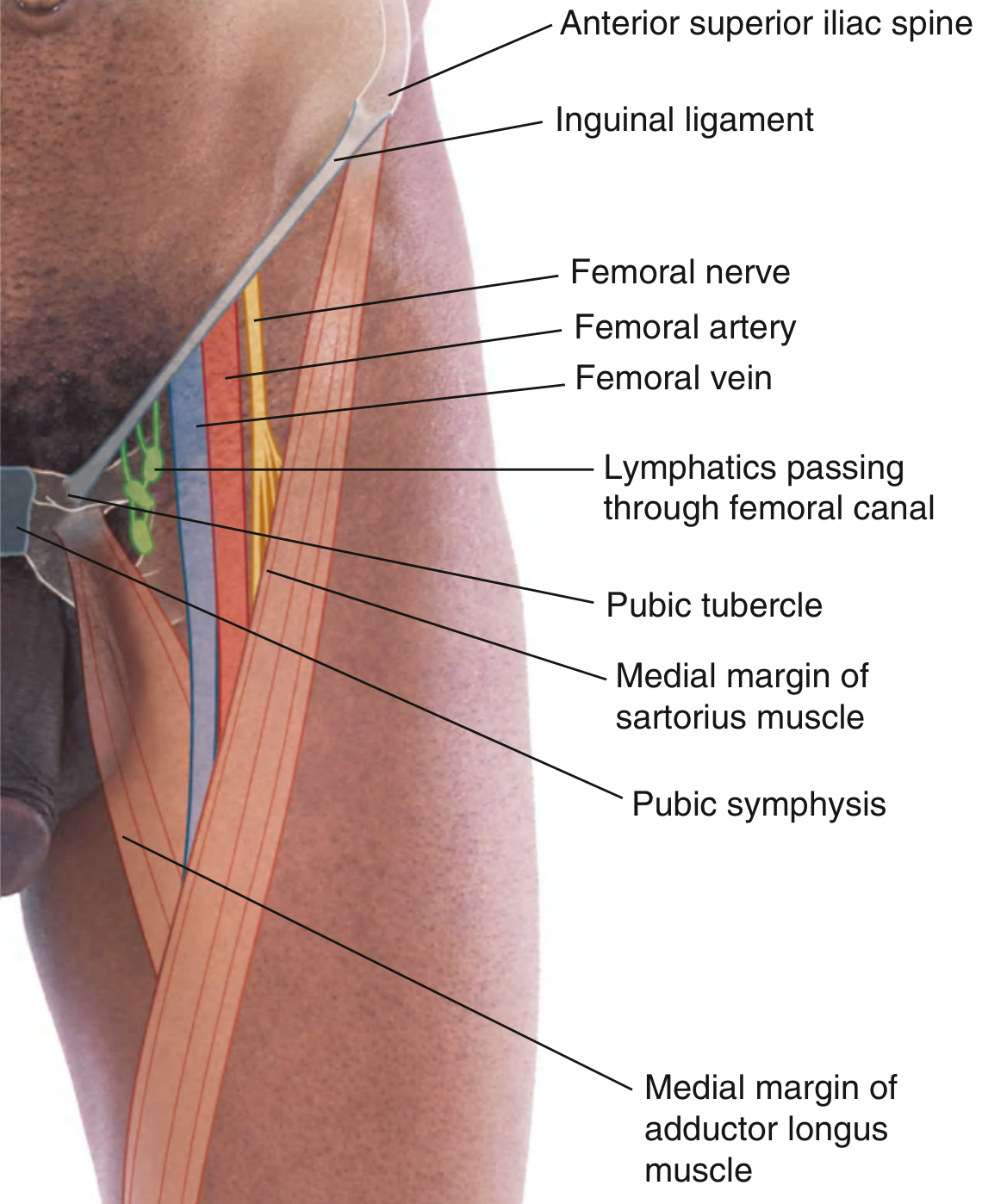
Iliopectineal Arch (Muscular vs. Vascular Compartments)
Below the inguinal ligament, the space is divided by the iliopectineal arch (a thickened band of the iliac fascia):
- Vascular compartment (medial to arch): femoral artery, femoral vein, femoral branch of genitofemoral nerve, deep inguinal lymphatics
- Muscular compartment (lateral to arch): iliopsoas muscle, femoral nerve, lateral femoral cutaneous nerve
Key Relations and Clinical Points
Femoral Pulse
The femoral artery pulse is palpable just inferior to the inguinal ligament, midway between the ASIS and pubic symphysis. This is the standard landmark for femoral artery access.
Vascular Access
The femoral triangle is the primary site for:
- Arterial catheterization - coronary angiography, angioplasty, peripheral vascular interventions
- Venous access - right heart catheterization, pulmonary artery access, renal/gonadal vein sampling
Femoral Hernia
- More common in women (due to wider pelvis/femoral ring)
- Passes through the femoral canal, inferior and lateral to the pubic tubercle (distinguishes it from an inguinal hernia, which lies superior and medial to the pubic tubercle)
- High risk of strangulation due to the rigid boundaries of the femoral ring
Femoral Nerve Injury
- The femoral nerve (L2-L4) lies lateral to the femoral sheath and artery
- Injury causes weakness of hip flexion and knee extension, loss of knee jerk, and sensory loss over the anteromedial thigh and medial leg (via saphenous nerve)
Adductor Canal (Subsartorial/Hunter's Canal)
At the apex of the femoral triangle, the femoral vessels enter the adductor canal, accompanied by the saphenous nerve and the nerve to vastus medialis. The canal runs under sartorius between vastus medialis and adductor longus/magnus, terminating at the adductor hiatus.
Summary Table
| Feature | Detail |
|---|---|
| Shape | Wedge/pyramid, apex pointing inferiorly |
| Base | Inguinal ligament |
| Lateral border | Medial margin of sartorius |
| Medial border | Medial margin of adductor longus |
| Floor (lateral) | Iliopsoas |
| Floor (medial) | Pectineus (+adductor longus below) |
| Roof | Fascia lata + skin |
| Apex leads to | Adductor canal |
| Contents (L to M) | Nerve, Artery, Vein, Lymphatics (NAVL) |
| Femoral nerve | Outside femoral sheath |
| Femoral hernia | Through femoral canal (most medial) |
Sources: Gray's Anatomy for Students, pp. 675-676 | THIEME Atlas of Anatomy - General Anatomy and Musculoskeletal System, p. 573 | Fischer's Mastery of Surgery - 8th ed., p. 6566
Lumber plexus
Finding Sources
Finding Sources
Reading File
Reading File
Reading File
Reading File
Reading File
Loading Image
Loading Image
Reading File
Now I have comprehensive information. Here is the full overview:
Lumbar Plexus
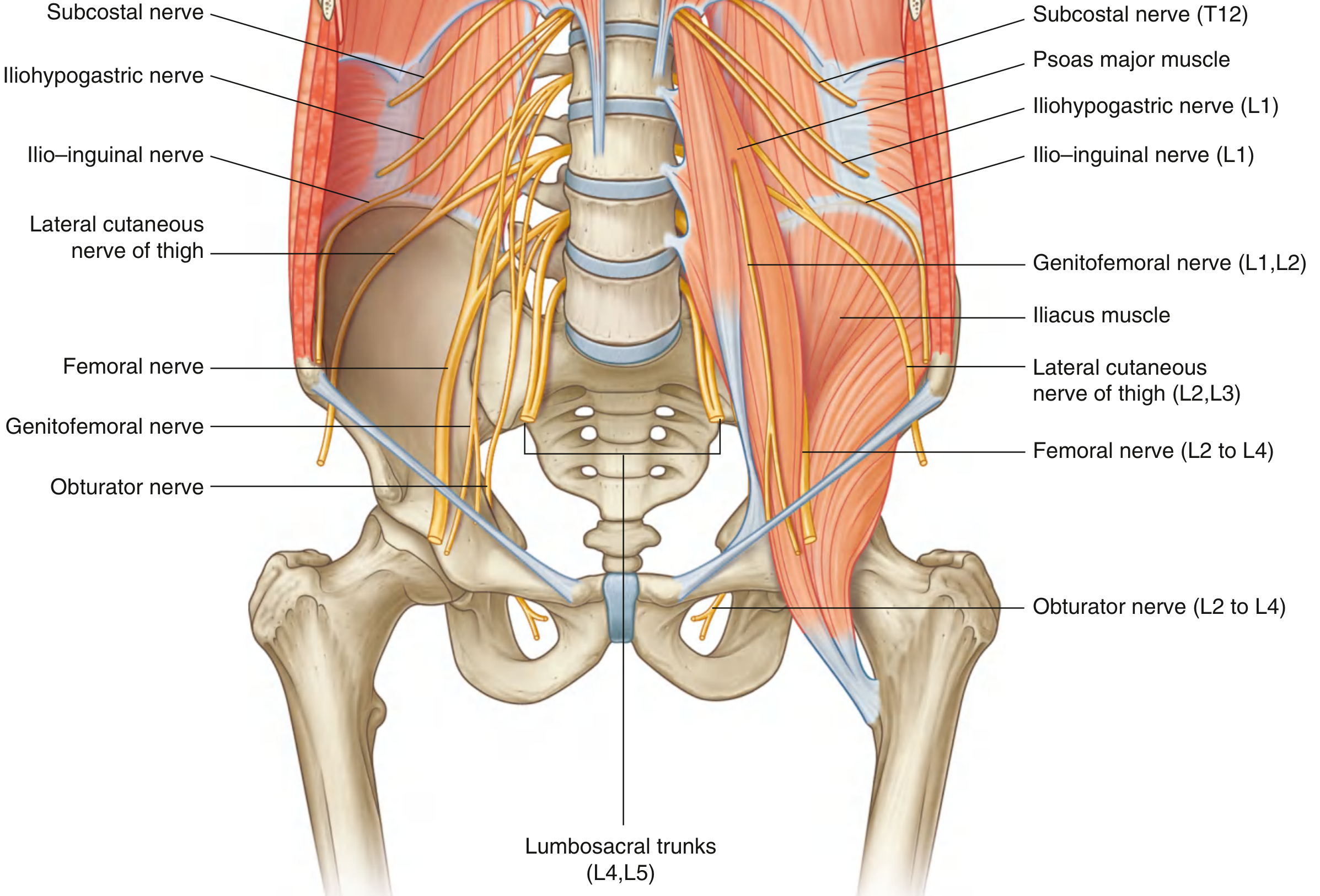
The lumbar plexus is a network of nerves formed by the anterior primary rami (ventral rami) of spinal levels T12 (subcostal), L1, L2, L3, and most of L4, located within the posterior abdominal wall.
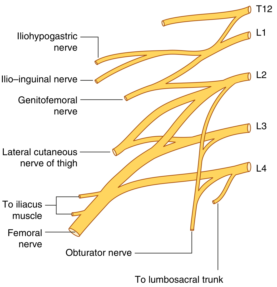
Formation and Location
- The plexus forms within the substance of the psoas major muscle, anterior to the transverse processes of the lumbar vertebrae.
- It receives a contribution from T12 (subcostal nerve).
- L4 is called the nervus furcalis because it splits - part goes to the lumbar plexus and part (with L5) forms the lumbosacral trunk, which continues into the sacral plexus.
- The L2, L3, L4 roots each divide into anterior and posterior divisions:
- Anterior divisions → Obturator nerve
- Posterior divisions → Femoral nerve + lateral femoral cutaneous nerve
Branches
The branches emerge from psoas major in three directions:
| Direction | Nerve |
|---|---|
| Anterior | Genitofemoral nerve |
| Medial | Obturator nerve |
| Lateral | Iliohypogastric, ilioinguinal, lateral cutaneous nerve of thigh (LCNT), femoral nerve |
Complete Branch Table
| Branch | Spinal Segments | Motor Supply | Sensory Supply |
|---|---|---|---|
| Iliohypogastric | L1 | Internal oblique, transversus abdominis | Posterolateral gluteal skin; skin above pubis |
| Ilioinguinal | L1 | Internal oblique, transversus abdominis | Upper medial thigh; anterior scrotum/labium majus, root of penis/mons pubis |
| Genitofemoral | L1, L2 | Genital branch: cremasteric muscle | Genital branch: anterior scrotum/labium majus; Femoral branch: upper anterior thigh |
| Lateral cutaneous nerve of thigh (LCNT) | L2, L3 | None (purely sensory) | Anterior and lateral thigh down to knee |
| Obturator | L2, L3, L4 | Adductor longus, brevis, magnus, gracilis, pectineus, obturator externus | Medial thigh; articular branches to hip and knee |
| Femoral | L2, L3, L4 | Iliacus, pectineus, sartorius, rectus femoris, vastus medialis, intermedius, lateralis | Anterior thigh; medial leg (via saphenous nerve) |
| Short muscular branches | L1-L4 | Psoas major, quadratus lumborum | - |
Individual Branches in Detail
1. Iliohypogastric Nerve (L1)
- Arises from the anterior ramus of L1 (together with ilioinguinal as a single trunk initially).
- Crosses anterior to quadratus lumborum, posterior to the kidney.
- Pierces transversus abdominis, then runs between it and internal oblique.
- Lateral cutaneous branch: supplies posterolateral gluteal skin.
- Anterior cutaneous branch: pierces external oblique above the superficial inguinal ring; supplies skin just above the pubis.
2. Ilioinguinal Nerve (L1)
- Travels parallel to and just below the iliohypogastric nerve.
- Enters the inguinal canal through the deep inguinal ring, exits through the superficial inguinal ring.
- Supplies skin of upper medial thigh, root/dorsal penis and upper scrotum (male), or mons pubis and labium majus (female).
- Surgical note: can be injured in herniorrhaphy, causing persistent groin neuralgia.
3. Genitofemoral Nerve (L1, L2)
- Pierces psoas major anteriorly and descends on its anterior surface.
- Divides into:
- Genital branch: enters the inguinal canal, supplies cremaster muscle and scrotal/labial skin.
- Femoral branch: passes under the inguinal ligament in the vascular compartment, supplies upper anterior thigh skin.
4. Lateral Femoral Cutaneous Nerve / LCNT (L2, L3)
- Formed from posterior divisions of L2 and L3.
- Exits the lateral border of psoas, crosses iliacus, passes under the inguinal ligament near the ASIS.
- Purely sensory: anterior and lateral thigh to the knee.
- Compression near the ASIS causes meralgia paresthetica - burning/numbness/dysesthesia over the lateral thigh. Associated with tight belts, obesity, pregnancy, or lumbar disc protrusion.
5. Obturator Nerve (L2, L3, L4) - Anterior Divisions
- Descends through psoas, exits medially, runs posterior to common iliac vessels.
- Exits the pelvis through the obturator foramen to enter the medial thigh.
- Divides into:
- Anterior division: adductor longus, gracilis, adductor brevis, pectineus; articular branch to hip.
- Posterior division: obturator externus, adductor magnus; articular branch to knee.
- Sensory: medial thigh, occasionally medial knee.
- Can be compressed by pelvic masses (tumor, gravid uterus) or injured in pelvic fractures.
6. Femoral Nerve (L2, L3, L4) - Posterior Divisions
- The largest branch of the lumbar plexus.
- Descends through psoas, exits at the lower lateral border, runs between psoas and iliacus under the iliacus fascia.
- Passes posterior to the inguinal ligament and enters the femoral triangle (lateral to femoral artery, outside the femoral sheath).
- Immediately divides into multiple branches in the thigh:
- Motor: iliacus, pectineus, sartorius, all four quadriceps muscles.
- Sensory (cutaneous): medial and intermediate cutaneous nerves of thigh (anterior thigh).
- Saphenous nerve (longest cutaneous branch): descends through the adductor canal, supplies medial leg and foot down to the medial malleolus.
- Articular: hip joint, knee joint.
Lumbosacral Trunk
The remaining part of L4 (not contributing to the lumbar plexus) joins with all of L5 to form the lumbosacral trunk, which descends over the pelvic brim to join the sacral plexus (S1-S3) and ultimately form the sciatic nerve.
Clinical Correlates
| Condition | Nerve Involved | Mechanism | Features |
|---|---|---|---|
| Meralgia paresthetica | LCNT (L2, L3) | Compression under inguinal ligament near ASIS | Burning/numbness on lateral thigh; no motor loss |
| Femoral nerve palsy | Femoral (L2-L4) | Pelvic hematoma (hemophilia, anticoagulants), hip surgery, inguinal region trauma | Weak hip flexion + knee extension; absent knee jerk (L3-L4); sensory loss on anteromedial thigh and medial leg |
| Obturator nerve palsy | Obturator (L2-L4) | Pelvic tumors, obturator hernia, pelvic fractures | Weak thigh adduction; sensory loss on medial thigh; pain radiating to knee |
| Ilioinguinal/iliohypogastric injury | L1 | Herniorrhaphy, appendicectomy, Pfannenstiel incision | Groin/scrotal/labial pain and numbness post-surgery |
| Lumbar plexopathy | Whole plexus | Retroperitoneal hematoma, tumor, diabetes, radiation | Combined femoral + obturator + LCNT deficits |
Key Memory Aid (Branches of Lumbar Plexus)
"I, I Get Laid On Fridays"
- Iliohypogastric (L1)
- Ilioinguinal (L1)
- Genitofemoral (L1, L2)
- Lateral cutaneous nerve of thigh (L2, L3)
- Obturator (L2-L4)
- Femoral (L2-L4)
Sources: Gray's Anatomy for Students, pp. 462-465 | Campbell's Operative Orthopaedics, 15th ed., p. 3876 | THIEME Atlas - General Anatomy and Musculoskeletal System, p. 555
This is a shared conversation. Sign in to Orris to start your own chat.