High yield facts on basal ganglia. Add usmle pearls

Reading File
Finding Sources
Searching the Web

basal ganglia USMLE high yield facts circuits disorders

Reading File
Reading File
Reading File
Reading File
Finding Sources
Finding Sources
Reading File
Reading File
Loading Image
Reading File
I now have comprehensive content. Let me compile the full high-yield answer.

Basal Ganglia - High Yield Facts + USMLE Pearls


1. Anatomy & Components

The basal ganglia are deep telencephalic gray matter nuclei. The main components:
StructureNotes
Caudate nucleusC-shaped, follows lateral ventricle; head + body + tail
PutamenLateral; fuses with caudate head anteriorly
Caudate + Putamen = Striatum (neostriatum)Receives ALL cortical inputs
Globus pallidus interna (GPi)Major output nucleus
Globus pallidus externa (GPe)Intrinsic relay (indirect pathway)
Putamen + Globus pallidus = Lenticular (lentiform) nucleus
Subthalamic nucleus (STN)Diencephalon; excitatory output via glutamate
Substantia nigra pars compacta (SNc)Dopamine source; midbrain
Substantia nigra pars reticulata (SNr)Output nucleus; functionally homologous to GPi
Pearl: The striatum is the INPUT station; GPi + SNr are the OUTPUT stations. The basal ganglia do NOT project directly to the spinal cord - everything goes via thalamus (VA/VL) back to cortex.
Nucleus accumbens = ventral striatum = key in reward/addiction circuits. Important in drug abuse questions.
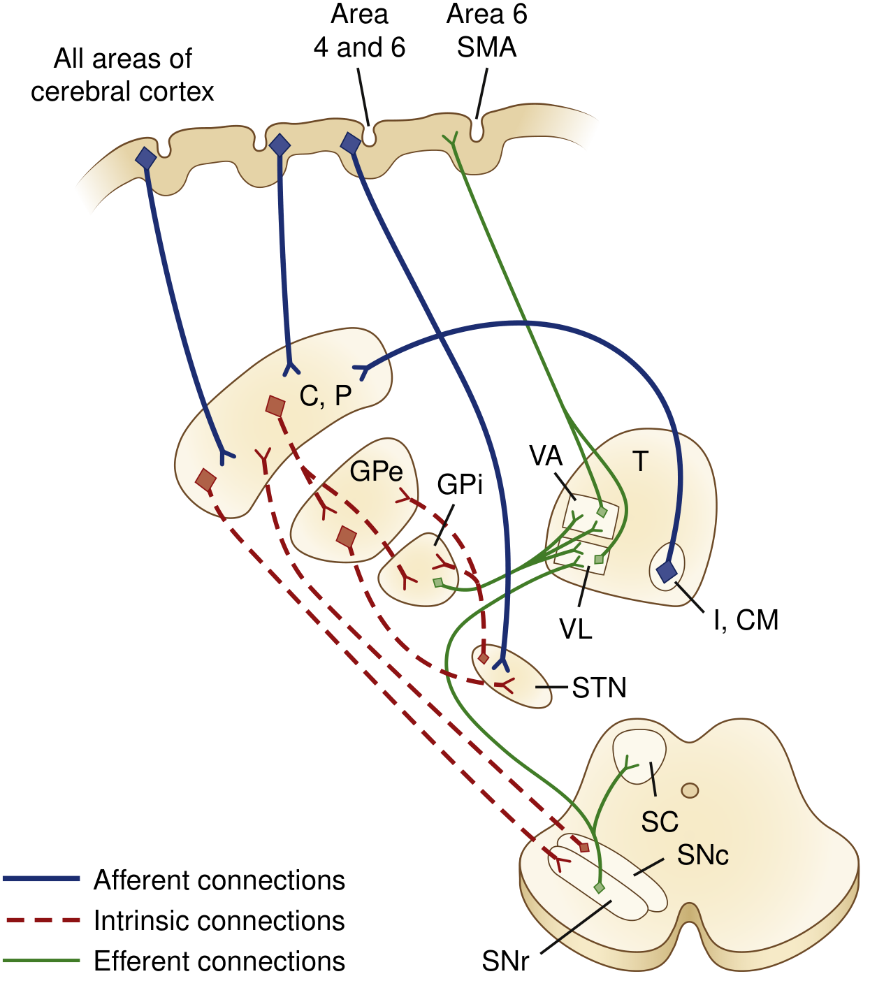
Basal ganglia connections diagram showing afferent (blue), intrinsic (red dashed), and efferent (green) connections

2. The Two Motor Pathways

Direct Pathway (net EXCITATORY to cortex - promotes movement)

Cortex → Striatum --(GABA)--> GPi/SNr --(GABA)--> Thalamus (VA/VL) --(Glu)--> Motor cortex
  • Striatum inhibits GPi/SNr → less inhibition of thalamus → thalamus activates cortex
  • Net effect: excitatory (two inhibitory synapses = net excitation)
  • Striatal neurons express D1 receptors - dopamine stimulates this pathway

Indirect Pathway (net INHIBITORY to cortex - suppresses movement)

Cortex → Striatum --(GABA)--> GPe --(GABA)--> STN --(Glu)--> GPi/SNr --(GABA)--> Thalamus → Cortex
  • Striatum inhibits GPe → less inhibition of STN → STN excites GPi/SNr → more inhibition of thalamus → less cortical activation
  • Net effect: inhibitory (three inhibitory synapses = net inhibition)
  • Striatal neurons express D2 receptors - dopamine inhibits this pathway
USMLE Pearl: Dopamine (via SNc) excites direct pathway (D1) AND inhibits indirect pathway (D2) - both actions result in net increased movement. Loss of dopamine = decreased movement (Parkinson's).
Neurotransmitter table:
PathwayTransmitter
Cortex → StriatumGlutamate
Cortex → STNGlutamate
Striatum → GPi (direct)GABA + substance P + dynorphin
Striatum → GPe (indirect)GABA + enkephalin
GPe → STNGABA
STN → GPi/SNrGlutamate
GPi/SNr → ThalamusGABA
SNc → StriatumDopamine
Pearl: Direct pathway neurons co-localize substance P; indirect pathway neurons co-localize enkephalin - histopathology can differentiate them.

3. Five Parallel Circuits

The basal ganglia run 5 parallel topographic loops:
  1. Motor (putamen) - voluntary movement
  2. Oculomotor (caudate body) - saccadic eye movements
  3. Dorsolateral prefrontal (caudate head) - executive function, working memory
  4. Lateral orbitofrontal - behavior, personality
  5. Limbic/anterior cingulate (ventral striatum/nucleus accumbens) - motivation, emotion, reward
Pearl: This explains why basal ganglia disorders have both motor AND psychiatric/cognitive features (e.g., OCD, depression, cognitive decline in Huntington's and Parkinson's).

4. Key Diseases

Parkinson's Disease

  • Pathology: Degeneration of SNc dopaminergic neurons; Lewy bodies (alpha-synuclein aggregates)
  • Effect on circuits: Loss of dopamine → reduced direct pathway excitation + reduced indirect pathway inhibition → net inhibition of thalamus → bradykinesia
  • Symptoms: Resting tremor ("pill-rolling"), rigidity (cogwheel), bradykinesia, postural instability
  • Treatment: L-DOPA (dopamine precursor), dopamine agonists (bromocriptine, pramipexole), MAO-B inhibitors (selegiline), anticholinergics (trihexyphenidyl - for tremor)
USMLE Pearls:
  • Parkinson's = hypokinetic disorder - substantia nigra loss
  • L-DOPA needs peripheral decarboxylase inhibitor (carbidopa) to prevent peripheral conversion
  • Anticholinergics work because ACh from striatal interneurons excites the indirect pathway - blocking ACh reduces excess indirect pathway activity
  • Deep brain stimulation targets the STN or GPi

Huntington's Disease

  • Pathology: CAG trinucleotide repeat expansion on chromosome 4p (huntingtin gene); autosomal dominant; anticipation
  • Neurons lost: Striatal GABAergic + cholinergic neurons of the indirect pathway affected FIRST (enkephalin-containing)
  • Effect on circuits: Loss of striatal inhibition of GPe → GPe inhibits STN → STN less active → less GPi inhibition → thalamus over-activated → hyperkinesis
  • Symptoms: Chorea (early), dementia, psychiatric symptoms (depression, personality change)
  • Late stage: Both pathways degenerate → rigid Parkinsonian state
USMLE Pearls:
  • Huntington's = hyperkinetic disorder - indirect pathway neurons lost first
  • Caudate atrophy on MRI → loss of caudate bulge → "box-car" lateral ventricles
  • No cure; tetrabenazine (VMAT2 inhibitor) for chorea - depletes dopamine/serotonin

Hemiballismus

  • Pathology: Lesion of the contralateral STN (most often lacunar stroke; also seen in hyperglycemia/hyperosmolar state)
  • Mechanism: Damaged STN → less excitation of GPi → less thalamic inhibition → excessive movement
  • Symptoms: Wild flinging/flailing movements of contralateral proximal limb (arm > leg)
  • Treatment: Haloperidol or other dopamine blockers
USMLE Pearl: Hemiballismus = contralateral STN lesion. Most common cause = lacunar infarct. Can also occur in non-ketotic hyperglycemia.

Wilson's Disease (Hepatolenticular Degeneration)

  • Copper accumulation in lenticular nucleus (putamen + globus pallidus) and liver
  • AR; ATP7B mutation (chromosome 13)
  • Presents with: dysarthria, tremor, choreoathetosis, psychiatric changes + liver disease + Kayser-Fleischer rings
  • Low ceruloplasmin, elevated urine copper
USMLE Pearl: Wilson's = basal ganglia + liver. Look for young patient with movement disorder + liver disease + psychiatric symptoms.

Other Disorders

DisorderKey FeatureBasal Ganglia Involvement
OCDIntrusive thoughts + compulsionsCaudate hyperactivity
Tourette syndromeMotor + vocal ticsStriatum dysfunction
ADHDInattention, impulsivityCaudate/striatum dopamine deficit
Drug addictionReward cravingNucleus accumbens (ventral striatum)

5. Neurotransmitter Quick Reference

NTRole in BGClinical relevance
DopamineExcites direct (D1), inhibits indirect (D2)Lost in Parkinson's
GABAInhibitory throughoutMajor intrinsic/output NT
GlutamateExcitatory inputs (cortex, STN)STN stimulation target
AcetylcholineStriatal interneurons - excites indirectAnticholinergics help parkinsonism
SerotoninRaphe → striatumDepression in PD/HD
NorepinephrineLC → striatum/SNAutonomic dysfunction in PD

6. USMLE Summary Table

DiseaseLesionPathway EffectMovement Type
Parkinson'sSNc (dopamine loss)Both pathways → thalamus inhibitedHypokinetic
Huntington'sStriatum (indirect >> direct)GPe → STN inhibited → thalamus excitedHyperkinetic (early)
HemiballismusSTNGPi less excited → thalamus excitedHyperkinetic (contralateral)
Wilson'sLenticular nucleusDisrupted circuitsMixed

7. High-Yield One-Liners

  • Input to BG = striatum (caudate + putamen), receives glutamatergic cortical input
  • Output of BG = GPi + SNr → VA/VL thalamus → motor cortex (all GABA)
  • Direct pathway = excitatory net effect on cortex (promotes movement)
  • Indirect pathway = inhibitory net effect on cortex (suppresses movement)
  • Dopamine pushes the balance toward movement (excites direct, inhibits indirect)
  • STN lesion = hemiballismus (hyperkinetic, contralateral)
  • SNc degeneration = Parkinson's (hypokinetic)
  • Caudate/putamen degeneration = Huntington's (hyperkinetic early, hypokinetic late)
  • Nucleus accumbens = reward, addiction
  • BG disorders show ipsilateral deficits? NO - basal ganglia disorders affect the contralateral side
  • Cerebellum vs BG: both modulate movement but never project directly to spinal cord - BG uses VA/VL thalamus, cerebellum uses VL/VPLc

Sources: Costanzo Physiology 7e | Neuroanatomy through Clinical Cases 3e (Blumenfeld) | Bradley & Daroff's Neurology in Clinical Practice

Generate usmle style high yield qustion from the topic basal ganglia

Here are 15 USMLE-style high-yield questions on the Basal Ganglia, covering all major testable concepts:

Basal Ganglia - USMLE Style Questions


Question 1

A 62-year-old man presents with a 2-year history of resting tremor in his right hand, shuffling gait, and difficulty initiating movements. On exam he has cogwheel rigidity and a masked facies. Which of the following best describes the net effect on the thalamus in this patient's condition?
A. Increased excitation via the direct pathway B. Decreased inhibition via the indirect pathway C. Increased inhibition due to overactive GPi/SNr D. Decreased excitation due to underactive STN E. Increased dopamine release from the nucleus accumbens

Answer & Explanation
Correct Answer: C
This is Parkinson's disease - degeneration of SNc dopaminergic neurons.
  • Loss of dopamine → direct pathway underactive (less inhibition of GPi) + indirect pathway overactive (less inhibition of GPe → less inhibition of STN → STN over-excites GPi)
  • Both effects → GPi/SNr fire excessively → increased GABA output → increased inhibition of VA/VL thalamus
  • Result: thalamus cannot activate motor cortex → bradykinesia
Why others are wrong:
  • A: Direct pathway is underactive in PD
  • B: Indirect pathway is overactive (more inhibition via GPe-STN-GPi axis)
  • D: STN is overactive in PD (GPe cannot inhibit it)
  • E: Nucleus accumbens dopamine is unrelated to the motor deficit here

Question 2

A 45-year-old woman is brought in by her family for progressive personality changes, irritability, and involuntary jerking movements of her arms and face over the past 8 months. Her father died of a similar illness in his 50s. MRI shows caudate nucleus atrophy. Which of the following receptor-bearing neurons are lost FIRST in this disease?
A. D1-bearing striatal neurons projecting to GPi B. D2-bearing striatal neurons projecting to GPe C. Dopaminergic neurons of the substantia nigra pars compacta D. Glutamatergic neurons of the subthalamic nucleus E. GABAergic neurons of the globus pallidus interna

Answer & Explanation
Correct Answer: B
This is Huntington's disease (AD, CAG repeat expansion, chromosome 4).
  • The indirect pathway striatal neurons (D2-bearing, enkephalin-containing, projecting to GPe) are lost first
  • Loss of striatal inhibition of GPe → GPe overactive → inhibits STN → STN less active → less excitation of GPi → less inhibition of thalamus → hyperkinesia (chorea)
  • Later, both pathways degenerate → rigid Parkinsonian state
A = direct pathway neurons (D1, substance P) - lost later C = Parkinson's disease pathology D = STN neurons are excitatory; not primarily lost in HD E = GPi output neurons; not the primary loss in HD

Question 3

A 70-year-old man with poorly controlled type 2 diabetes is brought to the ED after his wife noticed his left arm "flinging wildly" since this morning. Neurological exam shows large-amplitude, irregular, flinging movements of the left arm. MRI reveals a small hyperintense lesion on T1. Which structure is most likely involved?
A. Left subthalamic nucleus B. Right subthalamic nucleus C. Left globus pallidus interna D. Right caudate nucleus E. Right substantia nigra pars compacta

Answer & Explanation
Correct Answer: B
This is hemiballismus - a hyperkinetic movement disorder with contralateral proximal limb flinging.
  • Caused by a lesion of the contralateral STN
  • Right STN lesion → left arm hemiballismus
  • Mechanism: STN normally excites GPi → less STN activity → less GPi firing → less inhibition of thalamus → uncontrolled movement
  • The T1 hyperintense signal in non-ketotic hyperglycemia is a classic radiological finding (putamen/STN)
  • Most common cause overall: lacunar infarct; in this diabetic patient, non-ketotic hyperglycemia is a classic association
A = ipsilateral STN - would cause right-sided symptoms C, D, E = wrong structure or side

Question 4

A researcher is studying the basal ganglia and records a neuron that fires tonically and uses GABA as its neurotransmitter. When this neuron's activity decreases, the thalamus becomes more active. Where is this neuron located?
A. Striatum B. Subthalamic nucleus C. Globus pallidus interna D. Substantia nigra pars compacta E. Cerebral cortex

Answer & Explanation
Correct Answer: C
The GPi (and SNr) tonically fire GABA onto the thalamus (VA/VL nuclei), keeping it inhibited at baseline.
  • When GPi activity decreases → less GABA → thalamus disinhibited → more excitatory output to motor cortex → movement
  • This is the fundamental "release" mechanism in the direct pathway
A = Striatum - GABAergic, but projects TO GPi, not to thalamus B = STN - glutamatergic (excitatory), not GABAergic D = SNc - dopaminergic E = Cortex - glutamatergic

Question 5

A 28-year-old man is diagnosed with a tremor disorder and liver disease. Slit-lamp exam reveals golden-brown deposits at the corneal periphery. Serum ceruloplasmin is markedly low. Which of the following chromosomal abnormalities is associated with this condition?
A. Chromosome 4p - CAG repeat B. Chromosome 13 - ATP7B mutation C. Chromosome 6 - PARK2 mutation D. Chromosome 4q - HTT gene E. Chromosome 21 - APP gene

Answer & Explanation
Correct Answer: B
This is Wilson's disease (hepatolenticular degeneration):
  • AR inheritance, ATP7B gene on chromosome 13
  • Defective copper-transporting ATPase → copper accumulates in liver, lenticular nucleus (putamen + globus pallidus), and cornea
  • Kayser-Fleischer rings (corneal copper deposits)
  • Low ceruloplasmin, high urinary copper
  • Treatment: D-penicillamine or trientine (copper chelators)
A = Huntington's disease (chromosome 4p, CAG repeat) C = Parkin - autosomal recessive early-onset Parkinson's D = Huntingtin gene (same chromosome 4 but different locus than A) E = Alzheimer's (amyloid precursor protein)

Question 6

A medical student is reviewing the neurotransmitter pharmacology of the basal ganglia. She notes that a specific set of striatal projection neurons co-localizes GABA with enkephalin. Which pathway do these neurons belong to, and what is their projection target?
A. Direct pathway; projects to GPi B. Indirect pathway; projects to GPe C. Direct pathway; projects to SNr D. Indirect pathway; projects to STN E. Both pathways; project to VA/VL thalamus

Answer & Explanation
Correct Answer: B
Key co-localization facts:
  • Direct pathway striatal neurons: GABA + substance P + dynorphin → project to GPi/SNr; express D1 receptors
  • Indirect pathway striatal neurons: GABA + enkephalin → project to GPe; express D2 receptors
This is a classic USMLE histopathology/pharmacology pearl - enkephalin identifies the indirect pathway.

Question 7

A 66-year-old woman with Parkinson's disease continues to have debilitating tremor despite optimal L-DOPA/carbidopa therapy. Her neurologist considers adding an anticholinergic agent. What is the mechanism by which anticholinergic drugs reduce tremor in Parkinson's disease?
A. Block D2 receptors on indirect pathway neurons B. Prevent dopamine reuptake in the striatum C. Inhibit cholinergic excitation of the indirect pathway striatal neurons D. Enhance GABA release from GPi onto the thalamus E. Directly stimulate D1 receptors on direct pathway neurons

Answer & Explanation
Correct Answer: C
  • Striatal large aspiny interneurons release ACh and preferentially excite indirect pathway neurons
  • In PD, loss of dopamine reduces D2-mediated inhibition of the indirect pathway → indirect pathway is relatively overactive
  • Anticholinergics (e.g., trihexyphenidyl, benztropine) block muscarinic receptors → reduce ACh excitation of indirect pathway → decrease its overactivity → partial relief of symptoms (especially tremor)
  • Note: ACh and dopamine normally have opposing effects in the striatum - PD upsets this balance
A = D2 blockade would worsen PD symptoms B = Reuptake inhibition would increase dopamine effect (not the mechanism of anticholinergics) D = Enhancing GPi GABA → more thalamic inhibition → worse hypokinesia E = That's the mechanism of D1 agonists, not anticholinergics

Question 8

A 55-year-old man undergoes deep brain stimulation (DBS) for refractory Parkinson's disease. The surgical target is the structure whose hyperactivity drives excessive GPi firing and thus thalamocortical inhibition. Which structure is targeted?
A. Globus pallidus interna B. Substantia nigra pars compacta C. Subthalamic nucleus D. Ventral lateral nucleus of thalamus E. Nucleus accumbens

Answer & Explanation
Correct Answer: C
In PD:
  • Loss of dopamine → indirect pathway overactive → STN becomes overactive (GPe cannot suppress it)
  • Overactive STN over-excites GPi → GPi fires too much GABA → excessive thalamic inhibition → bradykinesia
DBS of the STN (or GPi) is the standard surgical target. High-frequency stimulation functionally inhibits the STN, reducing GPi overactivity.
A = GPi is also a DBS target but it is downstream of STN B = SNc is already degenerated; not a DBS target D = VL thalamus is the output recipient; not a target E = Nucleus accumbens is targeted in refractory OCD/depression, not PD

Question 9

Which of the following best describes the net effect of dopamine released from the substantia nigra pars compacta onto the striatum?
A. Inhibits both direct and indirect pathways equally B. Excites direct pathway (D1) and inhibits indirect pathway (D2) → net increased movement C. Inhibits direct pathway (D1) and excites indirect pathway (D2) → net decreased movement D. Only modulates the indirect pathway via D2 receptors E. Acts exclusively on GPi neurons to reduce GABA output

Answer & Explanation
Correct Answer: B
This is the most tested dopamine fact in USMLE basal ganglia questions:
  • D1 receptors on direct pathway neurons → dopamine excites these → more inhibition of GPi → less thalamic inhibition → more movement
  • D2 receptors on indirect pathway neurons → dopamine inhibits these → less inhibition of GPe → GPe inhibits STN → less GPi excitation → less thalamic inhibition → more movement
  • Both D1 and D2 effects point in the same direction: dopamine promotes movement

Question 10

A 19-year-old male has had multiple facial grimaces, repetitive throat-clearing vocalizations, and involuntary shoulder shrugging since age 8. He also has symptoms of OCD. Which of the following structures is most implicated in his motor symptoms?
A. Cerebellum, dentate nucleus B. Striatum (caudate and putamen) C. Red nucleus, midbrain tegmentum D. Hippocampus E. Anterior cingulate cortex exclusively

Answer & Explanation
Correct Answer: B
This is Tourette syndrome - characterized by motor + vocal tics beginning in childhood, often with OCD and ADHD comorbidities.
  • Implicated in striatal dysfunction (caudate + putamen), with disruption of cortico-striato-thalamo-cortical circuits
  • The co-occurrence with OCD reflects involvement of the orbitofrontal-caudate loop
  • Treatment: antipsychotics (haloperidol, fluphenazine), alpha-2 agonists (clonidine, guanfacine)
A = Cerebellar lesions cause ataxia and intention tremor, not tics C = Red nucleus = rubrospinal tract; not associated with tics E = Anterior cingulate is part of the limbic loop but alone insufficient

Question 11

A 34-year-old woman with a history of IV drug use presents with paranoia, hypersexuality, and obsessive drug-seeking behavior. Neuroimaging shows abnormal activation in a ventral striatal region critical for reward processing. Which structure is most likely affected?
A. Caudate head B. Putamen C. Nucleus accumbens D. Globus pallidus externa E. Subthalamic nucleus

Answer & Explanation
Correct Answer: C
The nucleus accumbens (ventral striatum) is the central structure of the brain's reward circuit:
  • Receives dopaminergic input from ventral tegmental area (VTA) via the mesolimbic pathway
  • Drugs of abuse flood this circuit with dopamine → reward, craving, addiction
  • Also involved in motivation, pleasure, reinforcement learning
A = Caudate head = dorsolateral prefrontal circuit (executive function) B = Putamen = motor circuit D = GPe = intrinsic BG relay E = STN = indirect pathway relay

Question 12

A 68-year-old man with Parkinson's disease is started on a medication that acts by inhibiting an enzyme responsible for peripheral conversion of L-DOPA to dopamine, thereby allowing more L-DOPA to cross the blood-brain barrier. What enzyme is being inhibited?
A. Monoamine oxidase B (MAO-B) B. Catechol-O-methyltransferase (COMT) C. Aromatic L-amino acid decarboxylase (AADC) D. Tyrosine hydroxylase E. Dopamine beta-hydroxylase

Answer & Explanation
Correct Answer: C
  • L-DOPA is given with carbidopa, a peripheral AADC (dopa decarboxylase) inhibitor
  • AADC converts L-DOPA → dopamine in the periphery
  • Carbidopa does NOT cross the BBB → peripheral dopamine conversion is blocked → more L-DOPA reaches the brain → more central dopamine synthesis → therapeutic effect
  • This also reduces peripheral side effects (nausea, hypotension)
A = MAO-B inhibitors (selegiline, rasagiline) - also used in PD but prevent dopamine breakdown centrally B = COMT inhibitors (entacapone, tolcapone) - also extend L-DOPA half-life but different mechanism D = Tyrosine hydroxylase converts tyrosine → L-DOPA (rate-limiting step); not targeted therapeutically E = DBH converts dopamine → norepinephrine

Question 13

In an advanced stage of Huntington's disease, a patient who previously had chorea now presents with rigidity and bradykinesia resembling Parkinson's disease. What best explains this transition?
A. Degeneration of the substantia nigra pars compacta B. Progressive degeneration of both direct and indirect pathway striatal neurons C. Compensatory upregulation of D2 receptors D. Loss of GPe neurons leading to STN overactivation E. Lewy body accumulation in the putamen

Answer & Explanation
Correct Answer: B
In Huntington's disease:
  • Early: Indirect pathway (enkephalin/D2) neurons degenerate first → hyperkinesia (chorea)
  • Late: Both direct AND indirect pathway striatal neurons degenerate → the net effect shifts - with loss of direct pathway input to GPi, GPi becomes overactive again, similar to Parkinson's → hypokinesia, rigidity
This explains why late-stage HD patients develop a Parkinsonian phenotype despite SNc being intact.
A = SNc is not the primary lesion in HD C = D2 upregulation would enhance indirect pathway sensitivity, not cause rigidity D = GPe neuron loss would increase STN activity → more GPi → more thalamic inhibition → hypokinesia (plausible mechanistically, but not the main explanation) E = Lewy bodies = Parkinson's/DLB, not HD

Question 14

A researcher lesions the globus pallidus interna (GPi) bilaterally in a primate. Which of the following is the most expected result?
A. Flaccid paralysis of all four limbs B. Spastic paraparesis C. Hyperkinetic involuntary movements D. Loss of all voluntary movement E. Cerebellar ataxia with intention tremor

Answer & Explanation
Correct Answer: C
  • GPi is the main inhibitory output of the basal ganglia to the thalamus
  • Lesioning GPi → removes tonic GABAergic inhibition from thalamus → thalamus over-activates cortex → hyperkinetic movements
  • This is the same mechanism as surgical pallidotomy (GPi lesioning), historically used to treat dyskinesias in PD
A/B = These imply UMN or LMN lesions - BG do not directly control the corticospinal tract D = No voluntary movement would require cortical or CST lesions E = Cerebellar signs require cerebellar lesions

Question 15

A patient is found to have a lesion in the structure that receives direct glutamatergic input from ALL areas of the cerebral cortex and serves as the primary INPUT nucleus of the basal ganglia. Which structure is described?
A. Globus pallidus interna B. Subthalamic nucleus C. Striatum (caudate + putamen) D. Substantia nigra pars reticulata E. Ventral anterior thalamic nucleus

Answer & Explanation
Correct Answer: C
  • The striatum (caudate + putamen) receives glutamatergic projections from virtually ALL areas of the cerebral cortex (topographically organized)
  • It is the primary INPUT station of the basal ganglia
  • Also receives dopaminergic input from SNc and serotonergic input from raphe nuclei
  • Key rule: Striatum = INPUT; GPi/SNr = OUTPUT
A = GPi is an OUTPUT nucleus B = STN receives direct cortical input too (hyperdirect pathway) but is an intrinsic/relay nucleus, not the primary input D = SNr is an OUTPUT nucleus (homologous to GPi) E = VA thalamus is downstream of BG output, projects back to cortex

Quick Reference: Tested Concepts Summary

ConceptKey Fact
Input nucleusStriatum (caudate + putamen)
Output nucleiGPi + SNr
Direct pathway net effectExcitatory to cortex (promotes movement)
Indirect pathway net effectInhibitory to cortex (suppresses movement)
Dopamine overall effectPromotes movement (D1 excites direct, D2 inhibits indirect)
PD pathologySNc degeneration → hypokinesia
HD earlyIndirect pathway (enkephalin/D2) → chorea
HD lateBoth pathways → Parkinsonism
HemiballismusContralateral STN lesion → hyperkinesia
Wilson'sLenticular nucleus + liver, Chr 13, ATP7B
Nucleus accumbensReward, addiction, limbic loop

Explain basal ganglia as if you are teaching a medical student who is learning it for the first time. Start with the big picture and purpose, then explain each component step-by-step in simple language. Define every technical term before using it. Explain the physiology and pathophysiology using cause-and-effect reasoning (‘because… therefore…’). Use analogies and visual descriptions whenever helpful. After the explanation, give high-yield exam points, common misconceptions, and USMLE pearls i must know

Finding Sources
Searching the Web

basal ganglia simple explanation direct indirect pathway analogy medical student

Reading File
Reading File
Reading File
Reading File
Reading File
Loading Image
Loading Image
I have everything I need. Now I'll write the full, student-friendly, deeply explained guide.

The Basal Ganglia - A Complete First-Principles Guide for Medical Students


PART 1: THE BIG PICTURE - What Are the Basal Ganglia and Why Do They Exist?

Before we name a single structure, let's understand the problem the basal ganglia solve.

The Problem

Your motor cortex (the part of your brain that sends "move!" signals to your muscles) is like a powerful car engine. But an engine without a gearbox, a brake, and a throttle is dangerous - it either runs flat out or stalls. The brain has the same challenge: how do you take a thought ("I want to pick up this cup") and turn it into a smooth, controlled, purposeful movement - while also suppressing all the other movements your muscles could be making at the same time?
That is exactly what the basal ganglia do. They act as the motor control supervisor - they don't create movement directly, they refine and filter movement signals coming from the cortex. They help you:
  1. Start the right movements
  2. Suppress unwanted or competing movements
  3. Scale the speed and force of movement appropriately
  4. Keep movements smooth and automatic once learned
Think of it like a music producer at a mixing board. The cortex is the raw music (loud, messy). The basal ganglia are the producer - turning certain tracks up, others down, so the final output is clean and coherent.
Critical first fact: The basal ganglia never directly talk to your muscles. They don't project to the spinal cord. They work entirely by modulating activity in the motor cortex through a relay station called the thalamus.

Beyond Movement

Here's something students often miss: the basal ganglia also handle cognition, emotion, and reward. Different parts of the same circuit process:
  • Motor control (movement)
  • Executive thinking (planning, decisions)
  • Emotional responses
  • Reward and motivation (including drug addiction)
This is why diseases of the basal ganglia (Parkinson's, Huntington's) don't just cause movement problems - they also cause depression, dementia, and personality changes. Same circuit, different channels.

PART 2: ANATOMY - Meet the Team

Term to know first - "Nucleus" (plural: nuclei): In neuroanatomy, a nucleus is a cluster of neuron cell bodies deep inside the brain (not the nucleus inside a cell). Think of it as a "neighborhood" of neurons grouped together because they work as a team.
Term to know - "Gray matter": Parts of the brain rich in neuron cell bodies look grayish. The basal ganglia are gray matter structures buried deep inside the white matter of the brain.
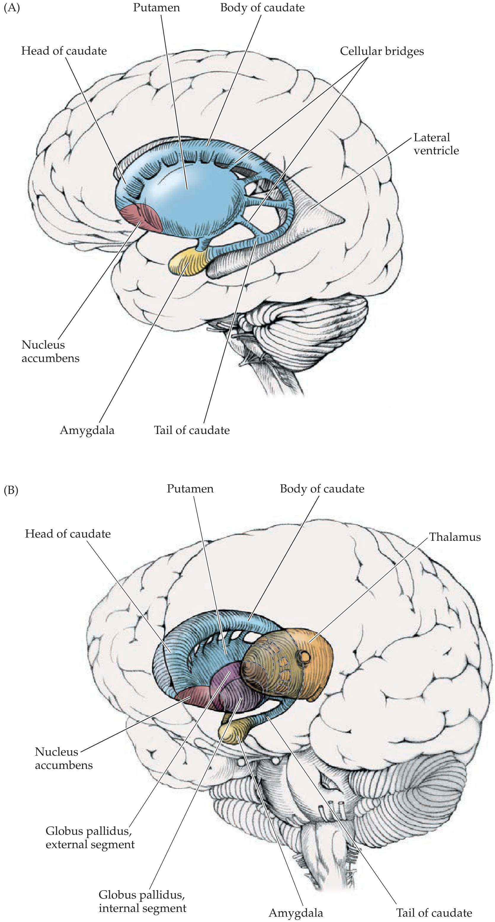
Here is your anatomy diagram showing the 3D positions:
3D anatomy of basal ganglia showing caudate (blue C-shape), putamen, globus pallidus internal and external segments, nucleus accumbens, amygdala, and thalamus
Now let's meet each player:

Structure 1: The Striatum (= Caudate + Putamen)

What it is: The striatum is the MAIN ENTRANCE of the basal ganglia. Almost all information flowing into the basal ganglia from outside enters here. It is made of two structures that are basically the same tissue split apart by a bundle of nerve fibers (the internal capsule):
  • Caudate nucleus - "Caudate" means "tail-bearing." It is a C-shaped structure that curves around the lateral ventricle (the fluid-filled space inside the brain), like a snake coiled around a pipe. It has a head, body, and tail.
  • Putamen - Sits just lateral (to the side) of the globus pallidus. "Putamen" means "shell" in Latin.
Together, the caudate + putamen are called the striatum (from Latin "striped") because thin bridges of neurons connect them through the internal capsule, making them look striped in cross-section.
Analogy: If the basal ganglia were a company, the striatum is the front desk - all incoming messages (from the cortex) arrive here first.
What neurotransmitter does it use? The striatum sends its outputs using GABA (gamma-aminobutyric acid) - an inhibitory neurotransmitter. This means when striatal neurons fire, they suppress activity in wherever they project.
Quick definition - Inhibitory vs. Excitatory: Inhibitory signals tell the receiving neuron to slow down or stop firing. Excitatory signals tell it to fire more. This distinction is the single most important concept for understanding the whole basal ganglia circuit.
Functional divisions of the striatum:
RegionInput fromLoop serves
PutamenMotor cortexMovement
Caudate headPrefrontal cortexCognition, executive function
Ventral striatum (nucleus accumbens)Limbic cortex, VTAReward, emotion, addiction

Structure 2: The Globus Pallidus (GP)

Term - "Globus pallidus": Latin for "pale globe." It appears pale on brain sections because many myelinated fibers run through it.
The globus pallidus has two segments - think of them as two completely different employees who happen to share an office:
  • Globus Pallidus externa (GPe) = the external or outer segment. This is an intrinsic relay station - it only talks to other parts within the basal ganglia.
  • Globus Pallidus interna (GPi) = the internal or inner segment. This is one of the two main OUTPUT stations of the basal ganglia. It sends signals out of the basal ganglia to the thalamus.
Together, the putamen + globus pallidus = "Lenticular nucleus" (lentil-shaped). You'll see this term in anatomy and stroke descriptions.

Structure 3: The Subthalamic Nucleus (STN)

What it is: A small, lens-shaped cluster of neurons sitting at the junction between the cerebrum and the brainstem (hence "sub-thalamic" = below the thalamus).
What neurotransmitter does it use? Glutamate - excitatory. The STN is one of the few excitatory neurons within the basal ganglia circuit.
Why does it matter clinically? Because the STN is the target of deep brain stimulation (DBS) in Parkinson's disease, and because a lesion here causes hemiballismus (wild flinging movements). Both of those are high-yield exam facts.

Structure 4: The Substantia Nigra

Term - "Substantia nigra": Latin for "black substance." It literally appears black on gross brain sections because the dopamine-producing neurons in it contain neuromelanin, a dark pigment. If you look at a brainstem section and see a dark band, that's the substantia nigra.
It has two parts:
  • Substantia nigra pars compacta (SNc) - the dopamine factory of the basal ganglia. These neurons synthesize dopamine and send it to the striatum via the nigrostriatal pathway. This is the part that degenerates in Parkinson's disease.
  • Substantia nigra pars reticulata (SNr) - structurally and functionally identical to the GPi. It acts as a second OUTPUT station of the basal ganglia, particularly for eye movements (projects to superior colliculus).

Structure 5: The Nucleus Accumbens

The ventral (lower) part of the striatum. It is the brain's pleasure/reward center. It receives dopamine from the ventral tegmental area (VTA) via the mesolimbic pathway. Drugs of abuse (cocaine, heroin, alcohol) hijack this circuit by flooding it with dopamine.

The Thalamus (not technically "in" the BG, but essential to understand)

Term - "Thalamus": The brain's great relay station, sitting in the center. Almost all sensory and motor information passes through it on the way to the cortex.
The relevant thalamic nuclei for the basal ganglia are the VA (ventral anterior) and VL (ventrolateral) nuclei. They receive inhibitory GABA from GPi/SNr, and when allowed to escape that inhibition, they fire excitatory glutamate back to the motor cortex.

PART 3: THE CIRCUIT - How It All Connects

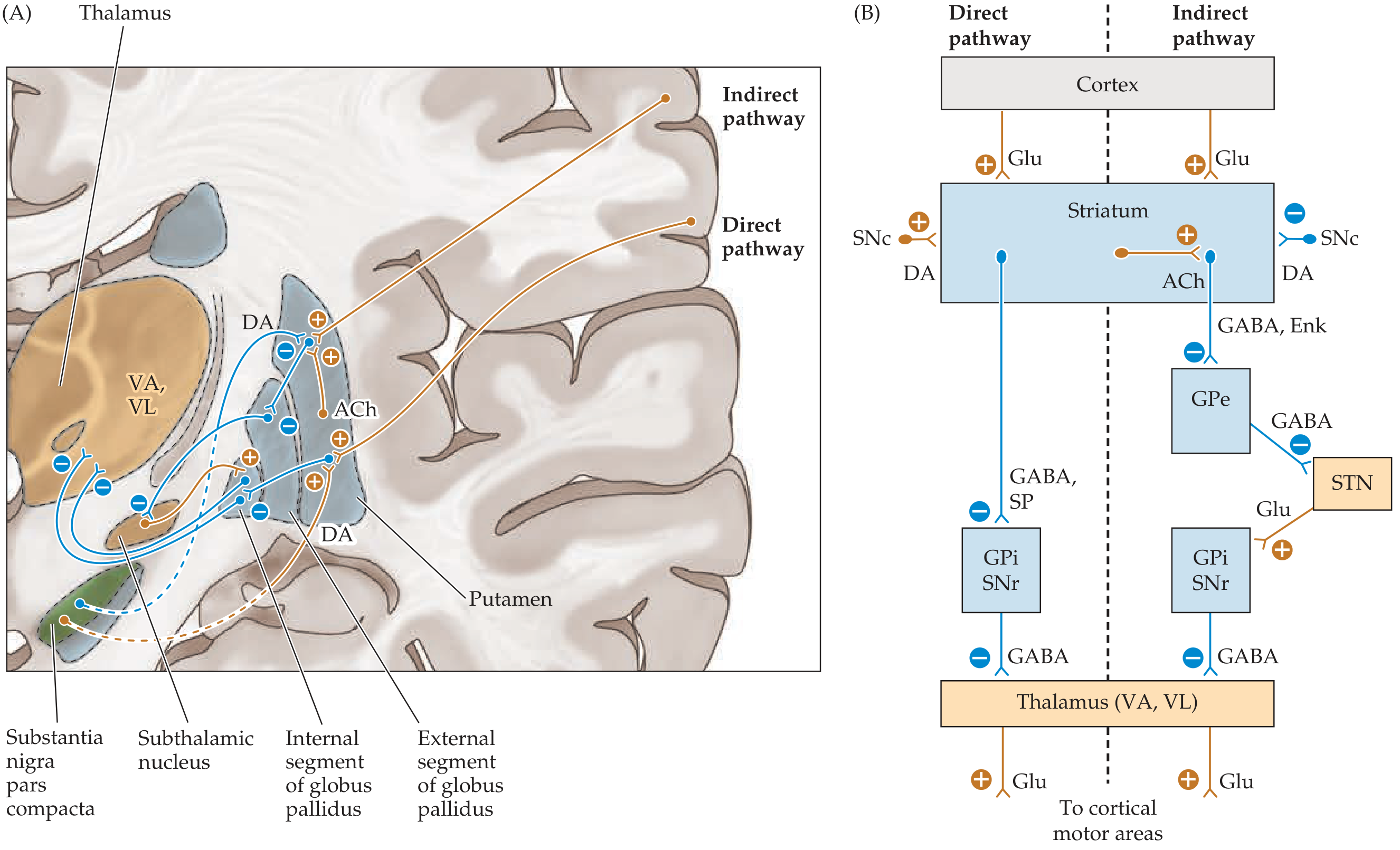
Here is the complete circuit diagram from your textbook:
Basal ganglia direct and indirect pathway circuit diagram showing cortex → striatum → GPi/SNr → thalamus → cortex with dopamine modulation from SNc

The Overarching Logic: "Tonic Inhibition with Selective Release"

Here is the single most important concept in understanding the basal ganglia:
At baseline (at rest), the GPi/SNr fires CONSTANTLY and TONICALLY. This constant firing releases GABA onto the thalamus, keeping the thalamus suppressed - preventing unwanted movements.
When you want to make a voluntary movement, the basal ganglia must reduce this tonic inhibition - it must "let the thalamus off the leash" so it can activate the motor cortex.
Analogy: Imagine the thalamus is a dog that wants to run (= make a movement). The GPi/SNr is a leash constantly holding it back. The basal ganglia's job is to selectively loosen or tighten that leash at the right moment.

PART 4: THE TWO PATHWAYS IN DEPTH

There are two routes from the striatum to the GPi/SNr - the direct pathway (shortcut, excites movement) and the indirect pathway (detour, suppresses movement). They work like a gas pedal and a brake on the same car.

Pathway 1: The DIRECT Pathway - "The Gas Pedal" - Promotes Movement

Neurons involved: D1-receptor-bearing striatal neurons (they carry substance P and dynorphin alongside GABA)
Step-by-step, with cause-and-effect:
Step 1: The motor cortex fires glutamate (excitatory) → activates striatal neurons via D1 receptors
Step 2: Because the striatum is now active, it fires GABA (inhibitory) → directly onto GPi/SNr
Step 3: Because GPi/SNr is now inhibited, it fires LESS GABA onto the thalamus
Step 4: Because the thalamus is no longer being held back, it fires glutamate (excitatory) → activates motor cortex
Net result: MOVEMENT IS FACILITATED ✓
Using the math trick from textbooks: two inhibitory synapses in a row = net excitation. (-) × (-) = (+)
Cortex (+) → Striatum (-) → GPi/SNr (-) → Thalamus (+) → Cortex
Counting the signs: (+) × (-) × (-) × (+) = net POSITIVE = excitation of cortex = movement

Pathway 2: The INDIRECT Pathway - "The Brake" - Suppresses Movement

Neurons involved: D2-receptor-bearing striatal neurons (they carry enkephalin alongside GABA)
Step-by-step, with cause-and-effect:
Step 1: The motor cortex fires glutamate (excitatory) → activates striatal neurons via D2 receptors
Step 2: Because the striatum is now active, it fires GABA (inhibitory) → onto GPe
Step 3: Because GPe is now inhibited, it fires LESS GABA onto the STN (GPe's normal job is to inhibit STN)
Step 4: Because the STN is no longer being suppressed by GPe, it fires more glutamate (excitatory) → onto GPi/SNr
Step 5: Because GPi/SNr is now MORE active, it fires MORE GABA → onto the thalamus
Step 6: The thalamus is now MORE suppressed → less glutamate to motor cortex
Net result: MOVEMENT IS SUPPRESSED ✗
Cortex (+) → Striatum (-) → GPe (-) → STN (+) → GPi/SNr (-) → Thalamus (+) → Cortex
Counting: (+) × (-) × (-) × (+) × (-) × (+) = net NEGATIVE = inhibition of cortex = movement suppressed

Why Have Both Pathways?

Analogy: Think about focusing a spotlight on a stage. You need both:
  • A bright light illuminating the target (direct pathway - activating the movement you want)
  • Darkness around everything else (indirect pathway - suppressing all the other movements you DON'T want)
The basal ganglia use this combination to produce precise, focused movements while simultaneously suppressing competing motor programs. This is called "center-surround inhibition" - excite the desired movement, inhibit everything else.

PART 5: DOPAMINE - The Master Regulator

Now here is where it all comes together beautifully. The SNc sends dopamine to the striatum and modulates BOTH pathways at the same time - and the genius is that it affects them in opposite directions, but both changes result in more movement.
Dopamine EffectReceptorPathwayWhat HappensResult
Dopamine → D1 receptorD1 (Gs-coupled)Direct pathwayExcites striatal neurons↑ Direct pathway → ↑ movement
Dopamine → D2 receptorD2 (Gi-coupled)Indirect pathwayInhibits striatal neurons↓ Indirect pathway → ↑ movement
Both effects push in the same direction: more dopamine = more movement.
Analogy: Dopamine is like a manager at work who both gives the "go" team a bonus (excites direct pathway) AND ties up the "stop" team in paperwork (inhibits indirect pathway). Either way, the end result is more work gets done (= more movement).
This is why losing dopamine (Parkinson's disease) causes decreased movement - you lose both the gas pedal AND lose the brake on the brake.

PART 6: PATHOPHYSIOLOGY - When Things Go Wrong

Now apply the circuit knowledge to diseases. This is where USMLE questions are born.

Disease 1: Parkinson's Disease

What is destroyed? SNc dopaminergic neurons. They degenerate, leaving dark empty spots in the substantia nigra (the "depigmentation" you see at autopsy - the black band becomes pale).
Why does this cause decreased movement?
Because dopamine is lost, the D1 receptors on direct pathway neurons don't get stimulated → direct pathway becomes underactive → GPi/SNr does not get inhibited properly → thalamus stays suppressed.
And at the same time: Because dopamine is lost, the D2 receptors on indirect pathway neurons don't get the inhibitory signal → indirect pathway becomes overactive → GPe gets over-inhibited → STN fires more → GPi/SNr gets over-excited → thalamus gets even MORE suppressed.
Therefore: Both the direct pathway failure AND the indirect pathway overactivity point to the same result - the thalamus cannot escape its inhibition, so the motor cortex cannot be activated properly.
Result: Bradykinesia (slow movement), akinesia (inability to initiate movement), rigidity, resting tremor.
Think of it as: the leash on the dog is pulled too tight from both ends.
↓ DA → ↓ Direct pathway → GPi/SNr over-fires
↓ DA → ↑ Indirect pathway → GPi/SNr over-fires
→ Thalamus chronically suppressed → ↓ cortical activation → HYPOKINESIA
Pathological hallmark: Lewy bodies - abnormal intracellular aggregates of alpha-synuclein protein inside surviving SNc neurons. On histology they appear as pink cytoplasmic inclusions with a pale halo.

Disease 2: Huntington's Disease

What is destroyed? Striatal neurons - specifically the indirect pathway neurons (D2, enkephalin-bearing) are lost FIRST. This is the key early-stage pathology.
Why does this cause INCREASED movement (chorea)?
Because indirect pathway striatal neurons are gone, GPe is no longer being inhibited → GPe becomes overactive → GPe over-inhibits STN → STN fires less → GPi/SNr gets less excitatory input → GPi/SNr fires less GABA → thalamus is no longer being held back → motor cortex is over-activated.
Therefore: Random, uncontrolled involuntary movements - chorea (from Greek "choros" = dance - writhing, dance-like movements).
↓ Striatal indirect neurons → GPe over-inhibits STN → ↓ STN → ↓ GPi/SNr → Thalamus unleashed → HYPERKINESIA
Think of it like: a lesion of the STN by another route. The net effect is the same - GPi/SNr is underactive, thalamus is unleashed.
Late stage of Huntington's: Eventually BOTH direct AND indirect pathway neurons die. When the direct pathway is also gone, the system loses all ability to activate movement. The patient becomes rigid and parkinsonian. This is why late-stage HD patients look like Parkinson's patients.
Genetics: Autosomal dominant, CAG trinucleotide repeat expansion in the huntingtin gene on chromosome 4p. Shows anticipation (repeat lengthens with each generation, disease worsens/earlier onset in children of affected parents).
MRI sign: Atrophy of the caudate nucleus head → loss of the normal caudate "bump" into the lateral ventricle → the lateral ventricle takes on a "box-car" shape (squared-off lateral ventricles).

Disease 3: Hemiballismus

What is destroyed/damaged? The subthalamic nucleus (STN) - usually from a contralateral lacunar infarct (small stroke), or in hyperglycemic states.
Why does this cause wild movements?
Because the STN is damaged, it cannot send its normal excitatory (glutamate) signal to the GPi/SNr → GPi/SNr fires less → thalamus is disinhibited → motor cortex is over-stimulated.
Therefore: Wild, large-amplitude, flinging (ballistic) movements of the contralateral proximal limbs.
Note: It's "hemi-" ballismus because only one STN is damaged → contralateral limb affected.
↓ STN (lesion) → ↓ GPi/SNr → ↑ Thalamus → ↑ Motor cortex → WILD FLINGING MOVEMENTS

PART 7: THE 5 PARALLEL LOOPS - Beyond Movement

The basal ganglia run five separate parallel channels, each using the same circuit logic but serving different functions:
LoopStriatal RegionCortical InputOutput Back ToFunction
MotorPutamenPrimary motor + premotor cortexSupplementary motor areaVoluntary movement
OculomotorCaudate bodyFrontal eye fieldsFrontal eye fieldsSaccadic eye movements
PrefrontalCaudate head (dorsal)Dorsolateral prefrontal cortexPrefrontal cortexExecutive function, working memory
OrbitofrontalCaudate head (ventral)Orbitofrontal cortexOrbitofrontal cortexBehavior, social judgment
LimbicNucleus accumbensLimbic cortex, amygdalaAnterior cingulateEmotion, motivation, reward
Why this matters clinically:
  • OCD = orbitofrontal-caudate circuit over-activity
  • Depression in PD = limbic loop dopamine loss
  • Cognitive decline in HD/PD = prefrontal loop involvement
  • Drug addiction = limbic loop (nucleus accumbens) hijacked by drugs

PART 8: NEUROTRANSMITTER MAP

Every synapse in the basal ganglia uses either GABA (inhibitory), glutamate (excitatory), or dopamine (modulatory). Here it is organized:
ConnectionNeurotransmitterEffect
Cortex → StriatumGlutamateExcitatory (+)
Cortex → STNGlutamateExcitatory (+)
Striatum → GPi (direct)GABA + Substance PInhibitory (-)
Striatum → GPe (indirect)GABA + EnkephalinInhibitory (-)
GPe → STNGABAInhibitory (-)
STN → GPi/SNrGlutamateExcitatory (+)
GPi/SNr → ThalamusGABAInhibitory (-)
Thalamus → CortexGlutamateExcitatory (+)
SNc → Striatum (D1)DopamineExcitatory (+) direct
SNc → Striatum (D2)DopamineInhibitory (-) indirect
Striatal interneuronsAcetylcholineExcites indirect pathway

PART 9: EXAM SECTION


HIGH-YIELD EXAM POINTS

  1. Striatum = INPUT nucleus. GPi + SNr = OUTPUT nuclei. Everything else is in between.
  2. Direct pathway = EXCITATORY net effect on motor cortex (promotes movement).
  3. Indirect pathway = INHIBITORY net effect on motor cortex (suppresses movement).
  4. Dopamine promotes movement via D1 (excites direct) AND D2 (inhibits indirect) - both push toward more movement.
  5. GPi/SNr fire TONICALLY at rest - this tonic GABA keeps the thalamus suppressed. Movement = releasing this brake.
  6. STN lesion → hemiballismus (contralateral). Cause: lacunar infarct, hyperosmolar hyperglycemia.
  7. SNc degeneration → Parkinson's (hypokinetic): TRAP - Tremor (resting), Rigidity, Akinesia/bradykinesia, Postural instability.
  8. Striatal indirect pathway neuron loss → Huntington's early (hyperkinetic/chorea). Late = both pathways → Parkinsonian.
  9. Huntington's genetics: AD, CAG repeat, chromosome 4p, anticipation, no cure. Tetrabenazine for chorea.
  10. Nucleus accumbens = reward, addiction (mesolimbic dopamine pathway from VTA).
  11. Basal ganglia disorders = contralateral symptoms (just like the corticospinal tract, the BG circuit is crossed).
  12. BG do NOT project to spinal cord. They modulate cortex via thalamus only.
  13. Carbidopa is given with L-DOPA to block peripheral decarboxylase (prevents L-DOPA from being converted to dopamine outside the brain before it crosses the BBB).
  14. Anticholinergics (trihexyphenidyl) help PD tremor by reducing ACh excitation of the overactive indirect pathway.
  15. Wilson's disease = copper in lenticular nucleus + liver (Chr 13, ATP7B). Low ceruloplasmin. Kayser-Fleischer rings.

COMMON MISCONCEPTIONS TO AVOID

Misconception 1: "Dopamine always inhibits"
  • Wrong for basal ganglia. Dopamine EXCITES direct pathway (D1) and INHIBITS indirect pathway (D2). The net result of dopamine is pro-movement.
Misconception 2: "Huntington's is just a movement disorder"
  • Wrong. Cognitive decline and psychiatric symptoms (depression, personality changes, psychosis) appear early and are often the presenting feature.
Misconception 3: "The basal ganglia control movement directly"
  • Wrong. They never project to the spinal cord or muscles. They only modulate the motor cortex via the thalamus.
Misconception 4: "Parkinson's and Huntington's are opposites, so one is too much dopamine and the other is too little"
  • Not exactly. Both can conceptually result in too little GPi/SNr inhibition (HD) or too much (PD), but the primary lesion in HD is striatal neuron loss, NOT a primary dopamine problem.
Misconception 5: "Late-stage Huntington's patients lose dopamine like Parkinson's"
  • The Parkinsonian appearance in late HD is NOT from SNc degeneration. It is from loss of BOTH pathway striatal neurons, leading to GPi overactivity through a different mechanism.
Misconception 6: "The thalamus is part of the basal ganglia"
  • The thalamus (VA/VL nuclei) is a relay station between the BG output and the cortex. It is not structurally part of the basal ganglia, even though it is part of the functional loop.
Misconception 7: "Hemiballismus is ipsilateral to the STN lesion"
  • Wrong. Because the STN sends its output through GPi → thalamus → cortex → contralateral spinal cord, the movements are CONTRALATERAL to the STN lesion.

USMLE PEARLS - THE ONES THAT ACTUALLY SHOW UP

Pearl 1 - The "double negative" trick When tracing the direct pathway, count how many inhibitory synapses: two minuses = plus (net excitation). For the indirect pathway: three minuses = minus (net inhibition). Never get confused by the pathway directions again.
Pearl 2 - Substance P vs. Enkephalin
  • Direct pathway neurons co-express substance P (think "D for Direct, Dynorphin, D1")
  • Indirect pathway neurons co-express enkephalin (think "E for Enkephalin, External GPe, D2") A question may show histopathology of striatum with enkephalin-positive neuron loss → that's early Huntington's.
Pearl 3 - Hemiballismus in non-ketotic hyperglycemia A diabetic patient with uncontrolled blood sugar who develops sudden unilateral flinging arm movements = hemiballismus from STN dysfunction due to metabolic derangement. T1 MRI shows hyperintensity in the basal ganglia/STN. This question appears regularly.
Pearl 4 - The DBS target Deep brain stimulation in PD targets the STN (most common) or GPi. Why STN? Because in PD, the STN is overactive (because GPe cannot inhibit it due to overactive indirect pathway). Stimulating/inhibiting the STN reduces GPi overactivity → reduces thalamic suppression → improves movement.
Pearl 5 - Carbidopa mechanism Carbidopa inhibits DOPA decarboxylase (= aromatic L-amino acid decarboxylase) in the periphery. It does NOT cross the BBB. Therefore it prevents peripheral conversion of L-DOPA to dopamine (which would cause nausea/hypotension) while allowing L-DOPA to cross the BBB and be converted centrally.
Pearl 6 - Tetrabenazine for Huntington's Treats chorea by depleting vesicular monoamine stores (VMAT2 inhibitor) → less dopamine released in striatum. Side effects: depression (caution - HD patients already at high suicide risk), Parkinsonism.
Pearl 7 - Wilson's disease = lenticular nucleus + liver "Lenticular" = putamen + globus pallidus. The copper depositing there causes movement disorder. Always check ceruloplasmin in young patients with movement disorder + liver disease + psychiatric symptoms.
Pearl 8 - The "boxcar" ventricles on MRI When the caudate nucleus head atrophies in Huntington's disease, the normal convex bulge into the lateral ventricle disappears. The lateral ventricle loses its natural curvature and looks squared off (like a boxcar). This is a specific radiological sign of HD on MRI.
Pearl 9 - ACh in the striatum Large aspiny (Tonically Active) interneurons in the striatum release ACh, which preferentially excites indirect pathway neurons. In PD, dopamine is lost BUT ACh is relatively preserved → ACh over-activates the indirect pathway. Anticholinergics (benztropine, trihexyphenidyl) restore the balance - particularly good for tremor (though NOT first-line in elderly due to cognitive side effects).
Pearl 10 - Anticipation in Huntington's The CAG repeat in HD expands with each generation, especially when passed from the FATHER (paternal transmission causes more severe anticipation). Therefore, children of affected fathers tend to develop HD earlier and more severely than if the mother was the carrier.

Sources: Neuroanatomy through Clinical Cases 3e (Blumenfeld) | Costanzo Physiology 7e | Bradley & Daroff's Neurology in Clinical Practice
This is a shared conversation. Sign in to Orris to start your own chat.