Which abnormalities correspond to the site of the lesion, a. Bitemporal hemianopia – Occipital lobe b. L/ superior homonymous quadrianopia – R/ temporal lobe c. Left homonymous hemianopia – R/ occipital lobe d. L/ inferior quadrianopia – Early pituitary tumor e. R/ homonymous hemianopia – R/ occipital lobe

Reading File
Finding Sources
Searching Images

visual field defects lesion site optic pathway hemianopia quadrantanopia diagram
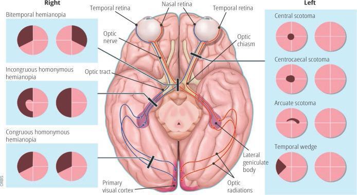
Anatomical diagram and educational infographic detailing the human visual pathway and associated visual field defects. The central illustration shows a superior (dorsal) view of the brain, highlighting the optic nerve, optic chiasm (decussation point), optic tract, lateral geniculate body, optic radiations, and the primary visual cortex in the occipital lobes. Pathways are color-coded (red and blue) to demonstrate the transition from nasal and temporal retinal fibers to the contralateral and ipsilateral hemispheres. Black lesion bars indicate specific sites of damage along this pathway. Flanking the central diagram are clinical representations of visual field loss patterns. The 'Right' panel displays bilateral defects including bitemporal hemianopia, incongruous homonymous hemianopia, and congruous homonymous hemianopia. The 'Left' panel displays unilateral or localized defects including central scotoma, centrocaecal scotoma, arcuate scotoma, and temporal wedge. This diagram serves as a neuro-ophthalmological guide for localizing lesions within the central nervous system based on perimetry findings.
Anatomical diagram and educational infographic detailing the human visual pathway and associated visual field defects. The central illustration shows a superior (dorsal) view of the brain, highlighting the optic nerve, optic chiasm (decussation point), optic tract, lateral geniculate body, optic radiations, and the primary visual cortex in the occipital lobes. Pathways are color-coded (red and blue) to demonstrate the transition from nasal and temporal retinal fibers to the contralateral and ipsilateral hemispheres. Black lesion bars indicate specific sites of damage along this pathway. Flanking the central diagram are clinical representations of visual field loss patterns. The 'Right' panel displays bilateral defects including bitemporal hemianopia, incongruous homonymous hemianopia, and congruous homonymous hemianopia. The 'Left' panel displays unilateral or localized defects including central scotoma, centrocaecal scotoma, arcuate scotoma, and temporal wedge. This diagram serves as a neuro-ophthalmological guide for localizing lesions within the central nervous system based on perimetry findings.

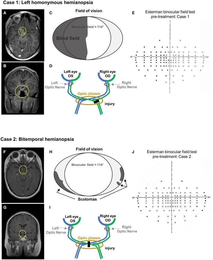
This composite educational image presents two clinical cases correlating neuroimaging findings with visual field defects. Case 1 (A-E) depicts left homonymous hemianopia. T1-weighted contrast-enhanced MRI in axial (A) and coronal (B) planes shows a heterogeneously enhancing mass in the suprasellar region (yellow circles), involving the optic chiasm and third ventricle floor. Schematic (C) and anatomical diagram (D) illustrate the corresponding lesion in the right optic tract and the resulting left hemifield blindness. The Esterman binocular field test (E) confirms significant unseen points (black dots) in the left visual field. Case 2 (F-J) depicts bitemporal hemianopia. MRI scans (F, G) reveal a residual suprasellar mass (yellow circles) inseparable from the hypothalamus and optic chiasm. The schematic (H) and optic pathway diagram (I) show a midline chiasmal injury, leading to peripheral scotomas. The Esterman test (J) demonstrates peripheral blind spots consistent with bitemporal vision loss. This comparison illustrates how specific anatomical injuries along the optic pathway correlate with distinct patterns of binocular visual impairment.
This composite educational image presents two clinical cases correlating neuroimaging findings with visual field defects. Case 1 (A-E) depicts left homonymous hemianopia. T1-weighted contrast-enhanced MRI in axial (A) and coronal (B) planes shows a heterogeneously enhancing mass in the suprasellar region (yellow circles), involving the optic chiasm and third ventricle floor. Schematic (C) and anatomical diagram (D) illustrate the corresponding lesion in the right optic tract and the resulting left hemifield blindness. The Esterman binocular field test (E) confirms significant unseen points (black dots) in the left visual field. Case 2 (F-J) depicts bitemporal hemianopia. MRI scans (F, G) reveal a residual suprasellar mass (yellow circles) inseparable from the hypothalamus and optic chiasm. The schematic (H) and optic pathway diagram (I) show a midline chiasmal injury, leading to peripheral scotomas. The Esterman test (J) demonstrates peripheral blind spots consistent with bitemporal vision loss. This comparison illustrates how specific anatomical injuries along the optic pathway correlate with distinct patterns of binocular visual impairment.

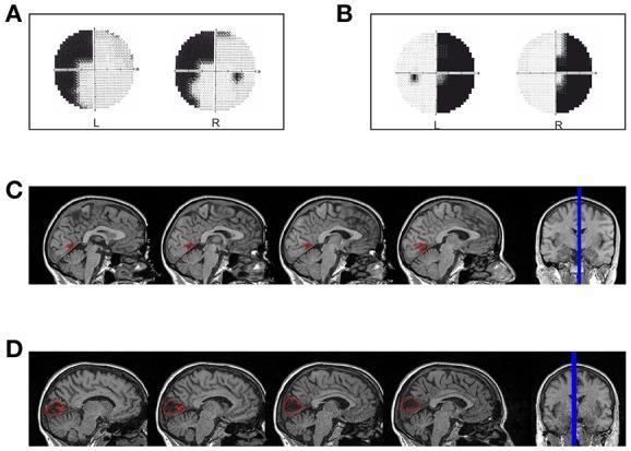
A multi-panel medical image illustrating the correlation between visual field defects and cortical lesions in two hemianopic patients. Panels A and B display Humphrey visual field perimetry results. Panel A shows a left superior quadrantanopia in patient LF, while Panel B shows a right homonymous hemianopia with foveal sparing in patient SL. Panels C and D provide diagnostic neuroimaging reconstructions via sagittal and coronal T1-weighted MRI slices. Panel C (Patient LF) highlights a focal red-labeled lesion in the anterior portion of the right calcarine fissure near the parieto-occipital fissure, consistent with the left-sided visual defect. Panel D (Patient SL) shows a larger red-labeled lesion in the left median para-sagittal occipital lobe involving the peri-calcarine cortex, corresponding to the right-sided hemianopia. The image serves as a clinical anatomical reference for mapping primary visual cortex (V1) damage to specific patterns of post-chiasmatic vision loss, such as quadrantanopia and hemianopia.

A multi-panel medical image illustrating the correlation between visual field defects and cortical lesions in two hemianopic patients. Panels A and B display Humphrey visual field perimetry results. Panel A shows a left superior quadrantanopia in patient LF, while Panel B shows a right homonymous hemianopia with foveal sparing in patient SL. Panels C and D provide diagnostic neuroimaging reconstructions via sagittal and coronal T1-weighted MRI slices. Panel C (Patient LF) highlights a focal red-labeled lesion in the anterior portion of the right calcarine fissure near the parieto-occipital fissure, consistent with the left-sided visual defect. Panel D (Patient SL) shows a larger red-labeled lesion in the left median para-sagittal occipital lobe involving the peri-calcarine cortex, corresponding to the right-sided hemianopia. The image serves as a clinical anatomical reference for mapping primary visual cortex (V1) damage to specific patterns of post-chiasmatic vision loss, such as quadrantanopia and hemianopia.

Finding Sources
Reading File
Reading File
Here is a thorough analysis of each option, backed by multiple authoritative textbooks:

Visual Field Defects - Lesion Localization

The key diagram from Goldman-Cecil Medicine (Figure 392-1) and the rules from Bradley & Daroff's Neurology summarize the entire optic pathway beautifully. Let's go through each option:
Visual pathway and field defects diagram

a. Bitemporal hemianopia - Occipital lobe ❌ INCORRECT

Bitemporal hemianopia is caused by a lesion at the optic chiasm (e.g., pituitary adenoma compressing the crossing nasal fibers). Bradley & Daroff's Neurology states explicitly: "Only a lesion of the optic chiasm causes true bitemporal hemianopia." The occipital lobe produces congruent homonymous defects, not bitemporal ones.

b. Left superior homonymous quadrantanopia - Right temporal lobe ✅ CORRECT

The temporal lobe carries Meyer's loop (the inferior fibers of the optic radiation, which swing forward into the temporal lobe). A lesion here produces a contralateral superior homonymous quadrantanopia - the classic "pie in the sky" defect. A right temporal lobe lesion → left superior homonymous quadrantanopia. Bradley & Daroff's confirms: "Temporal lobe lesions give slightly incongruent homonymous hemianopias involving the upper quadrant." Goldman-Cecil confirms: "Temporal (Meyer) loop of the optic radiation: congruous partial or complete (contralateral) homonymous superior quadrantanopia."

c. Left homonymous hemianopia - Right occipital lobe ✅ CORRECT

Post-chiasmal (retrochiasmal) lesions cause contralateral homonymous hemianopia. The right occipital (visual) cortex processes the left visual field. A lesion in the right occipital lobe → left homonymous hemianopia (usually with macular sparing if the infarct involves the tip). This is the most classic and reliable localization in the entire visual pathway.

d. Left inferior quadrantanopia - Early pituitary tumor ❌ INCORRECT

An early/small pituitary tumor compresses the inferior crossing nasal fibers of the optic chiasm first, producing a bitemporal superior quadrantanopia (upper outer fields go first), not an inferior one. Left inferior quadrantanopia would point to a right parietal lobe lesion (the superior fibers of the optic radiation run through the parietal lobe and carry the inferior visual field). Goldman-Cecil: "Parietal (superior) projection of the optic radiation: congruous partial or complete homonymous inferior quadrantanopia."

e. Right homonymous hemianopia - Right occipital lobe ❌ INCORRECT

This is backwards. The right occipital cortex receives input from the left visual field (nasal fibers of the right eye + temporal fibers of the left eye). A right occipital lesion produces left homonymous hemianopia, not right. For a right homonymous hemianopia, the lesion must be in the left retrochiasmal pathway (left optic tract, radiation, or occipital lobe).

Summary Table

OptionField DefectStated LesionCorrect LesionVerdict
aBitemporal hemianopiaOccipital lobeOptic chiasm
bL/ superior homonymous quadrantanopiaR/ temporal lobeR/ temporal lobe (Meyer's loop)
cLeft homonymous hemianopiaR/ occipital lobeR/ occipital lobe
dL/ inferior quadrantanopiaEarly pituitary tumorR/ parietal lobe
eR/ homonymous hemianopiaR/ occipital lobeL/ retrochiasmal pathway
The correct answers are b and c.
  • Goldman-Cecil Medicine, Fig. 392-1
  • Bradley & Daroff's Neurology in Clinical Practice, Box 16.2
This is a shared conversation. Sign in to Orris to start your own chat.