
interlobular tumor of brest
invasive lobular carcinoma breast histology
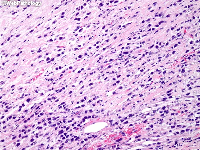
Breast histopathology image analyzed by bright-field light microscopy of formalin-fixed, paraffin-embedded tissue stained with Hematoxylin and Eosin (H&E). The lesion demonstrates invasive lobular carcinoma with a pleomorphic nuclear phenotype. Tumor cells are arranged in the classic lobular pattern with infiltrative, discohesive cells traversing a desmoplastic stroma, but display marked nuclear pleomorphism. Nuclei vary considerably in size and shape, show irregular contours, and are notably hyperchromatic. Cytoplasm is typically scant, and mitotic activity may be low to moderate depending on the field. The architectural pattern may include Indian-file strands, targetoid arrangements, and single-cell infiltration, consistent with lobular histology, but the prominent pleomorphism distinguishes it from conventional ILC. Background breast parenchyma often shows fibrous stroma and mild chronic inflammatory cells. No evident necrosis is observed in this field, though focal areas of cytoplasmic detail may be limited by processing. Clinically, pleomorphic lobular carcinoma is considered a high-grade variant with potential for more aggressive behavior than classic ILC, with implications for prognosis and treatment, including adjuvant endocrine therapy given the common hormone receptor positivity. This image is useful for pathology education, differential diagnosis training (distinguishing lobular from ductal carcinomas), and tumor grading in breast pathology.
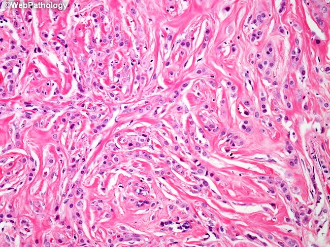
Histology-based description of invasive lobular carcinoma in breast tissue using light microscopy of Hematoxylin and Eosin stained section. The specimen demonstrates tumor cells arranged in discohesive, single‑file patterns within a dense, desmoplastic fibrous stroma. Neoplastic cells are relatively uniform with vesicular nuclei and small, conspicuous nucleoli, reflecting mild to moderate cytologic atypia typical of ILC. The stromal background is collagen-rich and extensively fibrotic, contributing to overall cellular scantiness and a characteristic sparse gland formation. Infiltration extends through the breast parenchyma with minimal formation of typical ductal structures; cells may be seen interspersed among adipocytes and along fibrous septa. The growth pattern and architectural arrangement support lobular lineage and diffuse spread, consistent with classic and mixed subtypes of ILC. Differential considerations include invasive ductal carcinoma with more cohesive nests, lobular neoplasia without invasion, and metastatic signet‑ring lesions, though mucin production is not evident here. Clinically, these histologic features inform tumor biology, suggesting potential estrogen receptor positivity and a propensity for multifocal/diffuse involvement, with implications for surgical planning and endocrine therapy. This image serves as a high‑yield educational reference for pathology trainees, researchers, and clinicians. Correlative immunohistochemistry may confirm lobular phenotype by E-cadherin loss.
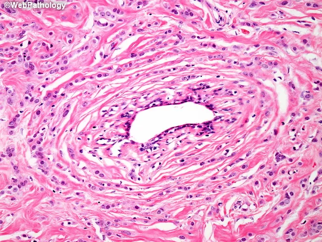
An H&E stained histology slide of breast tissue demonstrating invasive lobular carcinoma. The classic growth pattern shows small, uniform tumor cells infiltrating the fibrous stroma in single-file arrangements (Indian file) and, notably, concentric/periductal involvement around an uninvolved duct. This discohesive neoplastic population commonly lacks strong cytologic atypia, with bland round to oval nuclei and scant cytoplasm, set within a loose, fibromyxoid stroma. The architecture contrasts with the cohesive nests typical of invasive ductal carcinoma and highlights diffuse infiltration through the lobular unit rather than expansive mass formation. The infiltrative cells penetrate between native collagen bundles and adipose tissue, producing subtle architectural distortion. Although E-cadherin loss underlies lobular histogenesis, this immunophenotypic feature is not directly visible on H&E but is a well-established diagnostic correlate. Clinically, this pattern informs subtype classification, prognosis, and therapeutic planning, with implications for hormone receptor testing (ER/PR positive likelihood) and HER2 status guiding systemic therapy. Radiologic-pathologic correlation is important due to potential multifocality and satellite foci in lobular carcinoma. This image is valuable for education on lobular invasion, differential diagnosis from ductal carcinoma, and for research into targeted therapies addressing adhesion molecule pathways. Impact on surgical margin assessment and adjuvant planning should be considered clinically by teams.
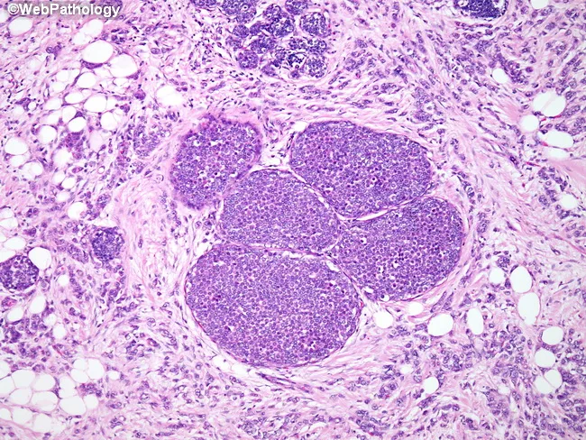
Low-power light microscopy of breast tissue stained with Hematoxylin and Eosin demonstrates invasive lobular carcinoma (ILC) with diffuse infiltration of the parenchyma and adipose fat by small uniform cells arranged in slender, linear cords and single-file patterns. The tumor cells exhibit scant cytoplasm and round to oval nuclei with mild atypia, forming non-cohesive strands that invade across the stroma, rather than forming cohesive nests typical of ductal carcinoma. In several lobules, there is accompanying lobular carcinoma in situ (LCIS) within expanded lobular units, highlighting a spectrum of lobular neoplasia. The top of the field shows a few uninvolved lobular structures, indicating focal preservation within a predominantly malignant milieu. The surrounding stroma shows mild desmoplasia in places and fat invasion by tumor cells is evident. This histology is characteristic of ILC and explains potential clinical features such as subtle breast thickening or mammographic underestimation of disease, because ILC often presents as diffuse infiltration rather than a discrete mass. Immunohistochemical confirmation (e-cadherin loss) supports diagnosis, while hormone receptor status (estrogen/progesterone receptor positive) frequently influences treatment planning. The image is valuable for educational purposes and for correlating morphology with management decisions, including surgical margins and systemic therapy considerations.
invasive lobular carcinoma breast treatment outcomes
Invasive Lobular Carcinoma (ILC) of the Breast
Note on terminology: "Interlobular" is likely a typo/variation of "Intralobular" (within the lobule). The key breast tumors arising from lobular units are Lobular Carcinoma In Situ (LCIS) and Invasive Lobular Carcinoma (ILC). This answer covers both, with focus on ILC.
Overview
- Robbins, Cotran & Kumar Pathologic Basis of Disease - p. 979
- Robbins & Kumar Basic Pathology - p. 715
Lobular Neoplasia: The Precursor Spectrum
| Lesion | Description | Relative Cancer Risk |
|---|---|---|
| Atypical Lobular Hyperplasia (ALH) | Monomorphic dyscohesive cells partially filling lobule; histologically identical to LCIS but more limited in extent | ~4x relative risk |
| Lobular Carcinoma In Situ (LCIS) | Same cells fully filling lobule; confined to ductal-lobular system, no invasion | ~8-10x relative risk |
| Invasive Lobular Carcinoma (ILC) | Cells breach basement membrane and invade stroma | Malignant |
- Robbins, Cotran & Kumar - p. 967
Molecular/Genetic Features
- E-cadherin (CDH1) loss: The hallmark of lobular lineage. Caused by CDH1 gene mutation or methylation silencing.
- ER-positive/PR-positive: Almost all lobular carcinomas express hormone receptors.
- HER2 overexpression: Rare.
- Germline CDH1 mutations: Associated with hereditary lobular breast cancer AND diffuse (signet ring) gastric cancer (hereditary diffuse gastric cancer syndrome).
- Molecularly, most ILCs fall in the luminal (ER+/HER2-) group.
Morphology / Histology
- "Indian file" / single-file pattern - Tumor cells invade stroma in single linear cords, one cell wide
- Targetoid/periductal pattern - Cells arrange concentrically around uninvolved ducts
- Signet ring cells - Intracytoplasmic mucin displacing the nucleus (seen in some subtypes)
- Minimal desmoplastic reaction - Many ILCs invade without producing fibrosis, making them harder to palpate and detect on imaging

Histologic Variants of ILC
| Variant | Key Features |
|---|---|
| Classic ILC | Uniform small cells in Indian-file pattern |
| Pleomorphic ILC | Marked nuclear pleomorphism, more aggressive; still discohesive |
| Solid ILC | Sheets/nests rather than single files |
| Alveolar ILC | Rounded nests of 20+ cells |
| Mixed ILC | Combined patterns |
Clinical Features
| Feature | Details |
|---|---|
| Presentation | Often palpable mass or mammographic density; a subgroup may be clinically and radiologically occult due to absent desmoplasia |
| Multifocality | Higher tendency than ductal carcinoma for multifocal/multicentric disease |
| Bilaterality | ~4% bilateral; sequential lesions common |
| Location | Upper outer quadrant most common (50%) |
| Imaging | May underestimate extent on mammography; MRI is superior for ILC staging |
Unique Metastatic Pattern
- Cerebrospinal fluid / leptomeninges (carcinomatous meningitis)
- Peritoneum and retroperitoneum
- Gastrointestinal tract (mimicking primary GI carcinoma)
- Ovaries and uterus
- Bone marrow
- Serosal surfaces
- Robbins & Kumar Basic Pathology - p. 715
Risk Stratification Table (Lobular Lesions)
| Pathologic Lesion | Relative Risk |
|---|---|
| Nonproliferative changes (cysts, apocrine metaplasia) | No increased risk |
| Atypical Lobular Hyperplasia (ALH) | ~4x |
| Lobular Carcinoma In Situ (LCIS) | ~8-10x |
Immunohistochemistry (IHC) Profile
| Marker | Typical Result |
|---|---|
| E-cadherin | Negative (lost) - key diagnostic marker |
| ER (Estrogen Receptor) | Positive in ~95% |
| PR (Progesterone Receptor) | Positive in ~70% |
| HER2 | Rarely overexpressed |
| Ki-67 | Usually low in classic ILC |
Treatment Principles
- Endocrine therapy (tamoxifen or aromatase inhibitors) is the mainstay of systemic treatment
- Chemotherapy: Less responsive compared to HER2+ or TNBC; neoadjuvant chemotherapy shows lower pCR rates
- Surgery: Mastectomy rates are higher due to multifocality and larger tumor extent; breast-conserving surgery is possible but requires wider margins
- CDK4/6 inhibitors (palbociclib, ribociclib, abemaciclib) combined with endocrine therapy for metastatic ER+ disease