
Excessive draining lacrimal gland or watering eye causes
epiphora watering eye causes lacrimal drainage
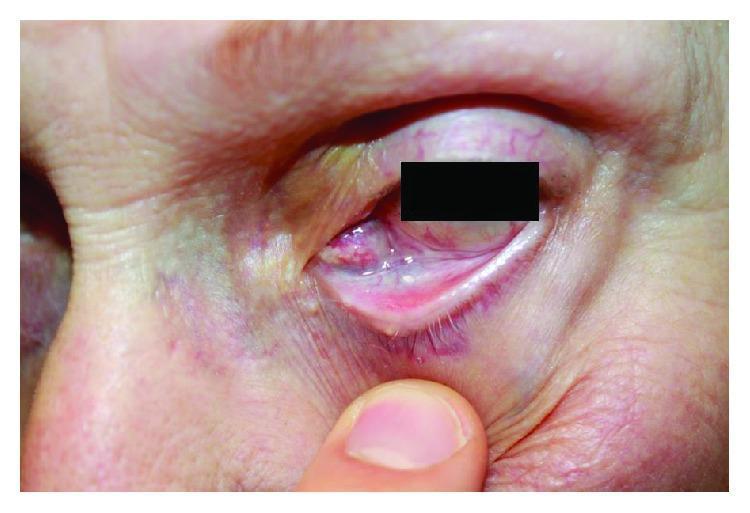
Clinical photograph of the left eye of an elderly patient demonstrating a significant medial orbital mass and secondary ocular changes. The image shows a prominent, fleshy, erythematous mass located at the medial canthus, which is causing significant mechanical ectropion (outward turning) of the lower eyelid. The affected lower eyelid appears inflamed, with visible conjunctival hyperaemia and swelling. Evidence of symptomatic epiphora is present, characterized by a glistening, wet appearance of the periocular skin and clumping of the lower eyelashes. The skin around the inner corner of the eye exhibits erythema and mild maceration, likely due to chronic tear exposure. This clinical presentation is characteristic of a rapidly growing orbital tumor, later diagnosed as epithelioid sarcoma, which physically displaces local adnexal structures and obstructs the lacrimal drainage system. The image serves as a high-level educational example of orbital pathology, the clinical signs of mechanical eyelid malposition, and the physical manifestations of lacrimal outflow obstruction.
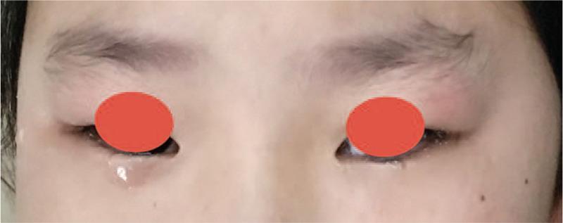
This clinical photograph displays a close-up frontal view of a pediatric patient's orbital region, demonstrating asymmetric epiphora (excessive tearing). The primary finding is a prominent tear drop and fluid pooling along the lower eyelid margin of the right eye, contrasting with the left eye, which shows minimal tear production. The facial skin is fair with a few scattered benign-appearing pigmented macules (freckles). Both eyes are partially obscured by red circles for privacy. This visual represents a clinical manifestation of unilateral nasolacrimal duct obstruction, often caused in pediatric cases by anatomical abnormalities such as a dentigerous cyst or ectopic tooth impinging on the lacrimal drainage system. The image highlights the objective difference in tear shedding (lacrimation) between a healthy eye and one with drainage impairment, serving as a diagnostic indicator for maxillofacial or ophthalmologic pathology.
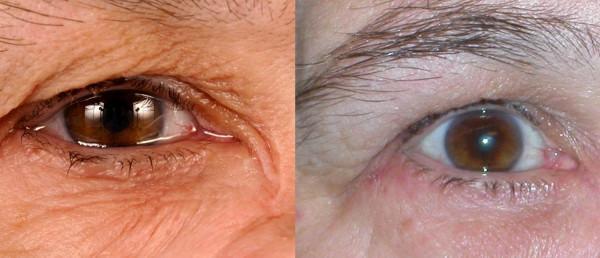
This clinical comparison photograph illustrates the management of severe hyperlacrimation (epiphora) using Botulinum Toxin A (BTA). The left frame shows the patient's eye with significant pathological tearing; visible features include a high tear meniscus, pooling of tears in the lower eyelid, and macerated, moist periorbital skin with pronounced wrinkling and mild erythema. This condition resulted from a lacrimal duct obstruction following maxillary sinus tumor resection. The right frame demonstrates the therapeutic effect after BTA injection into the palpebral part of the lacrimal gland. In this image, the tear volume is visibly normalized, the tear film is stable without overflow, and the surrounding skin appears drier and healthier. The comparison highlights BTA injection as a viable interim or alternative treatment for hyperlacrimation caused by ductal stenosis or 'crocodile tears' syndrome, effectively reducing lacrimal secretion without inducing xerophthalmia (dry eye).
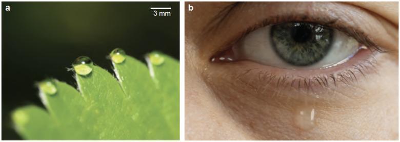
A comparison of gravitational fluid ejection mechanisms in biological systems. Image (a) is a macro photograph of plant guttation, showing spherical xylem fluid droplets adhering to the serrated margin (hydathodes) of a strawberry leaf, with a 3 mm scale bar for reference. Image (b) is a clinical photograph of a human eye demonstrating tear film dynamics and lacrimal drainage. The image highlights the ocular surface, the eyelid margin with eyelashes, and the lacrimal puncta (drainage openings) at the medial canthus. Excessive tearing (epiphora) is illustrated by a large, clear tear droplet suspended from the lower eyelid margin and tracking down the infraorbital skin. The comparison illustrates how both systems manage fluid overflow—via xylem exudation in plants and tear drainage/overflow in humans—governed by the interplay of surface tension, capillary adhesion, and gravitational forces.
causes of epiphora watering eye excessive lacrimation classification
Causes of a Watering Eye (Epiphora)
Mechanism 1: Excessive Tear Production (Hyperlacrimation / Reflex Lacrimation)
Ocular Surface Irritation
| Category | Specific Causes |
|---|---|
| Eyelid abnormalities | Trichiasis (inturned lashes touching cornea), distichiasis, entropion, epiblepharon, subtarsal foreign body |
| Conjunctival disease | Conjunctivitis (allergic, bacterial, viral - including chlamydial), blepharitis, meibomian gland dysfunction, pemphigoid |
| Corneal disease | Corneal abrasion/ulcer, keratitis (herpetic, bacterial, fungal), foreign body, calcific keratopathy |
| Intraocular disease | Uveitis (iritis), acute angle-closure glaucoma |
| Dry eye syndrome | Paradoxically, reduced basal tearing triggers reflex hypersecretion - the most common cause of epiphora in adults |
| Environmental | Wind, smoke, dust, cold air, bright light |
| Systemic/drugs | Epinephrine eye drops, cholinergic drugs, certain systemic medications |
| Neurological | "Crocodile tears" (aberrant facial nerve regeneration causing gustatory lacrimation), facial nerve irritation |
- Goldman-Cecil Medicine, p. 4123: "The most common cause of epiphora is chronic or acute irritation that results in the overproduction of reflex tearing from ocular dryness (dry eye syndrome). Ocular surface irritants, such as trichiatic eyelashes touching the cornea, also can stimulate excessive tear production."
Mechanism 2: Impaired Tear Drainage (True Epiphora)
A. Anatomical (Structural) Obstruction (~70% of drainage failures)
- Punctal stenosis or atresia (congenital)
- Punctal malposition (ectropion - outward turning of the lower lid displaces the punctum away from the tear lake)
- Kissing puncta syndrome (upper and lower puncta appose each other)
- Scarring from chronic inflammation (Stevens-Johnson syndrome, cicatricial pemphigoid)
- Canalicular stenosis from herpes simplex or zoster infection
- Drug toxicity (topical timolol, systemic docetaxel/5-fluorouracil)
- Trauma, sarcoidosis, actinomycotic canaliculitis
- Idiopathic acquired nasolacrimal duct obstruction - most common in middle-aged/elderly women
- Dacryocystitis (infection of the lacrimal sac, acute or chronic)
- Dacryolith (stone within the drainage system)
- Granulomatous disease: Wegener's granulomatosis, sarcoidosis
- Malignancy (lacrimal sac tumour, adjacent nasal or sinus tumour)
- Trauma (mid-face fracture, post-surgical after rhinoplasty, FESS, sinus surgery)
- Radiation exposure
- Bony remodelling with ageing (narrowing of the bony nasolacrimal canal)
- Present in up to 20% of neonates; caused by failure of the valve of Hasner to canalise
- Spontaneous resolution occurs in ~90% within the first year of life
- Differential in neonates: punctal atresia, congenital glaucoma, chlamydial conjunctivitis, keratitis, uveitis
B. Functional (Pump Failure) Obstruction (~30% of drainage failures)
-
Eyelid laxity / ectropion - the lacrimal pump (orbicularis oculi) fails to draw tears into the puncta
-
Facial nerve palsy (VII) - orbicularis weakness prevents lacrimal pump action; simultaneously, corneal exposure causes reflex hyperlacrimation
-
Lid malpositioning not amounting to full ectropion
-
Physiological pump failure in older patients
-
Scott-Brown's Otorhinolaryngology Vol. 1, p. 1293: "Epiphora from nasolacrimal system obstruction is either anatomic (70%) or functional (30%)... Functional obstructions are a result of either significant narrowing within the lacrimal system that delays normal lacrimal flow or a failure of the lacrimal pumping mechanism."
Summary Table (Children vs. Adults)
| Excess Tear Production | Drainage Failure |
|---|---|
| Allergic rhinitis | Congenital nasolacrimal duct obstruction |
| Upper respiratory tract infection | Skeletal/sinus abnormalities |
| Epiblepharon | Lid malposition |
| Subtarsal foreign body | Punctal malposition |
| Iritis | Punctal occlusion |
| Corneal abrasion/ulceration | Anomalous drainage system |
| Conjunctivitis | |
| Congenital glaucoma |
- Dry eye syndrome (reflex hyperlacrimation)
- Blepharitis / meibomian gland dysfunction
- Acquired nasolacrimal duct obstruction (idiopathic, age-related)
- Ectropion with lacrimal pump failure
- Allergic conjunctivitis
Clinical Pearl
Treatment for obstruction
dacryocystorhinostomy DCR lacrimal duct surgery technique
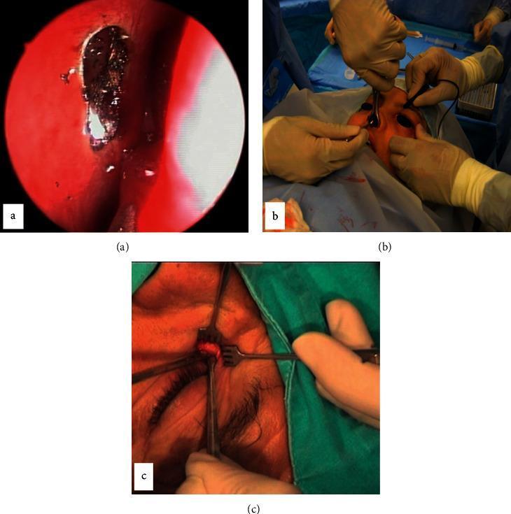
This composite of three intraoperative images illustrates different surgical approaches for dacryocystorhinostomy (DCR) to treat lacrimal duct obstruction. Image (a) displays a Transcanalicular Laser DCR (TCL-DCR), showing an endoscopic endonasal view with a focal area of laser-induced thermal ablation on the nasal mucosa, characterized by carbonization and localized hemorrhage. Image (b) depicts Non-Endoscopic Endonasal DCR (NEN-DCR), showing an external operating room perspective where surgeons use headlights and manual instruments to access the lacrimal sac through the nasal cavity. Image (c) shows External DCR (EXT-DCR), featuring a close-up of a cutaneous nasojugal incision. This view demonstrates the use of self-retaining retractors and forceps to expose the underlying lacrimal sac and periosteum for the creation of mucosal flaps. The sequence highlights the variation in invasiveness, from minimally invasive endoscopic laser techniques to traditional open external surgery.
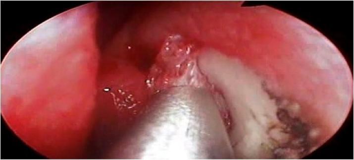
This endoscopic clinical photograph depicts an intraoperative view of the lateral nasal wall during a dacryocystorhinostomy (DCR). The visual field shows a pinkish-red, vascularized nasal mucosa where a curvilinear incision has been made along the maxillary line. A metallic surgical instrument, appearing to be straight Blakesley forceps, is actively lifting a mucosal flap to expose the underlying whitish-yellow bone, identified as the lacrimal bone. Minor bleeding and mucosal edema are visible at the incision margins, typical of surgical tissue disruption. This image serves as an educational representation of the initial steps in endonasal lacrimal surgery, specifically the creation of a mucosal entry point to access the lacrimal sac. The procedural focus is on anatomical landmark identification and the surgical technique for flap elevation in the management of nasolacrimal duct obstruction.
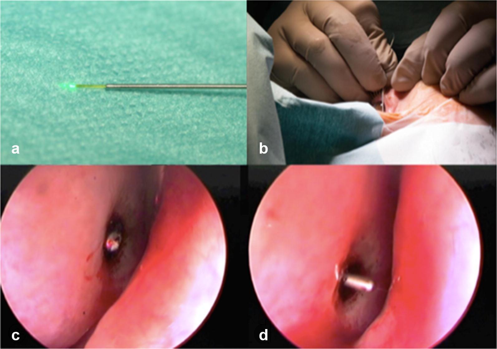
This four-panel image illustrates a laser-assisted dacryocystorhinostomy (DCR) procedure for treating nasolacrimal duct obstruction. Panel A shows a 300µm metallic laser fiber optic emitting a green aiming beam, used for precise tissue targeting. Panel B depicts the external surgical field where a surgeon, wearing sterile gloves, manipulates the laser fiber through the lacrimal canaliculus in a mediorostral orientation near the medial canthus. Panels C and D provide endonasal endoscopic views of the nasal cavity mucosa. Panel C demonstrates transillumination, where the green laser light is visible through the nasal mucosa at the anteroinferior rim of the middle turbinate just before breaching the bone to create a bony ostium. Panel D shows the subsequent step where a blunt metal probe is used to guide a silicone intubation tube through the newly formed ostium to ensure long-term patency of the bypass. This sequence highlights the minimally invasive transcanalicular approach to lacrimal surgery using 810-nm diode laser technology.
Treatment of Lacrimal Drainage Obstruction
1. Punctal Obstruction / Stenosis
Primary Punctal Stenosis (no eversion)
| Option | Detail |
|---|---|
| Dilatation alone | Simple punctal dilation with a dilator - rarely gives sustained benefit |
| Dilatation + mini-Monoka stent | Stent inserted after dilation, removed after 1 month - significantly better results |
| Punctoplasty | Surgical enlargement: one-, two-, or three-snip techniques; posterior ampulla wall excision; Kelly punch; laser; microsurgery. A temporary stent can be used alongside |
Secondary Punctal Stenosis (with eversion/ectropion)
-
Retropunctal (Ziegler) cautery - for pure punctal eversion; burns applied ~5 mm below punctum cause tissue shrinkage to invert the punctum
-
Medial conjunctivoplasty - diamond-shaped excision of tarsoconjunctiva for medial ectropion without horizontal laxity
-
Lower lid tightening (tarsal strip procedure) - for lower lid laxity; can be combined with conjunctivoplasty
-
Kanski's Clinical Ophthalmology 10th ed., p. 118-119
2. Canalicular Obstruction
| Degree | Treatment |
|---|---|
| Partial obstruction | Silicone stent intubation of one or both canaliculi for 6 weeks - 6 months |
| Total individual canalicular obstruction | Canalicular trephination with a minitrephine (Siser) + intubation; balloon canaliculoplasty; endoscopic laser canaliculoplasty |
| 6-8 mm of patent proximal canaliculus | Anastomosis of the patent segment into the lacrimal sac + intubation |
| Severe / anastomosis not possible | Conjunctivodacryocystorhinostomy (CDCR) + Lester Jones glass tube - caruncle excised, tube runs from lacus lacrimalis to nasal cavity |
The Lester Jones tube is also used when the lacrimal system is intact but the pump mechanism fails (e.g. facial nerve palsy).
- Kanski's Clinical Ophthalmology 10th ed., p. 119-120
3. Nasolacrimal Duct / Sac Obstruction
A. Congenital Nasolacrimal Duct Obstruction (CNLDO)
| Step | Timing | Intervention |
|---|---|---|
| 1. Watchful waiting | Birth - 12 months | Spontaneous resolution in ~90% by 1 year |
| 2. Lacrimal sac massage (Crigler technique) | First year | Index finger over common canaliculus, roll down over sac to generate hydrostatic pressure against the valve of Hasner |
| 3. Probing (Bowman probe) | 12-18 months (up to 24 months) | Passage of fine wire through canalicular system to rupture the membranous obstruction at the valve of Hasner; can be performed under topical anaesthesia in young infants as an outpatient; repeat probing if first attempt fails |
| 4. Endoscopic-guided probing | After failed blind probing | Gold standard for re-probing; visualises common causes of failure |
| 5. Silicone intubation ± balloon dilation | After probing failure | Tubes left for 1-6 months; removed under GA through the nose to prevent aspiration |
| 6. Dacryocystorhinostomy (DCR) | Persistent epiphora or complex anatomy | For upper duct obstruction, bony atresia, or after multiple probing failures |
Tip from Kanski's: "Because congenital nasolacrimal obstruction undergoes spontaneous resolution in 90% of babies by their first birthday, initial probing should be delayed until this age."
B. Acquired Nasolacrimal Duct Obstruction (Adults)
Indications
- Symptomatic distal nasolacrimal duct obstruction not relieved by probing and syringing
- NOT indicated for punctal or canalicular obstruction (DCR will not bypass these sites)
Two Main Approaches
| External DCR | Endoscopic (Endonasal) DCR | |
|---|---|---|
| Incision | Vertical skin incision 10 mm medial to inner canthus | No skin incision (endoscopic via nostril) |
| Bone removal | Anterior lacrimal crest + lacrimal fossa bone removed | Lacrimal bone and frontal process of maxilla removed with Kerrison punch or DCR drill |
| Sac opening | H-shaped incision creating anterior + posterior flaps; sutured to nasal mucosa flaps | Sac marsupialized with lacrimal probe tenting the sac; anterior + posterior flaps created |
| Stenting | Silicone Crawford tubes often placed | Crawford tubes placed; removed at 4-6 weeks |
| Advantages | Higher long-term success rate (~90%); allows sac tumour exclusion | No facial scar; simultaneous nasal pathology addressable; less bleeding |
| Post-op care | Saline irrigation, oral antibiotics 7 days, topical antibiotic drops 2 weeks | Same; endoscopic review + crust removal at 2 weeks |
DCR Surgical Steps (Endoscopic)
- Create posteriorly based mucosal flap over the nasolacrimal crest
- Remove lacrimal bone (soft) and frontal process of maxilla (harder, Kerrison punch or drill)
- Expose agger nasi cell (allows wider marsupialization)
- Tent lacrimal sac with probe through inferior canaliculus
- Marsupialize sac with H-incision (wide anterior + posterior flaps)
- Reapproximate mucosal flap over bone-sac junction
- Place Crawford tubes; pack with gelfoam
DCR Complications
-
Epistaxis (~2%)
-
Obstruction/rhinosinusitis of frontal or maxillary sinus
-
Orbital penetration / damage to extraocular muscles
-
CSF leak
-
Orbital haematoma
-
Failure (from scarring, granuloma formation, or unaddressed canalicular obstruction)
-
Scott-Brown's Otorhinolaryngology Vol. 1, p. 1293-1294; Kanski's Clinical Ophthalmology 10th ed., p. 120-122
4. Laser / Minimally Invasive Options
- Transcanalicular laser DCR - 810 nm diode laser fiber passed through canaliculus; creates bony ostium into nasal cavity under endonasal endoscopic visualization. Less invasive but lower long-term patency vs. external DCR.
- Balloon catheter dilation - for failed probing in CNLDO; dilates the nasolacrimal duct.
- Laser punctoplasty - for punctal stenosis.
Summary: Treatment by Site
Punctum  →  Dilation / Punctoplasty / Stent
Canaliculus  →  Silicone stenting / Trephination / CDCR + Jones tube
Nasolacrimal sac/duct  →  DCR (external or endoscopic)
Congenital  →  Massage → Probing → Intubation → DCR (step-up)
Any non oparative management
Non-Operative Management of Lacrimal Drainage Obstruction
1. Watchful Waiting (Observation)
- ~90% of affected neonates achieve spontaneous resolution by 12 months of age as the valve of Hasner canalises naturally
- Probing should be deferred until 12 months because the natural history is so favourable
- Even up to 24 months, evidence shows "little difference in final outcome at 24 months conferred by intervention versus non-intervention" - Kanski's Clinical Ophthalmology 10th ed.
2. Lacrimal Sac Massage (Crigler Technique)
- Place the index finger firmly over the common canaliculus (upper inner corner of the lid) to block reflux of fluid upward
- Roll the finger downward over the lacrimal sac, applying firm hydrostatic pressure
- This forces fluid downward against the obstructing membrane at the valve of Hasner, aiming to rupture it
- Taught to parents to perform multiple times daily
- Likelihood of success and optimal frequency remain undetermined from trials
- More useful for membranous (soft) obstruction than bony atresia
- Kanski's Clinical Ophthalmology 10th ed., p. 121
3. Topical Antibiotics
- Used for secondary bacterial conjunctivitis superimposed on CNLDO (e.g. mucopurulent discharge, eyelash matting)
- Broad-spectrum topical antibiotic drops (e.g. chloramphenicol, fusidic acid, tobramycin)
- Do not treat the underlying obstruction - only manage the complication
- In CNLDO: "Superimposed bacterial conjunctivitis may be treated with a broad-spectrum topical antibiotic" - Kanski's, p. 121
4. Systemic Antibiotics (for Acute Dacryocystitis)
| Setting | Antibiotic approach |
|---|---|
| Mild/outpatient | Oral antibiotics covering S. aureus, S. pneumoniae, H. influenzae - consider MRSA coverage (e.g. co-amoxiclav ± clindamycin/TMP-SMX) |
| Severe / infant | IV antibiotics (acute dacryocystitis in infants = medical emergency due to risk of orbital cellulitis and retrobulbar abscess) |
| Culture | Obtain by gentle pressure over the sac and expressing fluid through the punctum before starting antibiotics |
-
Do NOT probe during active dacryocystitis - congested epithelium risks false passage formation, orbital cellulitis, and fistula formation
-
Do NOT make skin incisions during the acute phase - risks external fistula
-
Surgery (DCR) is deferred until infection is controlled - arrange ophthalmology follow-up within 24-48 hours
-
If a mass persists after resolution or pus is inspissated/loculated, needle aspiration through the skin or urgent endonasal DCR may then be required
-
Rosen's Emergency Medicine, p. 894; Scott-Brown's Vol. 2, p. 325
5. Warm Compresses
- Applied for 10-15 minutes, 3-5 times daily
- Useful as adjunct in dacryocystitis and blepharitis-related epiphora
- Help normalise flow of obstructed glands, reduce inflammation, and soften any inspissated secretions
- Rosen's Emergency Medicine, p. 894
6. Treatment of the Underlying Cause
| Underlying Cause | Non-operative Treatment |
|---|---|
| Chronic allergic rhinitis / URTI | Antihistamines, intranasal steroids, decongestants |
| Sarcoidosis / Wegener's granulomatosis | Systemic immunosuppression (corticosteroids, methotrexate) |
| Tumour causing extrinsic compression | Chemotherapy / radiotherapy (though surgery usually needed) |
| Nasal polyps | Intranasal corticosteroids, endoscopic polypectomy |
| Bony remodelling with age | Conservative observation if mild; surgery if symptomatic |
7. Congenital Dacryocele
- Resolution is common with conservative treatment alone (observation + massage)
- Probing only if conservative management fails
- Systemic antibiotics if secondary dacryocystitis occurs (common in dacryoceles)
Summary
| Scenario | Non-operative Approach |
|---|---|
| CNLDO in infant (<12 months) | Watchful waiting + lacrimal sac massage |
| Secondary conjunctivitis | Topical antibiotics |
| Acute dacryocystitis | Warm compresses + systemic antibiotics (no probing/surgery until infection resolves) |
| Dacryocele | Observation + massage |
| Allergic rhinitis-related | Antihistamines, intranasal steroids |
| Systemic granulomatous disease | Treat underlying condition |