
give me clinical features , causes , diagnosis and mangement of drug induced lupus erythamatosis
"drug-induced lupus erythematosus"[MeSH Terms]
drug induced lupus erythematosus clinical features rash
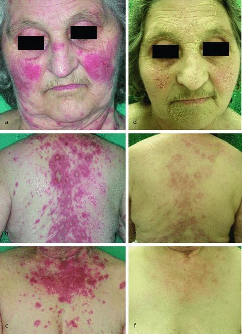
This composite clinical photograph series illustrates the progression of drug-induced subacute cutaneous lupus erythematosus (SCLE) in a 70-year-old female. Images (a-c) demonstrate the baseline presentation: (a) shows a prominent, symmetric, bright red malar erythema across the cheeks and bridge of the nose, characteristic of a 'butterfly rash'; (b) and (c) display extensive macular erythematous lesions on the upper back, chest, and arms, with some lesions exhibiting an annular or polycyclic morphology. These lesions are well-demarcated, non-scarring, and primarily distributed in sun-exposed areas. Images (d-f) show the clinical outcome one month following the discontinuation of the offending agent (hydrochlorothiazide) and treatment with low-dose systemic steroids. There is a marked reduction in the intensity and distribution of the erythema; the malar rash has significantly faded, and the truncal macular lesions have diminished to faint, sparse pigmented patches. This comparison serves as an educational tool for identifying photosensitive drug-induced cutaneous reactions and monitoring clinical response to drug withdrawal and corticosteroid therapy.
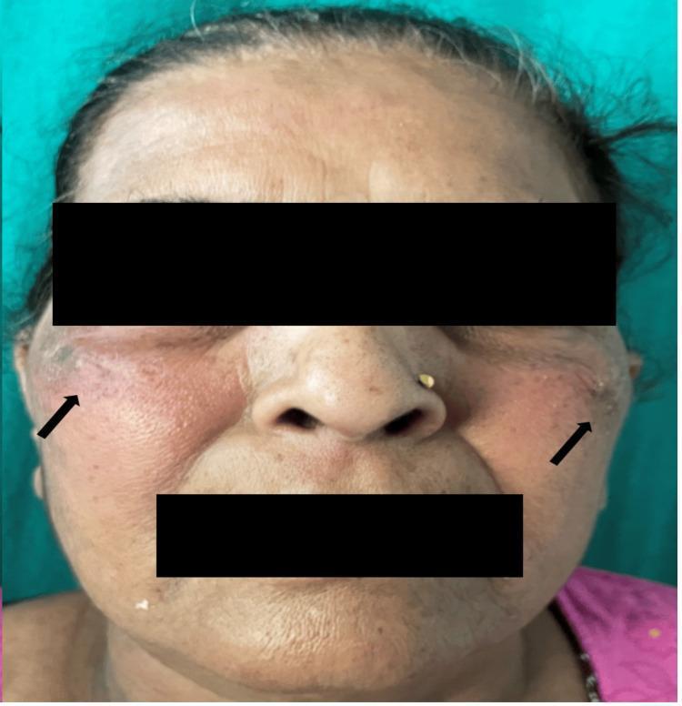
Clinical photograph of a 59-year-old female patient showing a characteristic malar rash. The image displays confluent, erythematous patches distributed over the bilateral cheeks and extending towards the temporal regions, as indicated by black arrows. The rash exhibits a symmetric, butterfly-like distribution, though the nasolabial folds appear relatively spared. The skin within the affected areas shows noticeable texture changes and erythema. Additional visible features include forehead rhytids, perioral lines consistent with age-related skin changes, and uneven pigmentation. The patient has a nose ring in the left nostril. The clinical presentation is consistent with cutaneous lupus erythematosus, specifically a drug-induced manifestation. The educational focus is on identifying classic dermatological signs of systemic inflammatory or drug-induced conditions through facial skin morphology and distribution patterns.
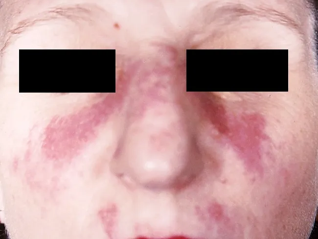
This clinical dermatology photograph displays the classic malar or butterfly rash of systemic lupus erythematosus (SLE) on the central face. Modality is clinical photography using standard white-light illumination; frontal/anterior view; color-balanced, high-resolution capture to depict superficial erythema and patchy hyperemia across the malar eminences and nasal bridge. The rash forms a bilateral, 'butterfly' distribution that typically reaches the cheeks and bridge of the nose while sparing the nasolabial folds. The observed features include confluent to patchy, erythematous macules and plaques with uniform erythema, mild perivascular edema, and subtle textural change without overt crusting or scaling in this image. The clinical morphology is characteristic for acute cutaneous lupus erythematosus; photosensitivity may exacerbate lesions. This cutaneous finding is one of the diagnostic criteria for SLE when aligned with serologic abnormalities (ANA, anti-dsDNA) and systemic features; its presence increases diagnostic probability in a compatible patient. Differential considerations include rosacea, seborrheic dermatitis, contact dermatitis, and dermatomyositis rash; however, the malar distribution and nasal bridge involvement help distinguish lupus. Clinically, this image supports SLE workup and educational reference for recognizing lupus-associated facial rash in medical students, residents, and researchers; useful for pattern-recognition training and multimodal data repository indexing. This image emphasizes clinical-context interpretation and education.
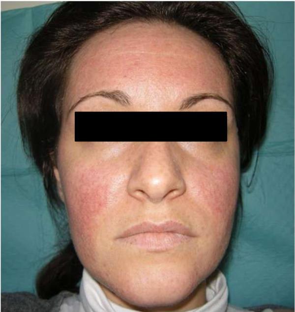
This clinical photograph displays a frontal view of a female patient's face, illustrating a drug-induced cutaneous eruption. The image demonstrates a diffuse, erythematous facial rash with a predominant distribution over the forehead and the malar (butterfly) regions. The erythema is characterized by a pink-to-red hue with an associated fine papular texture, most notably on the bilateral cheeks. Significant sparing is observed in the periorbital (orbicularis oculi) and lower perioral areas. The photograph captures the clinical state seven days following the cessation of a suspected causative agent (metformin), showing a partial resolution with decreased intensity of hyperemia and papular prominence compared to acute presentation. This visual is useful for teaching the morphological characteristics of drug-induced exanthems and their differentiation from dermatological conditions like rosacea or subacute cutaneous lupus erythematosus.
"drug-induced lupus erythematosus" diagnosis management
Drug-Induced Lupus Erythematosus (DILE)
1. Causes (Implicated Drugs)
High-Risk / Classic Agents
| Drug | Class | Notes |
|---|---|---|
| Procainamide | Antiarrhythmic | Highest risk; up to 30% develop DILE; 80% develop ANA positivity |
| Hydralazine | Antihypertensive | First agent identified (1954); dose- and acetylator-dependent |
| Isoniazid | Antitubercular | Slow acetylators at higher risk |
| D-penicillamine | DMARD | Classic trigger |
| Quinidine | Antiarrhythmic | Less common now |
| Minocycline | Antibiotic | Increasingly recognized |
Moderate/Expanding List
- Anti-TNF biologics: Infliximab, etanercept (paradoxically cause DILE despite use in autoimmune disease)
- Anticonvulsants: Phenytoin, carbamazepine, ethosuximide
- Antihypertensives: Methyldopa, ACE inhibitors, beta-blockers
- Antibiotics: Rifampin, nitrofurantoin, tetracyclines
- Antithyroid: Propylthiouracil
- Antipsychotics: Chlorpromazine, lithium
- Others: Sulfasalazine, hydrochlorothiazide (DILE-SCLE), proton pump inhibitors, IFN-alpha, immune checkpoint inhibitors (nivolumab, pembrolizumab), trastuzumab, aromatase inhibitors, vedolizumab
2. Pathogenesis / Risk Factors
- Hapten mechanism: Drugs bind to self-proteins and trigger an immune response
- DNA hypomethylation: Hydralazine and procainamide cause epigenetic alterations in T-cells, leading to overexpression of immune genes and autoimmunity
- Slow acetylator phenotype: Individuals with low N-acetyltransferase activity metabolize drugs more slowly, accumulating reactive intermediates. Slow acetylators develop ANA faster and at lower cumulative doses (confirmed for hydralazine, procainamide, isoniazid). Rapid acetylators are not protected but require higher cumulative doses
- HLA-DR4 association: Genetic susceptibility
- Direct cytotoxicity of drug metabolites
3. Clinical Features
Systemic Symptoms (most common)
- Fever
- Arthralgia / arthritis (most consistent finding)
- Myalgia
- Malaise, fatigue
Serositis
- Pleuritis (pleuritic chest pain, pleural effusion)
- Pericarditis (chest pain, pericardial effusion)
Skin Manifestations
- Malar (butterfly) rash
- Photosensitivity
- In DILE-SCLE: widespread, superficial, non-scarring annular or polycyclic lesions on sun-exposed areas
Hepatic Involvement
- More common in DILE than in idiopathic SLE; elevated liver enzymes
Notably ABSENT or Rare
- Renal involvement - rare (distinguishes from SLE)
- CNS involvement - rare
- Oral ulcers - uncommon
- Severe cytopenias
4. Diagnosis
Diagnostic Approach
| Test | Findings in DILE |
|---|---|
| ANA | Positive (homogeneous pattern most common); often precedes symptoms |
| Anti-histone antibodies | Hallmark of DILE; positive in 75-95% of cases; directed against H2A-H2B dimer complex |
| Anti-dsDNA | Rare in DILE (helps distinguish from SLE, where it is common) |
| Anti-Sm | Absent in DILE (specific to SLE) |
| Anti-ssDNA | Can be positive |
| Complement (C3, C4) | Hypocomplementemia less common than in SLE |
| CBC | Mild cytopenias may occur |
| ESR/CRP | Elevated (nonspecific) |
| Renal function, urinalysis | Usually normal |
- DILE: Anti-histone+ / Anti-dsDNA- / Anti-Sm-
- SLE: Anti-dsDNA+ / Anti-Sm+ / Anti-histone (can be positive too)
Note: Skin biopsy may be indistinguishable from idiopathic SLE, limiting its diagnostic utility.
5. Management
A. Primary Treatment - Drug Withdrawal
- Discontinue the causative drug - this is the cornerstone of treatment
- Symptoms typically resolve within weeks to several months after cessation
- Resolution of clinical findings may occasionally take months to years
- ANA may persist for longer periods even after symptom resolution
B. Symptomatic / Supportive Treatment
- NSAIDs - for arthralgia, myalgia, fever, mild serositis
- Low-dose corticosteroids - for moderate symptoms (e.g., pleuritis, pericarditis, rash)
- Example: Prednisolone 0.5 mg/kg/day, tapered as symptoms resolve
- Hydroxychloroquine - may be used for persistent skin or joint manifestations
C. Immunosuppression
- Reserved for severe or refractory cases
- Azathioprine or other immunosuppressants may be used if symptoms persist despite drug withdrawal and steroids
D. Follow-up
- Monitor ANA and clinical status
- Avoid restarting the offending drug or substituting a structurally related agent
- Choose an alternative drug from a different class
Summary Comparison: DILE vs. Idiopathic SLE
| Feature | DILE | Idiopathic SLE |
|---|---|---|
| Gender ratio | Males = Females | Female >> Male |
| Renal disease | Rare | Common |
| CNS disease | Rare | Common |
| Anti-histone Ab | 75-95% positive | ~50-70% positive |
| Anti-dsDNA | Rare | Common (60-80%) |
| Anti-Sm | Absent | Present |
| Complement | Usually normal | Often low |
| Prognosis | Resolves on drug stop | Chronic, relapsing |