How to measure anteroposterior to transverse thoracic diameter in females

Reading File
Finding Sources
Finding Sources
Searching PubMed

thoracic index anteroposterior transverse diameter ratio

Reading File
Reading File
Reading File
Searching PubMed

Haller index pectus excavatum

Searching Images

Haller index CT transverse and anteroposterior chest diameter measurement
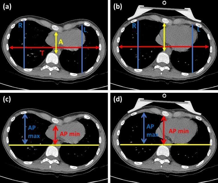
This composite diagnostic image features four axial chest CT scans (a-d) demonstrating the measurement of thoracic indices used to evaluate pectus excavatum (PE). Panels (a) and (b) illustrate the parameters for the Haller Index (HI) and Asymmetry Index (AI). The Haller Index is derived from the ratio of the maximum internal transverse diameter (T, red horizontal line) to the shortest anteroposterior diameter between the sternum and vertebrae (A, yellow vertical line). The Asymmetry Index is calculated using the largest sagittal distances of the right (R) and left (L) hemithorax (blue vertical lines). Panels (c) and (d) demonstrate the Correction Index (CI). This involves measuring the minimum distance between the posterior mediastinum and anterior spine (AP min, red arrow) and the maximum distance between the anterior spine and the inner anterior chest wall (AP max, blue arrow) relative to a horizontal reference line (yellow). Panels (b) and (d) show these measurements while a vacuum bell (VB) device is applied to the anterior chest wall, illustrating the acute change in thoracic morphology and sternal position.
This composite diagnostic image features four axial chest CT scans (a-d) demonstrating the measurement of thoracic indices used to evaluate pectus excavatum (PE). Panels (a) and (b) illustrate the parameters for the Haller Index (HI) and Asymmetry Index (AI). The Haller Index is derived from the ratio of the maximum internal transverse diameter (T, red horizontal line) to the shortest anteroposterior diameter between the sternum and vertebrae (A, yellow vertical line). The Asymmetry Index is calculated using the largest sagittal distances of the right (R) and left (L) hemithorax (blue vertical lines). Panels (c) and (d) demonstrate the Correction Index (CI). This involves measuring the minimum distance between the posterior mediastinum and anterior spine (AP min, red arrow) and the maximum distance between the anterior spine and the inner anterior chest wall (AP max, blue arrow) relative to a horizontal reference line (yellow). Panels (b) and (d) show these measurements while a vacuum bell (VB) device is applied to the anterior chest wall, illustrating the acute change in thoracic morphology and sternal position.

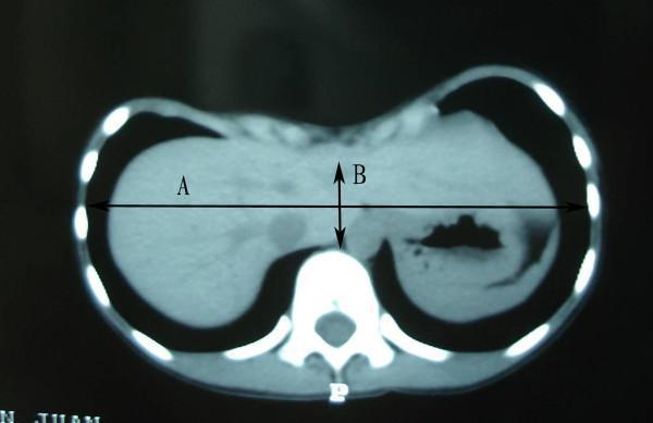
This diagnostic axial CT scan of the lower thorax demonstrates the methodology for calculating the Haller Index in a patient with pectus excavatum. The image displays a clear posterior displacement of the sternum toward the vertebral column, resulting in a significant depression of the anterior chest wall. Two measurement lines are superimposed on the cross-sectional view: Line 'A' represents the maximum internal transverse diameter of the rib cage, measured from the inner aspect of the lateral ribs. Line 'B' represents the minimum anteroposterior diameter at the level of maximum sternal depression, measuring the shortest distance between the posterior surface of the sternum and the anterior surface of the vertebral body. The Haller Index is the mathematical ratio of A divided by B. Visible internal structures include the liver, stomach bubble, and lower lung parenchyma, which appear displaced due to the thoracic cage deformity. This imaging is clinically used to objectively assess the severity of chest wall malformations and guide surgical intervention decisions.
This diagnostic axial CT scan of the lower thorax demonstrates the methodology for calculating the Haller Index in a patient with pectus excavatum. The image displays a clear posterior displacement of the sternum toward the vertebral column, resulting in a significant depression of the anterior chest wall. Two measurement lines are superimposed on the cross-sectional view: Line 'A' represents the maximum internal transverse diameter of the rib cage, measured from the inner aspect of the lateral ribs. Line 'B' represents the minimum anteroposterior diameter at the level of maximum sternal depression, measuring the shortest distance between the posterior surface of the sternum and the anterior surface of the vertebral body. The Haller Index is the mathematical ratio of A divided by B. Visible internal structures include the liver, stomach bubble, and lower lung parenchyma, which appear displaced due to the thoracic cage deformity. This imaging is clinically used to objectively assess the severity of chest wall malformations and guide surgical intervention decisions.

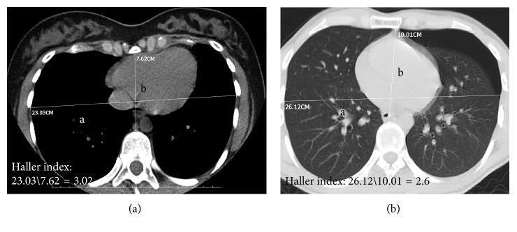
This diagnostic image displays two axial thoracic CT scans illustrating the calculation of the Haller index, a radiological measurement used to assess the severity of chest wall deformities like pectus excavatum. Panel (a) shows a mediastinal window, and panel (b) shows a lung (parenchymal) window. In both images, the Haller index is calculated by dividing the transverse diameter of the thoracic cage (labeled with values 23.03 cm and 26.12 cm) by the shortest anteroposterior distance from the posterior surface of the sternum to the anterior surface of the vertebral body (labeled 'b' with values 7.62 cm and 10.01 cm). The calculated Haller indices are 3.02 and 2.6, respectively. Anatomical landmarks include the heart, vertebral body, and ribs. Additionally, panel (b) demonstrates the lung parenchyma, where a left-sided pneumothorax is noted, visible as a peripheral area devoid of bronchovascular markings. This comparison highlights how different CT windows and patient pathologies, such as pneumothorax, can influence clinical measurements of thoracic morphology.

This diagnostic image displays two axial thoracic CT scans illustrating the calculation of the Haller index, a radiological measurement used to assess the severity of chest wall deformities like pectus excavatum. Panel (a) shows a mediastinal window, and panel (b) shows a lung (parenchymal) window. In both images, the Haller index is calculated by dividing the transverse diameter of the thoracic cage (labeled with values 23.03 cm and 26.12 cm) by the shortest anteroposterior distance from the posterior surface of the sternum to the anterior surface of the vertebral body (labeled 'b' with values 7.62 cm and 10.01 cm). The calculated Haller indices are 3.02 and 2.6, respectively. Anatomical landmarks include the heart, vertebral body, and ribs. Additionally, panel (b) demonstrates the lung parenchyma, where a left-sided pneumothorax is noted, visible as a peripheral area devoid of bronchovascular markings. This comparison highlights how different CT windows and patient pathologies, such as pneumothorax, can influence clinical measurements of thoracic morphology.

The method is the same in females as in males. I found no sex-specific technique in the library. The right approach depends on why you want the ratio.

1. On CT (the standard method, the Haller index)

This is the textbook method for pectus excavatum and other chest wall deformities.
  1. Use an axial chest CT slice at the level of the deepest sternal depression.
  2. Measure the transverse diameter as the maximum internal width of the rib cage, from the inner margin of the ribs on each side.
  3. Measure the AP diameter as the shortest distance from the posterior surface of the sternum (or xiphoid) to the anterior surface of the vertebral body. Drawing a horizontal line across the front of the vertebral body helps.
  4. Divide the transverse diameter by the AP diameter.
Murray & Nadel's textbook says the diameters are "measured from the internal margins of the ribs, vertebra, and sternum." It gives a normal ratio of 2.5 or less. A value above 3.25 indicates a significant deformity that may need surgical correction (Murray & Nadel's Textbook of Respiratory Medicine, Ch. 131, Pectus Excavatum, p. 3067). Current Surgical Therapy 14e also uses 3.25 or more as the usual threshold for operative repair. Goldman-Cecil quotes a ratio above 3.5 as significant (Chapter 86, Clinical manifestations and diagnosis).
Haller index CT measurement
In this image, line A is the maximum internal transverse diameter. Line B is the minimum AP diameter, from the sternum to the vertebral body. The index is A divided by B.
The Haller index can underestimate severity. Current Surgical Therapy 14e suggests the correction index, which measures the percentage of sternal depression, may be better. A value of 28% or more is considered significant.

2. On a chest radiograph

Use a PA film for the transverse (maximum internal rib cage) width. Use the lateral film for the AP depth, from the posterior sternum to the anterior vertebral body, at the same level. Radiographs are less accurate, and CT is preferred for severity.

3. Clinical or bedside estimate (my general knowledge, not from the library)

  • Use calipers or a pelvimeter. Measure the AP diameter at the level of the sternal angle or mid-sternum, and the lateral diameter at the same level.
  • Take both measurements at end-expiration, with the patient relaxed and upright.
  • In women, keep the caliper arm above the breast tissue, at the sternal angle (the level of T4 and the 2nd rib). Otherwise breast volume inflates the AP value.
  • The normal adult AP:transverse ratio is about 0.7 to 0.75. A ratio approaching 1.0 suggests a barrel chest. A low ratio suggests a flat chest or pectus excavatum.

Points for females

  • Pectus excavatum is about four times more common in males than females (Murray & Nadel's).
  • I found no female-specific Haller index cutoffs in the library. Studies may report slightly different normal values by sex and age, so check them if you need exact figures.
  • A narrow AP diameter can also cause innocent flow murmurs (Fuster and Hurst's The Heart).
A search of PubMed for the last two years found only recent pectus excavatum meta-analyses, on surgical technique, fitness and pain. None changes how the index is measured.
If you meant something else, such as a research anthropometry protocol, a pregnancy-related change, or a barrel chest assessment in COPD, tell me and I'll adjust.
This is a shared conversation. Sign in to Orris to start your own chat.