I now have all the content needed. Let me compile the comprehensive answer with all images from Robbins.
Colorectal Carcinoma: Pathogenesis, Morphology & Molecular Correlation (with HNPCC/Lynch Syndrome)
Sources: Robbins & Kumar Basic Pathology (9780323790185) and Robbins, Cotran & Kumar Pathologic Basis of Disease (9780443264528)
Overview and Epidemiology
Colorectal adenocarcinoma is the most common malignancy of the GI tract and responsible for nearly 10% of all cancer deaths worldwide. Approximately 1.2 million new cases occur annually. Incidence peaks at 60-70 years of age. The United States accounts for ~10% of worldwide cases, representing ~15% of all US cancer deaths - second only to lung cancer.
Risk factors:
- Low dietary fiber, high refined carbohydrate and fat intake
- Obesity, tobacco smoking, alcohol consumption
- Protective: aspirin and NSAIDs (via COX-2 inhibition - COX-2 is overexpressed in 90% of colorectal carcinomas and 40-90% of adenomas)
Three Major Molecular Pathways to Colorectal Carcinoma
Robbins, Cotran & Kumar Pathologic Basis of Disease (Table 17.10) describes three pathways:
| Etiology | Molecular Defect | Target Genes | Transmission | Predominant Site | Histology |
|---|
| Familial adenomatous polyposis | APC/Wnt pathway | APC | Autosomal dominant | None (pancolonic) | Tubular, villous; typical adenocarcinoma |
| HNPCC (Lynch syndrome) | DNA mismatch repair | MSH2, MLH1 | Autosomal dominant | Right colon | Sessile serrated polyp; mucinous adenocarcinoma |
| Sporadic (70-80%) | APC/Wnt pathway | APC | None | Left colon | Tubular, villous; typical adenocarcinoma |
| Sporadic (10%) | DNA mismatch repair | MSH2, MLH1 | None | Right colon | Sessile serrated polyp; mucinous adenocarcinoma |
| Sporadic (5-10%) | Hypermethylation (CIMP) | MLH1, BRAF | None | Right colon | Sessile serrated polyp; mucinous adenocarcinoma |
PATHWAY 1: APC/β-catenin Pathway (Classic Adenoma-Carcinoma Sequence)
Accounts for up to 80% of sporadic colon cancers.
The Adenoma-Carcinoma Sequence - Robbins Diagram
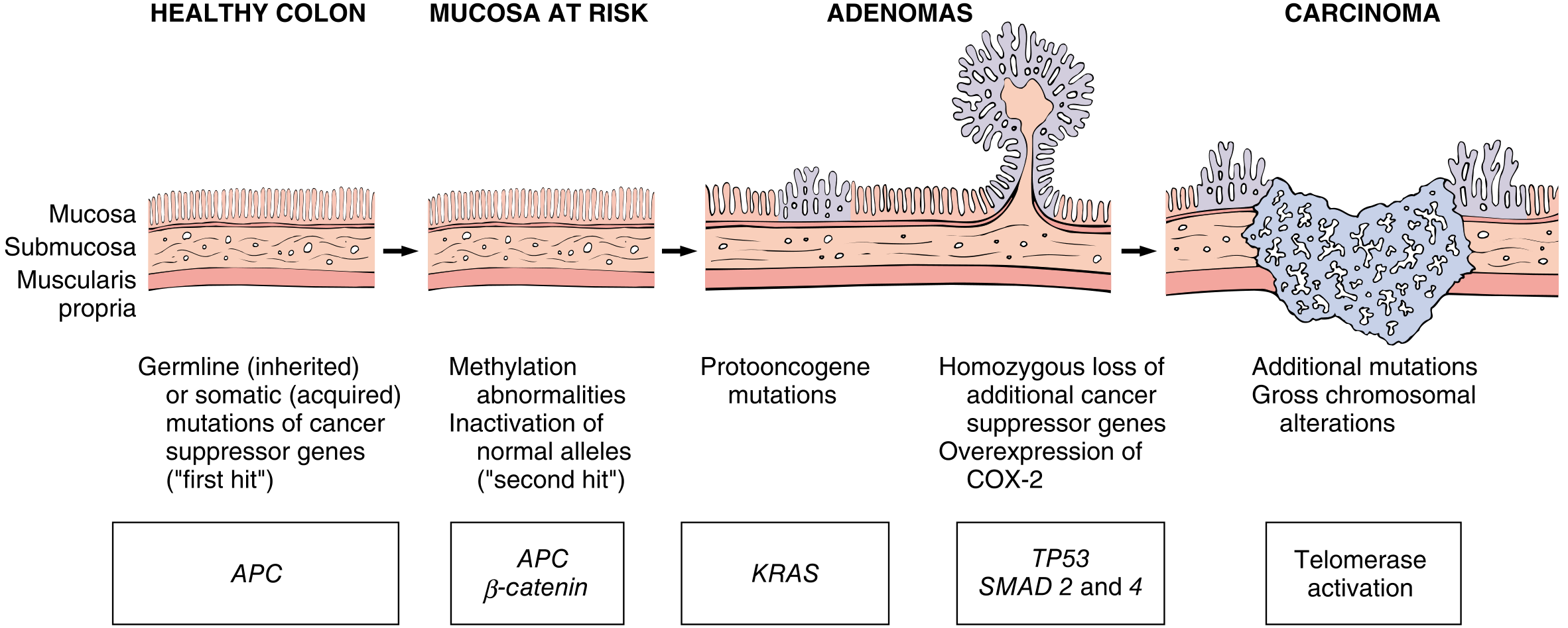
Fig. 17.52 - Robbins, Cotran & Kumar: Morphologic and molecular changes in the adenoma-carcinoma sequence. Loss of one normal copy of the tumor suppressor gene APC occurs early (first hit per Knudson hypothesis), followed by loss of the second copy (second hit). Additional mutations in KRAS, losses at 18q21 (SMAD2 and SMAD4), and inactivation of TP53 lead to carcinoma.
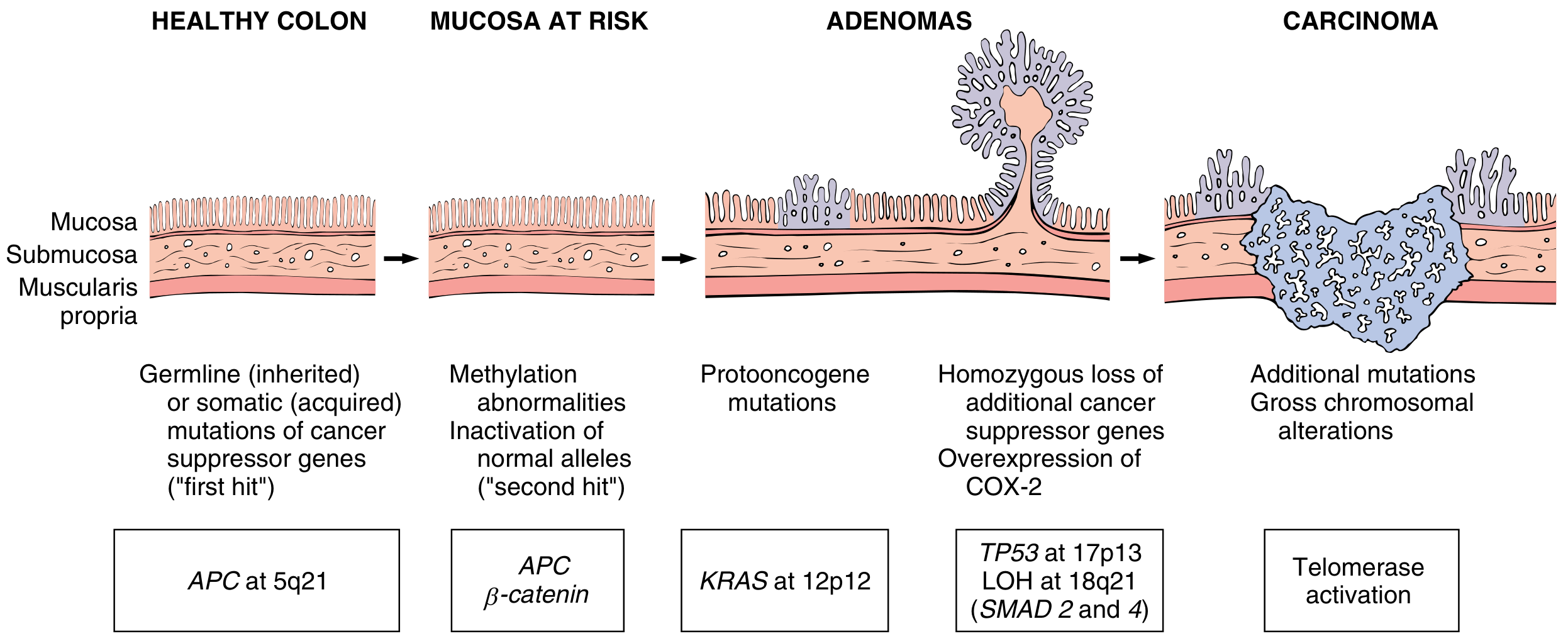
Fig. 13.36 - Robbins & Kumar Basic Pathology: The APC at 5q21 → APC/β-catenin → KRAS at 12p12 → TP53 (17p13), LOH at 18q21 (SMAD2 and 4) → Telomerase activation sequence. COX-2 overexpression occurs at the adenoma stage.
Molecular Steps in APC/β-catenin Pathway
Step 1 - APC loss (chromosome 5q21):
- APC is a key negative regulator of β-catenin in the WNT signaling pathway
- APC normally binds to and promotes degradation of β-catenin
- With APC loss: β-catenin accumulates → translocates to nucleus → forms complex with DNA-binding factor TCF → activates transcription of MYC and cyclin D1 → promotes proliferation
- Both APC alleles must be lost (mutation or epigenetic silencing) for adenoma formation
- In FAP: patients are born with one mutant allele; somatic loss of the second allele triggers adenoma development
- Key note: some colon cancers without APC mutations harbor β-catenin mutations that prevent APC-dependent degradation - same net effect
Step 2 - KRAS mutation (chromosome 12p12):
- Activating mutations promote growth and prevent apoptosis
- Present in <10% of adenomas <1 cm, but 50% of adenomas >1 cm and 50% of invasive adenocarcinomas
- This temporal pattern confirms KRAS mutation is a late event
Step 3 - SMAD2 and SMAD4 loss (18q21):
- These are effectors of TGF-β signaling
- TGF-β normally inhibits the cell cycle; loss allows unrestrained cell growth
Step 4 - TP53 inactivation (17p13):
- Mutated in 70-80% of colon cancers but uncommonly in adenomas
- Confirms this is a very late event in tumor progression
- Often caused by chromosomal deletions (chromosomal instability, CIN)
Step 5 - Telomerase activation:
- Allows indefinite replication without telomere shortening
- Marks transition to frank carcinoma
PATHWAY 2: Microsatellite Instability (MSI) Pathway - The HNPCC/Lynch Syndrome Pathway
HNPCC / Lynch Syndrome
Definition and Genetics
Hereditary nonpolyposis colorectal cancer (HNPCC), also known as Lynch syndrome, is an autosomal dominant condition caused by inherited mutations in DNA mismatch repair (MMR) genes - proteins responsible for detection, excision, and repair of errors occurring during DNA replication.
- HNPCC accounts for 2-4% of all colorectal cancers - the most common syndromic form
- Most patients have mutations in MSH2 or MLH1; at least 5 MMR genes are known
- Patients inherit one mutant gene and one normal allele
- When the second copy is lost (mutation or epigenetic silencing): defects in mismatch repair → mutation rates up to 1000 times higher than normal
Cancer Spectrum in Lynch Syndrome
Cancer risk extends well beyond the colon:
- Colorectum (most common - right colon predominance)
- Endometrium (second most common)
- Stomach, ovary, ureters, brain, small bowel, hepatobiliary tract, pancreas, skin
Molecular Mechanism: Microsatellite Instability
The human genome contains approximately 50,000-100,000 microsatellites - short repeating sequences prone to expansion during DNA replication. These are the most frequent mutation sites in HNPCC.
When MMR is deficient:
- Errors at microsatellite repeats are not corrected
- Accumulation of mutations at rates 1000x higher than normal
- This phenomenon is called microsatellite instability (MSI)
- Key tumor suppressor genes within microsatellite-containing regions are silenced - including TGF-β receptor II and BAX (pro-apoptotic protein)
- Cancer develops through a sessile serrated adenoma → mucinous adenocarcinoma sequence
Distinguishing Features of HNPCC-Associated Cancers vs. Sporadic
| Feature | HNPCC/Lynch | Sporadic (APC pathway) |
|---|
| Age at onset | Younger | Older (peak 60-70) |
| Location | Right colon (proximal) | Left colon (distal) |
| Polyp precursor | Sessile serrated adenoma | Tubular/villous adenoma |
| Histology | Mucinous, often with signet ring cells | Typical glandular adenocarcinoma |
| Molecular defect | MMR deficiency, MSI | CIN, APC/β-catenin mutations |
| Number of precursor polyps | Few | Many (in FAP) or moderate |
PATHWAY 3: CpG Island Methylator Phenotype (CIMP)
- Marked by silencing of genes via promoter hypermethylation - most commonly MLH1
- MLH1 silencing → microsatellite instability without a germline MMR mutation
- Often also involves BRAF mutations
- This explains the ~5-10% of sporadic MSI-high colon cancers that occur via epigenetic rather than mutational MMR gene loss
- Predominantly right-sided, mucinous adenocarcinomas
MORPHOLOGY
Gross Appearances
Right-sided (cecum/ascending colon) tumors:
- Tend to grow as polypoid, exophytic masses that extend along one wall
- Rarely cause obstruction (wide luminal diameter)
- Clinical presentation: occult blood loss → iron deficiency anemia
Left-sided (descending colon/sigmoid/rectum) tumors:
- Tend to be annular, encircling lesions producing "napkin-ring" constriction
- Produce early obstruction
- Clinical features: change in bowel habits, obstipation, cramping
Robbins Gross Photos:
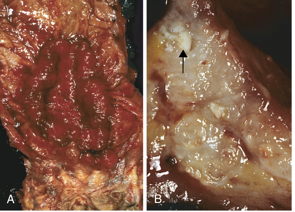
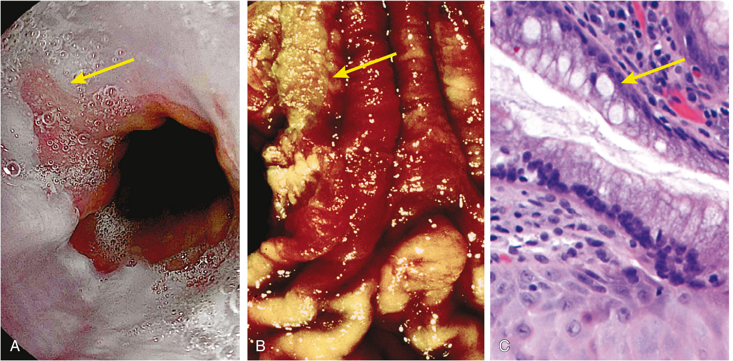
Fig. 17.54 - Robbins, Cotran & Kumar: (A) Circumferential, ulcerated rectal cancer - note the anal mucosa at the bottom. (B) Cancer of the sigmoid colon that has invaded through the muscularis propria into subserosal adipose tissue; areas of chalky necrosis (arrow) within the colon wall.
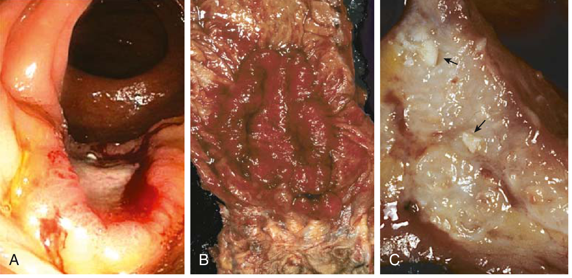
Fig. 13.38 - Robbins & Kumar Basic Pathology: (A) Endoscopic view of ulcerated ascending colon adenocarcinoma. (B) Resected rectum showing circumferential adenocarcinoma with anal mucosa at bottom. (C) Sigmoid colon cancer invading through muscularis propria into subserosal fat (left), with chalky necrosis (arrows).
Histologic Appearances
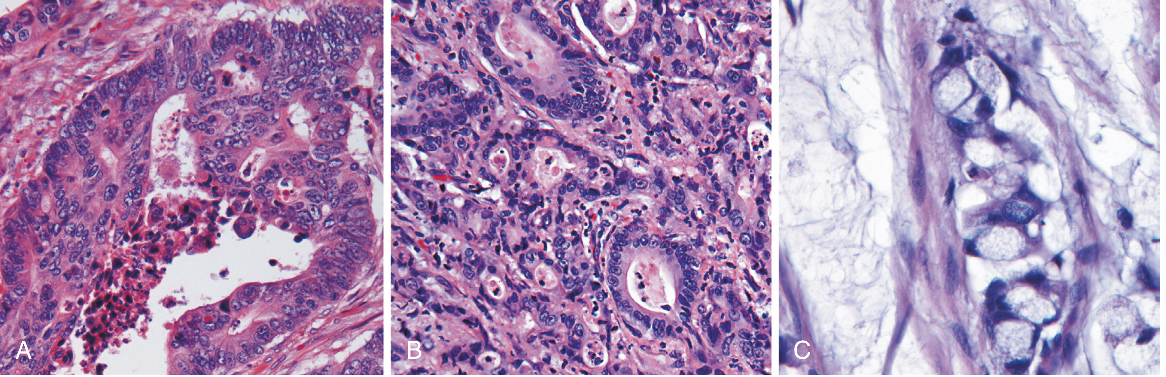
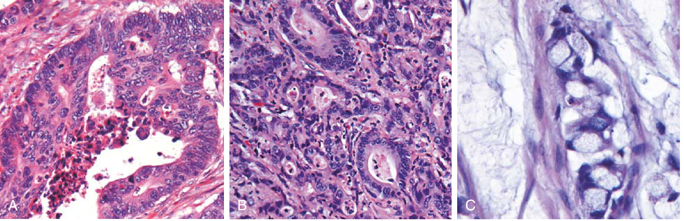
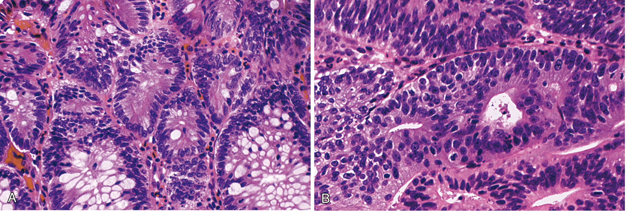
Fig. 17.55 - Robbins, Cotran & Kumar: (A) Well-differentiated adenocarcinoma - elongated hyperchromatic nuclei; necrotic debris in gland lumen is typical. (B) Poorly differentiated adenocarcinoma - forms few glands, largely infiltrating nests. (C) Mucinous adenocarcinoma with signet-ring cells and extracellular mucin pools (characteristic of HNPCC-associated tumors).
Fig. 13.39 - Robbins & Kumar Basic Pathology: Same panel - (A) Well-differentiated with elongated hyperchromatic nuclei and luminal necrotic debris. (B) Poorly differentiated, infiltrating nests. (C) Mucinous adenocarcinoma with signet-ring cells and extracellular mucin pools.
Key histologic subtypes:
- Well-differentiated adenocarcinoma - recognizable glandular structures, elongated hyperchromatic nuclei, luminal necrotic debris ("dirty necrosis")
- Poorly differentiated adenocarcinoma - few glands, predominantly infiltrating solid nests
- Mucinous (colloid) adenocarcinoma - extracellular mucin pools with floating signet-ring cells - characteristic of MSI/HNPCC pathway, right colon, worse prognosis
Metastatic Disease
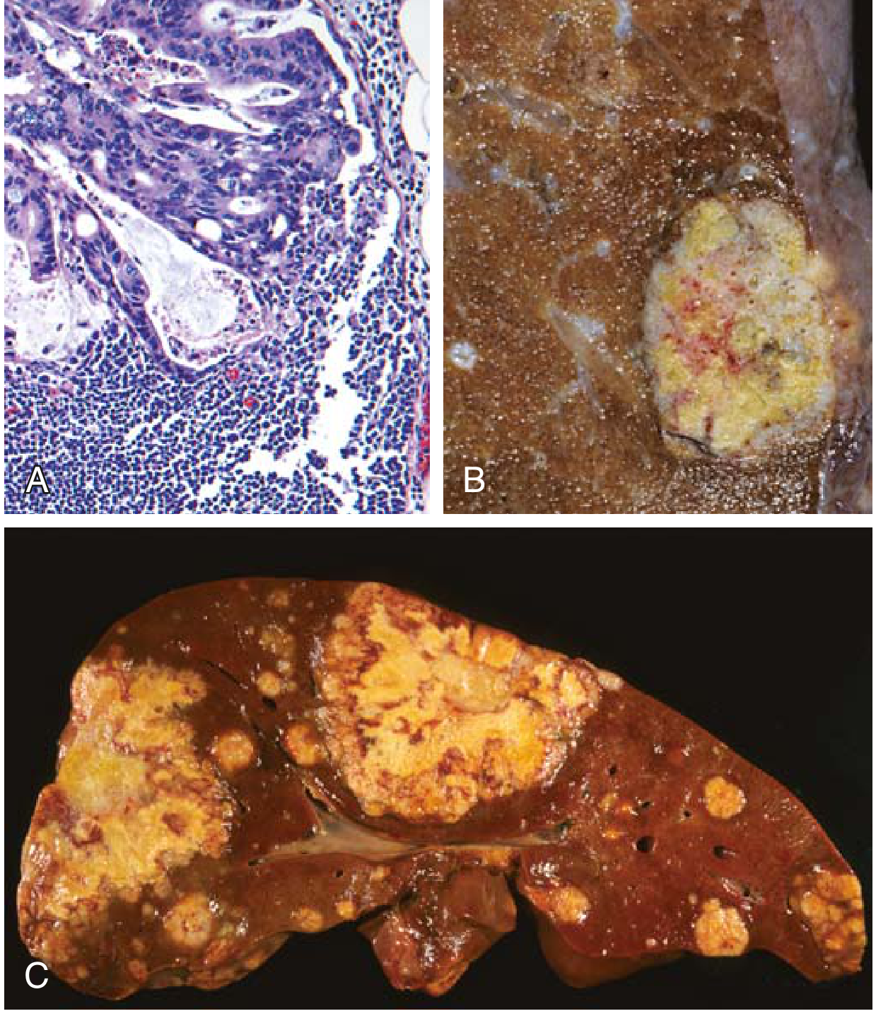
Because of portal drainage of the colon, the liver is the most common site of distant metastasis.
The rectum does not drain via portal circulation - rectal and anorectal carcinomas can bypass the liver and metastasize to the lung first.
Fig. 13.40 - Robbins & Kumar Basic Pathology: (A) Lymph node metastasis - glandular structures in subcapsular sinus. (B) Solitary subpleural lung metastasis. (C) Liver with multiple metastatic deposits - note central necrosis.
Prognosis and Staging
The two most important prognostic factors are:
-
Depth of invasion (T stage):
- Confined to mucosa (no crossing of muscularis mucosae): ~100% 5-year survival
- Into submucosa or muscularis propria: 95% and 70-90% respectively
- Through visceral serosa or into adjacent organs: further reduction
-
Lymph node metastases (N stage): significantly reduces survival; most LN+ cases receive adjuvant chemotherapy ± radiation (neoadjuvant approach common for rectal cancer)
-
Distant metastasis (M stage): only 15% or fewer alive at 5 years
This staging correlates with the historic Dukes classification (A-D) and is now standardized as the TNM system.
Summary: Molecular Correlation Table
PATHWAY GENE DEFECT MECHANISM CANCER TYPE
─────────────────────────────────────────────────────────────────────────────
APC/Wnt APC (5q21) β-catenin accumulation Left colon, tubular/villous
(80% sporadic) KRAS (12p12) Proliferation/survival adenocarcinoma
SMAD2/4 (18q21) Loss TGF-β brake (Chromosomal instability)
TP53 (17p13) Loss apoptosis
MSI Pathway MSH2 / MLH1 MMR deficiency → MSI Right colon, mucinous
(HNPCC, 2-4%; (germline mut.) 1000x mutation rate adenocarcinoma
sporadic 10%) TGFβRII, BAX silenced (MSI-high)
CIMP Pathway MLH1 (epigenetic) Promoter hypermethylation Right colon, mucinous
(sporadic 5-10%) BRAF mutation → MLH1 silencing → MSI adenocarcinoma
Clinical Pearls for HNPCC/Lynch Syndrome
- Amsterdam criteria (clinical diagnosis): 3 relatives with Lynch syndrome-associated cancers, across 2 generations, one diagnosed <50 years, one being first-degree relative of the other two
- Testing: MMR protein immunohistochemistry (loss of MLH1, MSH2, MSH6, PMS2) + MSI PCR testing
- MSI-high tumors (whether sporadic or Lynch-associated) tend to respond better to immune checkpoint inhibitors (pembrolizumab) - this is a major clinical implication
- Colonoscopic surveillance every 1-2 years from age 20-25 for Lynch syndrome carriers
All content and images sourced directly from: Robbins & Kumar Basic Pathology (Basic Pathology, 9780323790185) and Robbins, Cotran & Kumar Pathologic Basis of Disease (9780443264528)