
Hemoglobinopathies
What level of detail and scope do you need?
sickle cell disease hemoglobin polymerization microscopy blood cells
thalassemia red blood cells target cells
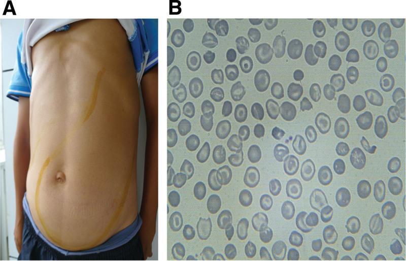
This composite educational image illustrates the clinical signs and laboratory findings of thalassemia intermedia. Panel A is a clinical photograph of a patient's torso showing massive abdominal distention. Yellow skin markings delineate the margins of significant hepatomegaly and massive splenomegaly, with the spleen extending from the left upper quadrant across the midline and deep into the pelvic region, occupying nearly the entire abdominal cavity. Panel B is a peripheral blood smear (light microscopy) demonstrating classic hematological abnormalities associated with thalassemia. The red blood cells exhibit marked microcytosis (small size) and hypochromia (central pallor), along with numerous target cells (codocytes) and irregularly contracted cells (schistocytes). These findings collectively highlight the physical manifestations of extramedullary hematopoiesis and chronic hemolytic anemia characteristic of advanced hemoglobinopathies.
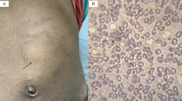
Educational medical image panel consisting of a clinical photograph and a diagnostic pathology image illustrating physical and hematological findings of a hemoglobinopathy such as Hb E/beta-thalassemia. Panel A is a clinical photograph of a patient's abdomen showing a prominent bulge in the left upper quadrant and mid-abdomen, with a black arrow indicating the anterior notch of a massively enlarged spleen (splenomegaly). The overlying skin shows subtle striae or discoloration. Panel B is a peripheral blood smear (Leishman stain, 200x) showing significant red blood cell (RBC) dysmorphology. Key findings include target cells (codocytes) marked by black arrows, teardrop cells (dacrocytes) marked by blue arrows, and basophilic stippling within microcytic, hypochromic RBCs marked by yellow arrows. These visual markers are classic indicators of disordered erythropoiesis and hemoglobin synthesis abnormalities, providing a diagnostic bridge between clinical examination (splenomegaly) and laboratory hematopathology.
Classification
- Sickle Cell Disease (SCD) - most common and most severe
- Other Variant Hemoglobins - HbC, HbD, HbE, unstable hemoglobins
- β-Thalassemia - reduced β-globin chain synthesis
- α-Thalassemia - reduced α-globin chain synthesis
Sickle Cell Disease
Molecular Basis and Pathophysiology
- Higher intracellular hemoglobin concentration
- Lower pH (acidosis)
- Increased temperature
- Decreased oxygen tension
- Fetal hemoglobin (HbF)
- Other hemoglobins (such as HbF or HbC)
Consequences of Polymerization
- Red Cell Damage - polymer formation injures the erythrocyte membrane and cytoskeleton, reducing cation and water content and altering membrane lipid distribution
- Intravascular Hemolysis - damaged red cells rupture, releasing heme into plasma, which:
- Scavenges nitric oxide (NO)
- Reduces NO bioavailability, impairing vasodilation and increasing vasoconstriction
- Generates reactive oxygen species, causing endothelial dysfunction
- Vaso-occlusion - sickled cells interact with endothelium, leukocytes, and platelets to occlude small vessels, causing ischemia-reperfusion injury and pain crises
- Endothelial Activation - reduced NO availability increases expression of adhesion molecules and promotes inflammation
Clinical Features
- Vaso-occlusive crisis (acute pain crisis) - bone and soft tissue pain from microvascular occlusion; most common complication
- Acute chest syndrome - pulmonary infiltrates, chest pain, dyspnea; major cause of mortality
- Acute splenic sequestration - sudden pooling of blood in spleen with shock
- Stroke - from large vessel disease
- Hemolytic anemia (Hb typically 6-9 g/dL)
- Jaundice and gallstones
- Pulmonary hypertension
- Kidney disease and proteinuria
- Avascular necrosis of femoral head
- Retinopathy and vision loss
- Priapism
- Leg ulcers
Diagnosis
- Hemoglobin electrophoresis or HPLC - demonstrates HbS pattern
- Peripheral blood smear - sickled red cells
- Newborn screening - detects HbS in cord blood
- Genetic testing - confirms β6 codon mutation
Management
- Pain management during crises
- Hydration and oxygenation
- Prophylactic penicillin (children < 5 years) and vaccinations (pneumococcal, meningococcal, H. influenzae)
-
Hydroxyurea - "gentle" DNA synthesis inhibitor that:
- Increases fetal hemoglobin (HbF) levels
- Reduces white cell production (anti-inflammatory)
- Increases red cell size, lowering intracellular HbS concentration
- Metabolizes to nitric oxide (NO), a potent vasodilator
- Reduces pain crises and decreases need for transfusions
-
Allogeneic bone marrow transplantation - potentially curative, especially in children with matched donors
-
Gene therapy - emerging corrective approach showing encouraging results
Thalassemias
Molecular Basis and Pathophysiology
- Mutations reduce synthesis of one globin chain type
- The unpaired normal globin chains accumulate and precipitate intracellularly
- These precipitates damage red cells, causing hemolysis and ineffective erythropoiesis
- Result: anemia, tissue hypoxia, and chronic hemolytic disease
Classification and Clinical Severity
β-Thalassemia
| Form | Genotype | Clinical Features |
|---|---|---|
| β-Thalassemia Major | Homozygous (B⁺/B⁻, B⁺/B⁻, or B⁺/B⁺) | Severe anemia; regular blood transfusions required from early childhood; typically presents by 3-6 months |
| β-Thalassemia Intermedia | Variable (B⁺/B⁻, B⁻/B⁻, or mixed) | Moderately severe anemia; transfusions not regularly required; intermediate severity |
| β-Thalassemia Minor (Trait) | Heterozygous (B⁺/B⁻) | Asymptomatic with mild or absent anemia; red cell abnormalities present on smear |
α-Thalassemia
| Form | Genotype | Clinical Features |
|---|---|---|
| Silent Carrier | −/α, α/α | Asymptomatic; no red cell abnormality |
| α-Thalassemia Trait | −/−, α/α or −/α, −/α | Asymptomatic; resembles β-thalassemia minor |
| HbH Disease | −/−, −/α | Moderately severe; moderate hemolytic anemia |
| Hb Bart Hydrops Fetalis | −/−, −/− | In utero hemolytic anemia; severe fetal hydrops; usually fatal in utero |
Laboratory and Clinical Features
- Severe microcytic, hypochromic anemia
- Target cells (codocytes)
- Teardrrop cells (dacrocytes)
- Nucleated red blood cells
- Basophilic stippling
- Hemoglobin electrophoresis shows abnormal patterns
- Presents 3-6 months after birth when HbF naturally declines and adult HbA synthesis normally increases
- Severe hemolytic anemia requiring chronic transfusions
- Massive hepatomegaly and splenomegaly from extramedullary hematopoiesis
- Bone deformities (frontal bossing, maxillary prominence) from marrow expansion
- Growth retardation
- Endocrine complications (hypogonadism, diabetes from iron overload)
- Iron overload from chronic transfusions causing cirrhosis, cardiomyopathy
- Secondary hemochromatosis requiring iron chelation therapy
Diagnosis
- Hemoglobin electrophoresis or HPLC showing reduced HbA with elevated HbF and HbA2
- DNA sequencing to identify specific mutations
- Newborn screening in endemic regions
- Prenatal diagnosis available via amniocentesis or chorionic villus sampling
Management
- Chronic transfusion therapy (maintaining Hb 9-10 g/dL) to suppress ineffective erythropoiesis
- Iron chelation therapy (deferasirox, deferoxamine, deferiprone) to prevent iron overload complications
- Folic acid supplementation
- Spleen removal if transfusion requirements increase
- Hematopoietic stem cell transplantation (curative in 80-90% of matched siblings)
- Gene therapy (emerging, showing promise)
- Usually managed without regular transfusions
- May require transfusions during illness or pregnancy
- Similar monitoring for iron overload and complications
Clinical Images
Other Hemoglobinopathies
- Hemoglobin C - structural variant causing mild hemolytic anemia; when combined with HbS (SC disease) produces moderate disease
- Hemoglobin E - common in Southeast Asia; usually benign as heterozygote, but HbE/β-thalassemia resembles β-thalassemia intermedia
- Hemoglobin D, G, and others - rare variants with variable clinical significance
- Unstable Hemoglobins - structurally abnormal variants prone to precipitation; cause hemolytic anemia
Summary: Key Distinguishing Features
| Feature | Sickle Cell Disease | β-Thalassemia Major |
|---|---|---|
| Defect Type | Qualitative (HbS) | Quantitative (↓ β-globin) |
| Mechanism | HbS polymerization under hypoxia | Excess unpaired α-chains |
| Age of Onset | Symptomatic after 3-4 months | 3-6 months (after HbF decline) |
| RBC Morphology | Sickled cells | Target cells, microcytic, hypochromic |
| Primary Pathology | Vaso-occlusion + hemolysis | Ineffective erythropoiesis + hemolysis |
| Main Complications | Pain crises, stroke, ACS | Hepatosplenomegaly, iron overload, cardiomyopathy |
| Management | Hydroxyurea, transfusions (as needed), bone marrow transplant | Chronic transfusions, iron chelation, transplant |
- Classification - qualitative (structural) vs. quantitative (synthesis) defects
- Sickle Cell Disease - molecular basis, polymerization mechanism, pathophysiology of vaso-occlusion, clinical features, diagnosis, and management
- Thalassemias - α and β types with clinical severity classification (major, intermedia, minor/trait), pathophysiology, laboratory findings, and management
- Other variants - HbC, HbE, unstable hemoglobins
- Clinical images - showing hepatosplenomegaly and characteristic blood smear findings
- Comparison table - key distinguishing features between SCD and β-thalassemia major
- Detailed pathophysiology of a specific complication
- Recent treatment advances or emerging therapies
- Pregnancy management in hemoglobinopathies
- Genetic counseling and prenatal diagnosis
- A particular hemoglobin variant