
Metabolic response to injury chapter 1 in unit 1 of barley and love text book with clear explaining in exam point of view
Reading File
Now let me search the library for Bailey & Love and the metabolic response to injury chapter.
Finding Sources
Finding Sources
Found Bailey and Love. Now let me get the full TOC for Chapter 1 and read the metabolic response to injury section.
Finding Sources
Reading File
Excellent! Found Chapter 1. Let me read the full chapter content in parallel sections.
Reading File
Finding Sources
Finding Sources
Reading File
Reading File
Reading File
Reading File
Reading File
Reading File
Reading File
I now have comprehensive content from Chapter 1 of Bailey & Love. Let me compile the full exam-focused explanation.
Loading Image
Loading Image
I now have all the content needed for a complete, exam-focused explanation. Here is the full chapter breakdown:
Metabolic Response to Injury - Chapter 1, Bailey & Love's Short Practice of Surgery (28th Edition)
1. INTRODUCTION - What Is the Metabolic Response to Injury?
When the body suffers significant tissue damage (from surgery or accidental trauma), it responds with a series of systemic changes affecting vital organ function. This is called the surgical stress response or metabolic response to injury.
Pathways involved:
- Hormonal (neuroendocrine)
- Inflammatory (cytokines)
- Neural circuits
Effects include:
- Altered body metabolism
- Changes in wound healing and immunity
- Specific organ dysfunction
Exam point: If uncontrolled, this response leads to sepsis → MODS (multiple organ dysfunction syndrome). MODS carries ~25% mortality even in modern trauma systems.
2. HOMEOSTASIS AND THE TWO PHASES
Homeostasis = maintaining a constant internal environment for optimal cellular function.
The metabolic response is classically divided into two phases:
| Feature | Catabolic (Ebb) Phase | Anabolic (Flow) Phase |
|---|---|---|
| Timing | Immediately after injury (0-48 hrs) | After resuscitation |
| Metabolic rate | Decreased | Increased (hypermetabolism) |
| Cardiac output | Reduced | Increased |
| Temperature | Hypothermia | Fever |
| Key hormones | Catecholamines, cortisol, aldosterone | Insulin (with resistance) |
| Purpose | Conserve volume and energy | Mobilise stores for repair |
Exam point: The catabolic phase is also called the "ebb" phase and the anabolic flow phase corresponds to SIRS (systemic inflammatory response syndrome).
3. MAGNITUDE OF THE INJURY RESPONSE
The metabolic response is graded - more severe injury = greater response.
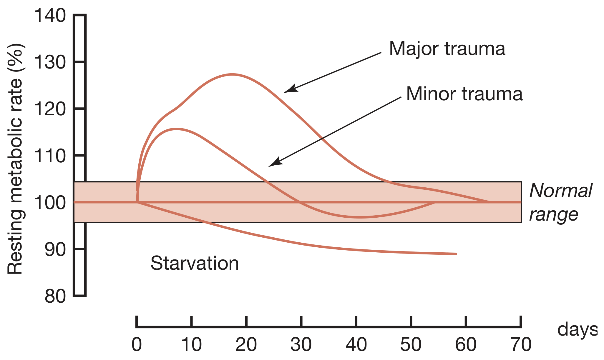
Resting metabolic rate rises proportionally with injury severity. Major trauma peaks around day 10-15 at ~128% of normal.
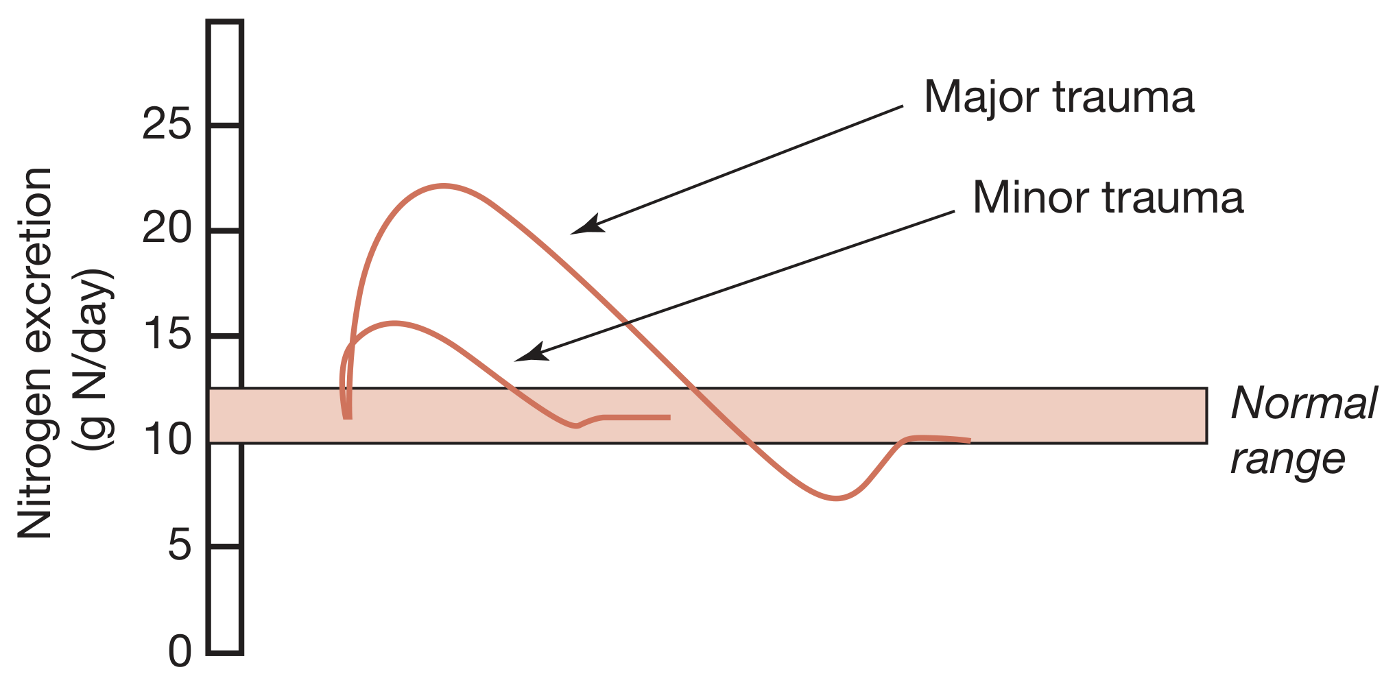
Urinary nitrogen excretion rises to ~22 g N/day in major trauma vs ~15 g N/day in minor trauma, reflecting protein catabolism.
Examples of graded responses:
- Minor elective surgery: transient modest rise in temperature, HR, RR, WBC, energy expenditure
- Major trauma/burns/sepsis: SIRS, hypermetabolism, marked catabolism, shock, MODS
Exam point: Genetic variability also determines response intensity - some patients respond much more dramatically than others to similar injuries.
4. MEDIATORS OF THE METABOLIC RESPONSE
A. Tissue Damage and Inflammation (DAMPs Pathway)
Step-by-step pathway:
- Tissue injury releases DAMPs (damage-associated molecular patterns), also called alarmins
- Examples: heat shock proteins, HMGB1 (high mobility group protein B1), S100 proteins, nucleic acid fragments
- DAMPs activate PRRs (pattern recognition receptors):
- Toll-like receptors (TLRs)
- NOD-like receptors (NLRs)
- Found on: macrophages, neutrophils, dendritic cells
- PRR activation triggers formation of inflammasomes (complex intracellular proteins)
- Inflammasomes activate caspases → activate key cytokines:
- IL-1, IL-6, TNF-alpha, IL-8, interferons, chemokines
- Result: sterile systemic inflammatory cascade → local inflammation → SIRS
Key consequences of DAMPs on endothelium and platelets:
- Leaky capillaries (oedema)
- Coagulopathy
- Local ischaemia → more DAMPs (self-perpetuating cycle)
Secondary triggers that amplify/prolong the response (Table 1.1):
- Sepsis
- Haemorrhage
- Massive transfusion
- Acidosis
- Surgery itself
- Crush syndrome
- Ischaemia-reperfusion injury
Exam point: The "redundancy" of DAMPs (ability to activate multiple receptors/pathways) is why therapeutic blockade of inflammation has been so difficult clinically.
B. Neuroendocrine Response to Injury
The classic pathway:
Tissue injury/pain
       ↓
Afferent nociceptive neurones
       ↓
Spinal cord → Thalamus → Hypothalamus
       ↓
CRF (corticotropin-releasing factor) released
       ↓
ACTH from anterior pituitary
       ↓
Adrenal cortex → CORTISOL (within hours)

Hypothalamus also activates:
→ Sympathetic nervous system → ADRENALINE (epinephrine)
→ Pancreas → GLUCAGON
The "counter-regulatory hormones" triad:
- Glucocorticoids (cortisol)
- Glucagon
- Catecholamines (adrenaline)
High-yield exam fact: IV infusion of these three hormones reproduces many aspects of the metabolic response to injury.
Other hormonal changes:
- Growth hormone (GH) ↑ - but paradoxically with low IGF-1; GH has direct lipolytic, insulin-antagonising and pro-inflammatory properties
- Prolactin ↑
- Peripheral thyroid hormones inactivated
- Gonadal function suppressed
The neuroendocrine response is BIPHASIC (Summary Box 1.2):
| Phase | Timing | Features |
|---|---|---|
| Acute | Hours | Elevated counter-regulatory hormones (cortisol, glucagon, adrenaline) - beneficial for short-term survival |
| Chronic | Days | Hypothalamic suppression, low serum target organ hormones - contributes to chronic wasting |
C. Cytokines and Their Actions
Proinflammatory cytokines (IL-1, TNFα, IL-6, IL-8) produced within first 24 hours:
| Cytokine | Key Action |
|---|---|
| IL-1 | Pyrexia (acts on hypothalamus), augments stress response |
| TNFα | Pyrexia, promotes proteolysis in muscle |
| IL-6 | Major driver of hepatic acute-phase protein response |
| IL-8 | Neutrophil chemotaxis |
Exam point: IL-6 is the primary driver of the hepatic acute-phase protein response.
D. Agonists and Antagonists: An Uncertain Balance
The inflammatory response involves counter-regulatory (anti-inflammatory) mediators as well:
- IL-10, IL-4, TGF-beta (anti-inflammatory)
- Glucocorticoids also have anti-inflammatory effects
This balance between pro- and anti-inflammatory mediators determines whether SIRS progresses to MODS or resolves.
Exam point: CARS (Compensatory Anti-inflammatory Response Syndrome) can lead to immune suppression if the anti-inflammatory arm is too dominant, predisposing to secondary infections.
5. METABOLIC CHANGES AFTER SURGERY AND TRAUMA
Catabolic Phase (0-48 hours)
- Hypovolaemia, ↓ BMR, ↓ cardiac output, hypothermia, lactic acidosis
- Dominant hormones: catecholamines, cortisol, aldosterone
- Insulin levels fall or do not rise appropriately → hyperglycaemia
Flow Phase (after resuscitation - corresponds to SIRS)
Features:
- Tissue oedema (vasodilatation + ↑ capillary permeability)
- Hypermetabolism (↑ BMR)
- ↑ Cardiac output
- Raised body temperature
- Leukocytosis
- ↑ Oxygen consumption
- ↑ Gluconeogenesis
Purpose of neuroendocrine changes (Summary Box 1.4):
- Provide essential substrates for survival via tissue breakdown
- Postpone anabolism
- Optimise host defence
Exam point: These changes are helpful short-term but harmful long-term, especially in the critically ill.
The vicious catabolic cycle:
Catabolism + insulin resistance → hyperglycaemia → ↑ risk of sepsis → further neuroendocrine/inflammatory stimulation → worsened catabolism
6. MANAGING THE CATABOLIC RESPONSE
The body does not catabolise uniformly - it reprioritises resources:
- Catabolic (peripheral) tissues: skeletal muscle, adipose tissue, skin
- Anabolic (central) tissues: liver, immune system, wound
A. Hypermetabolism
- Most trauma patients: energy expenditure 15-25% above predicted resting values
- Burns patients can go even higher
- Caused by: central thermodysregulation (cytokine cascade), ↑ sympathetic activity, ↑ protein turnover, wound circulation abnormalities (Cori cycle)
B. Skeletal Muscle Protein Metabolism
Normal: protein synthesis = breakdown (turnover 1-2%/day)
After injury: breakdown > synthesis → net muscle wasting
Mechanism of muscle wasting:
- ↑ Protein degradation via the ubiquitin-proteasome pathway (key exam fact)
- ↓ Protein synthesis
Consequences of muscle wasting:
- Loss of respiratory muscle → hypoventilation, chest infections
- Loss of gut muscle → ↓ gut motility
- Amino acids (especially glutamine and alanine) exported from muscle to liver (for gluconeogenesis) and wound
- Net catabolism occurs because alanine and glutamine are derived partly from irreversible degradation of branched-chain amino acids
Summary Box 1.5 - Skeletal muscle wasting:
- Provides amino acids for central organs/tissues
- Mediated mainly by ubiquitin-proteasome pathway
- Inevitable to some degree, prolonged by sepsis
- Can cause immobility, poor healing, hypostatic pneumonia, death if excessive
C. Acute-Phase Protein Response (Hepatic)
IL-6 (primarily) drives the liver to reprioritise protein synthesis:
| Acute-Phase Proteins | Direction | Examples |
|---|---|---|
| Positive reactants | ↑ | CRP, fibrinogen, alpha-1 antitrypsin |
| Negative reactants | ↓ | Albumin, transferrin, pre-albumin |
Exam point: Albumin falls after injury NOT mainly due to reduced synthesis, but due to increased transcapillary escape (3x increase in TER) secondary to microvascular permeability changes. Normal albumin TER is already ~10x the synthesis rate.
The acute-phase response is a "double-edged sword" - provides proteins needed for repair but at the cost of lean tissue and energy.
D. Insulin Resistance
After injury:
- Glucose production ↑ (gluconeogenesis)
- Glucose uptake in peripheral tissues ↓
- Mechanism: proinflammatory cytokines + decreased responsiveness of insulin-regulated glucose transporter proteins
- Degree of resistance is proportional to injury magnitude
Clinically:
- After routine upper abdominal surgery: insulin resistance persists ~2 weeks
- With prolonged sepsis: extends further
- Behaves like Type 2 diabetes mellitus
- Management in ICU: IV insulin infusion to maintain blood glucose within limits
- Caution: tight control risks significant hypoglycaemia
7. CHANGES IN BODY COMPOSITION FOLLOWING INJURY
A standard 70 kg male has:
- Fat: 13 kg
- Fat-free mass (protein + water): ~57 kg
After major surgery/critical illness:
- Fat mass ↓
- Skeletal muscle mass ↓
- Body weight may paradoxically increase due to expansion of extracellular fluid (oedema)
Summary Box 1.7:
- Catabolism decreases fat mass and skeletal muscle mass
- Body weight may increase paradoxically due to extracellular fluid expansion
8. AVOIDABLE FACTORS THAT COMPOUND THE RESPONSE
Summary Box 1.8 - Avoidable factors (must know for exams):
| Factor | Mechanism |
|---|---|
| Volume loss / haemorrhage | Activates ADH, RAAS → salt/water retention |
| Hypothermia | Amplifies catabolism, impairs coagulation |
| Tissue oedema | Impairs oxygen delivery, delays healing |
| Systemic inflammation / underperfusion | Perpetuates SIRS, promotes MODS |
| Starvation | Muscle breakdown for energy, immune compromise |
| Immobility | Muscle wasting, DVT, pneumonia |
Volume loss in detail:
- Baroreceptors (carotid, aortic arch) + volume receptors (left atrium) → CNS → ADH + aldosterone release
- ADH → fluid retention at kidney
- ↓ Pulse pressure → juxtaglomerular apparatus → renin-angiotensin → ↑ aldosterone
- Aldosterone → Na+ reabsorption → water conservation
- Net effect: oliguria after surgery (normal/expected)
- Excess saline resuscitation worsens salt/water retention → peripheral AND visceral oedema → delayed gastric emptying, prolonged hospital stay
9. ENHANCED RECOVERY AFTER SURGERY (ERAS)
Modern understanding of the metabolic response has driven ERAS (Enhanced Recovery After Surgery) protocols.
Goal: Actively reduce the need for a homeostatic response by minimising the primary insult.
Summary Box 1.9 - ERAS key elements:
- Minimal access techniques (laparoscopic surgery)
- Blockade of afferent pain stimuli (epidural analgesia, spinal, wound catheters)
- Note: epidural analgesia is no longer recommended for laparoscopic surgery (patient-controlled analgesia is sufficient)
- Minimal periods of starvation (avoid prolonged preoperative fasting)
- Early mobilisation
Benefits of ERAS:
- Reductions in length of hospital stay of 30-50%
- Improved outcomes
- Reduced healthcare costs
Other modulating strategies:
- Beta-blockers: associated with improved short- and long-term survival after major surgery (by modulating the hyper-adrenergic state)
- Epidural analgesia in open surgery: reduces pain, blocks cortisol stress response, attenuates postoperative insulin resistance
- IV lidocaine infusion (6-12 hr): opioid-sparing, improves gut function
- 'One-shot' spinal diamorphine: opioid-sparing effect
HIGH-YIELD EXAM SUMMARY
| Topic | Key Point |
|---|---|
| Two phases of metabolic response | Catabolic (ebb) → Anabolic (flow/SIRS) |
| Initiating molecules | DAMPs activate PRRs → inflammasomes → caspases → IL-1, IL-6, TNFα |
| Counter-regulatory hormone triad | Cortisol + Glucagon + Catecholamines |
| Muscle wasting pathway | Ubiquitin-proteasome pathway |
| Why albumin falls | Increased transcapillary escape (NOT reduced synthesis) |
| Driver of acute-phase response | IL-6 (mainly) |
| Insulin resistance duration | ~2 weeks after upper abdominal surgery |
| Paradox of body weight | Weight may increase despite catabolism (due to oedema) |
| MODS mortality | ~25% |
| ERAS hospital stay benefit | 30-50% reduction |
| GH after injury | ↑ GH but low IGF-1; GH is lipolytic and insulin-antagonising |
| Why DAMP blockade fails clinically | Redundancy - DAMPs activate multiple receptors/pathways |
| Epidural - when NOT used | Not recommended for laparoscopic surgery |
Bailey and Love's Short Practice of Surgery, 28th Edition, Chapter 1, pp. 22-35
Unit 2 of basic principles of surgery from bailey and love tb with detail explanation in exam pov
Finding Sources
Finding Sources
The blocks don't directly correspond to units. Let me look at the actual chapter structure and find what constitutes "Unit 2" (chapters grouped under basic surgical principles).
Finding Sources
Finding Sources
Let me check block1's first page again and look for chapter headings with chapter numbers to find where Unit 2 starts.
Reading File
Finding Sources
Block 1 contains many chapters. Let me read the section around line 600 to find what chapter 2 is, and check the chapter headings in block1.
Reading File
Bailey & Love doesn't use "Units" as a structural division - it uses Chapters. "Unit 2" likely refers to the 2nd major section of the book. In Bailey & Love 28th edition, the early chapters (1-10) cover basic surgical principles. The user likely wants Chapter 2 (Shock and Blood Transfusion) which follows Chapter 1. Let me also check what chapters 3, 4 etc. are and confirm the scope, then read Chapter 2 fully.
Finding Sources
Reading File
Good. Now let me get ALL the headings for Chapter 2 (lines 600-1979) and read key sections in parallel.
Finding Sources
Reading File
Finding Sources
Reading File
Reading File
Reading File
Reading File
Reading File
Reading File
Reading File
Now let me get the transfusion reactions, blood products and key remaining sections.
Reading File
Reading File
I now have comprehensive content for Chapter 2 (Shock & Blood Transfusion) and the start of Chapter 3 (Wound Healing). Bailey & Love doesn't actually label "Units" - it organises by chapters. The first several chapters (1-5) cover basic surgical principles. The user's "Unit 2" most likely refers to Chapter 2: Shock, Haemorrhage and Blood Transfusion. Let me now compile the full, exam-focused answer.> Note: Bailey & Love (28th Edition) does not use a "Unit" structure - it organises by chapters. The early chapters covering Basic Principles of Surgery are Chapters 1-5. "Unit 2" most likely refers to Chapter 2: Shock, Haemorrhage and Blood Transfusion (which immediately follows Chapter 1 on Metabolic Response to Injury). Here is the complete, exam-focused breakdown:
Chapter 2: Shock, Haemorrhage and Blood Transfusion
Bailey & Love's Short Practice of Surgery, 28th Edition - Unit 2 (Basic Principles)
LEARNING OBJECTIVES (Exam Checklist)
- Pathophysiology of shock
- Different patterns of shock + principles/priorities of resuscitation
- Monitoring and end points of resuscitation
- Recognition and management of bleeding
- Blood and blood products, benefits and risks of transfusion
1. SHOCK - DEFINITION
Shock is a systemic state of low tissue perfusion that is inadequate for normal cellular respiration.
With insufficient delivery of oxygen and glucose:
- Cells switch from aerobic → anaerobic metabolism
- If not restored promptly → cell death
2. PATHOPHYSIOLOGY OF SHOCK
A. Cellular Level
- Reduced perfusion → oxygen deprivation
- Anaerobic metabolism → lactic acid production → metabolic acidosis
- Glucose exhaustion → anaerobic respiration ceases
- Failure of Na+/K+ pumps in cell membrane and organelles
- Lysosome rupture → release of autodigestive enzymes → cell lysis
- Intracellular contents (K+) released into bloodstream → hyperkalaemia
B. Microvascular Level
- Hypoxia + acidosis → complement activation + leukocyte priming
- Oxygen free radicals + cytokine release → capillary endothelial injury
- Damaged endothelium becomes "leaky" → fluid extravasation → tissue oedema
- Oedema exacerbates cellular hypoxia (vicious cycle)
- Cell death releases more K+ → hyperkalaemia + acidosis → further immune activation
C. Systemic Level
| System | Response |
|---|---|
| Cardiovascular | Baroreceptor response → ↑ sympathetic activity → catecholamine release → tachycardia + vasoconstriction (EXCEPT in distributive/septic shock) |
| Respiratory | Metabolic acidosis + ↑ sympathetic drive → ↑ RR + ↑ minute ventilation → compensatory respiratory alkalosis |
| Renal | ↓ perfusion pressure → ↓ GFR → ↓ urine output; RAAS activation → further vasoconstriction + Na+/water retention |
| Endocrine | ADH released (in response to ↓ preload) → water retention; RAAS + adrenal systems activated |
3. CLASSIFICATION OF SHOCK (Summary Box 2.1)
Must Know: 5 Types
| Type | Mechanism | Key Features |
|---|---|---|
| Haemorrhagic / Hypovolaemic | Reduced circulating volume | Most common type; basis of all shock |
| Cardiogenic | Primary pump failure | Low CO, high SVR, high venous pressure |
| Obstructive | Mechanical obstruction of cardiac filling | Cardiac tamponade, tension pneumothorax, PE, air embolus |
| Distributive | Vasodilatation + maldistribution of flow | Sepsis, anaphylaxis, neurogenic (spinal injury) |
| Endocrine | Disordered hormonal response | Addison's, hypo/hyperthyroidism |
KEY TABLE 2.1 - Cardiovascular Characteristics of Shock
| Parameter | Hypovolaemic | Cardiogenic | Obstructive | Distributive |
|---|---|---|---|---|
| Cardiac output | Low | Low | Low | High |
| Systemic vascular resistance | High | High | High | Low |
| Venous pressure | Low | High | High | Low |
| Mixed venous saturation | Low | Low | Low | High |
| Base deficit | High | High | High | High |
Exam tip: Distributive (septic) shock is the ONLY type with HIGH cardiac output and LOW SVR. Mixed venous O₂ is HIGH in distributive shock because cells cannot utilise oxygen.
Hypovolaemic Shock - Non-haemorrhagic causes:
- Poor fluid intake (dehydration)
- Vomiting, diarrhoea
- Urinary loss (diabetes insipidus)
- Third-spacing (bowel obstruction, pancreatitis)
Obstructive Shock - Causes (must know):
- Cardiac tamponade - fluid in pericardium compresses heart
- Tension pneumothorax - mediastinal shift, compressed IVC
- Massive pulmonary embolus - obstructs right heart outflow
- Air embolus
Distributive Shock - Three sub-types:
- Septic shock - bacterial/humoral mediators → vasodilatation + arteriovenous shunting
- Anaphylaxis - histamine release → vasodilatation
- Neurogenic shock - high spinal cord injury → loss of sympathetic outflow → loss of vascular tone
4. CLINICAL CONSEQUENCES OF SHOCK
A. Unresuscitatable Shock
- Profound shock for prolonged period → cells pass "point of no return"
- Myocardial cell death from poor coronary perfusion + severe acidaemia + hyperkalaemia
- Loss of ability to maintain SVR → peripheries no longer respond to vasopressors
B. Ischaemia-Reperfusion Injury
- When blood flow is restored after ischaemia, paradoxically worsened injury occurs
- Mechanism: reperfusion activates neutrophils + complement → oxygen free radicals
- Results in capillary endothelial injury, increased permeability, organ dysfunction
C. Multiple Organ Failure (MOF/MODS)
- Sustained ischaemia → sequential organ failure
- Sequence: lungs → kidneys → liver → gut → heart
- Each failing organ amplifies the systemic inflammatory response
Effects of organ failure (Summary Box 2.2):
| Organ | Consequence of Failure |
|---|---|
| Lung | ARDS (acute respiratory distress syndrome) |
| Kidney | Acute kidney injury, oliguria, uraemia |
| Liver | Jaundice, coagulopathy, encephalopathy |
| Gut | Ileus, bacterial translocation, sepsis |
| Heart | Low cardiac output, dysrhythmias |
| Brain | Confusion, coma |
5. RECOGNITION AND DIAGNOSIS OF SHOCK
Stages of Shock
| Stage | Description | Key Features |
|---|---|---|
| Compensated (mild) | Compensatory mechanisms maintain perfusion | Tachycardia, cool peripheries, prolonged CRT, normal BP |
| Decompensation (moderate) | Compensation begins to fail | Hypotension appears, ↑ RR, reduced urine output |
| Severe | Mechanisms overwhelmed | Marked hypotension, altered GCS, oliguria/anuria |
Clinical Signs - KEY EXAM TABLE (Table 2.2)
| Parameter | Class 1 (<15%) | Class 2 (15-30%) | Class 3 (30-40%) | Class 4 (>40%) |
|---|---|---|---|---|
| Blood loss | <750 mL | 750-1500 mL | 1500-2000 mL | >2000 mL |
| Heart rate | Normal | >100 | >120 | >140 |
| Blood pressure | Normal | Normal | Decreased | Very low |
| Respiratory rate | Normal | 20-30 | 30-40 | >35 |
| Urine output | >30 mL/hr | 20-30 mL/hr | 5-15 mL/hr | Negligible |
| CNS/mental status | Slightly anxious | Mildly anxious | Confused | Lethargic |
Important Clinical Pitfalls for Exams:
- Hypotension is a LATE sign - children and fit young adults maintain BP until final stages
- Tachycardia is NOT always present:
- Beta-blocker users cannot mount tachycardia
- Pacemaker patients cannot mount tachycardia
- Young patients with penetrating trauma may have paradoxical bradycardia
- Capillary refill is unreliable in adults - variable; in septic shock peripheries are WARM with BRISK CRT despite profound shock
- Elderly hypertensives may appear normotensive but are actually hypotensive relative to their baseline
6. HAEMORRHAGE
Definition and Key Distinction
Haemorrhage and shock are not the same thing:
- Actively bleeding patient may not yet be in shock
- Shocked patient may no longer be actively bleeding
This distinction is critical for resuscitation:
- Actively bleeding → priority = STOP THE BLEEDING (damage control)
- Not actively bleeding → priority = CORRECT THE SHOCK STATE (restore perfusion)
Types of Haemorrhage
| Type | Description |
|---|---|
| Primary | Occurs at time of injury/surgery |
| Reactionary | Within 24 hours - clot dislodgement as BP rises or patient warms up |
| Secondary | >24 hours (usually 7-14 days) - due to infection eroding vessel wall |
| Revealed | Visible externally |
| Concealed | Hidden internally (e.g. haemoperitoneum, haemothorax, retroperitoneal) |
Exam point: Secondary haemorrhage is almost always due to infection. Reactionary haemorrhage occurs within 24 hours.
Common sites of concealed haemorrhage (must know):
- Chest (haemothorax)
- Abdomen/pelvis (haemoperitoneum, retroperitoneal)
- Long bone fractures (femur fracture: up to 2L blood loss)
- Scalp wounds
Assessment of Blood Loss
- Haemoglobin is a POOR early indicator - in rapid haemorrhage, Hb concentration is UNCHANGED initially (whole blood lost, concentration same)
- Hb falls later as interstitial/intracellular fluid shifts into the vascular compartment
7. HAEMORRHAGE RESUSCITATION (Damage Control Resuscitation)
Principle
In actively bleeding patients → Damage Control Resuscitation (DCR):
- Rapid haemostasis is the priority
- Maintain the blood's ability to clot
Steps:
1. Identify and control haemorrhage:
- External: direct pressure, tourniquet, wound packing
- Internal: operative or interventional radiological control
2. Permissive hypotension:
- In penetrating trauma with active bleeding: accept lower BP (systolic ~80-90 mmHg) to avoid dislodging clots
- NOT indicated in traumatic brain injury (where adequate CPP must be maintained)
3. Haemostatic resuscitation:
- Use blood products in 1:1:1 ratio (packed RBC : FFP : platelets)
- Minimise crystalloid (large volumes worsen coagulopathy, acidosis, hypothermia = "lethal triad")
The Lethal Triad of Trauma:
- Acidosis + Hypothermia + Coagulopathy
- Each worsens the others; together they are frequently fatal
8. SHOCK RESUSCITATION
Fluid Therapy
Rule: Correct hypovolaemia BEFORE vasopressors/inotropes
"Administration of inotropic or chronotropic agents to an empty heart will rapidly and permanently deplete the myocardium of oxygen stores."
Vascular access:
- Short, wide-bore peripheral cannulae (e.g. 14G or 16G)
- Long, narrow central venous catheters have too much resistance for rapid infusion - more appropriate for monitoring
Types of Fluids:
| Fluid Type | Examples | Use |
|---|---|---|
| Crystalloid | Normal saline, Hartmann's (Ringer's lactate) | Standard resuscitation |
| Colloid | Albumin, Gelofusine | No proven advantage over crystalloid; more expensive with worse side effects |
| Blood products | Packed RBC, FFP, platelets | Haemorrhagic shock |
| Hypotonic (avoid) | Dextrose 5% | Poor volume expander; only for free water loss (diabetes insipidus) or Na+ overload |
Exam point: Crystalloid vs. colloid - studies show no clinically significant difference in outcomes. Colloids provide only 1.3x more volume expansion than crystalloid (less than previously thought). No evidence to support routine colloids - more expensive and worse side effects.
Vasopressor and Inotropic Support
- NOT first-line in hypovolaemia
- Only after adequate preload is established
- Dobutamine - for cardiogenic shock (inotrope)
- Noradrenaline - for distributive shock (vasopressor)
- Vasopressin - adjunct vasopressor in refractory septic shock
9. MONITORING IN SHOCK (Table 2.4)
Standard Clinical Monitoring
- Pulse rate, BP, RR
- Temperature
- Urine output (catheter) - most reliable bedside indicator of renal perfusion
- Conscious level (GCS)
- Capillary refill time
Laboratory / Advanced Monitoring
| Parameter | Significance |
|---|---|
| Base deficit | Degree of metabolic acidosis; reflects severity of hypoperfusion |
| Lactate | Product of anaerobic metabolism; >2 mmol/L = significant hypoperfusion; >4 = severe shock |
| Mixed venous O₂ saturation (SvO₂) | Normal 50-70%; <50% = hypovolaemic/cardiogenic; >70% = septic shock |
Mixed venous O₂ in sepsis - exam trap:
- In sepsis, SvO₂ is HIGH (>70%) because cells cannot utilise oxygen + arteriovenous shunting
- If septic patient has SvO₂ <70% → they are in COMBINED septic + hypovolaemic/cardiogenic shock
- Treatment: correct hypovolaemia + inotropes (dobutamine) to get SvO₂ >70%
End Points of Resuscitation
- Lactate clearance - most reliable end point; target lactate <2 mmol/L
- Urine output >0.5 mL/kg/hr
- Normalisation of base deficit
- Normalisation of GCS
Exam point: It is much easier to know when to START resuscitation than when to STOP. Over-resuscitation causes fluid overload, oedema, and worsens outcomes.
10. TRANSFUSION
History Highlights (Table 2.5 - for exam MCQs)
- 1818: James Blundell - first successful human-to-human transfusion (post-partum haemorrhage)
- 1901: Karl Landsteiner - discovered ABO system
- 1914: Albert Hustin - first non-direct transfusion using sodium citrate as anticoagulant
- 1939: Rhesus system identified
Blood Collection
- UK: up to 450 mL drawn, maximum 3 times/year
- Tested for: Hepatitis B, Hepatitis C, HIV-1, HIV-2, Syphilis
- Leukodepleted (precaution against variant CJD; also reduces immunogenicity)
- ABO + Rh(D) typed; irregular red cell antibodies screened
Blood Products
| Product | Content | Indication |
|---|---|---|
| Whole blood | All components | Rarely available in civilian practice; ideal in major haemorrhage |
| Packed red blood cells (pRBC) | RBCs, minimal plasma | Anaemia, haemorrhage |
| Fresh frozen plasma (FFP) | All coagulation factors + protein C/S | Coagulopathy, massive transfusion |
| Platelets | Platelet concentrates | Thrombocytopenia, massive transfusion |
| Cryoprecipitate | Fibrinogen, factor VIII, vWF, factor XIII | Hypofibrinogenaemia, haemophilia A, vWD |
11. BLOOD GROUPS
ABO System (Table 2.7)
| Phenotype | Genotype | Antigens | Antibodies | Frequency |
|---|---|---|---|---|
| O | OO | O | Anti-A, Anti-B | 46% |
| A | AA or AO | A | Anti-B | 42% |
| B | BB or BO | B | Anti-A | 9% |
| AB | AB | AB | None | 3% |
- Group O = Universal donor (no antigens to provoke reaction)
- Group AB = Universal recipient (no circulating antibodies)
Rhesus System
- Rh(D) antigen present in ~85% of UK population
- Rh(D)+ people: develop anti-D antibodies only if exposed to Rh(D)+ blood
- Rh(D)- women who receive Rh(D)+ blood → may form anti-D antibodies → risk of haemolytic disease of the newborn in subsequent pregnancies
- Prevention: anti-D immunoglobulin given to Rh(D)- women after potential sensitisation
Cross-Matching Timeline (critical for exams)
| Urgency | Test | Time |
|---|---|---|
| Immediate/emergency | Group O Rh(D)- blood (females), O+ (males) | Immediately |
| Urgent | Type-specific (ABO/Rh only) | 10-15 minutes |
| Standard | Full cross-match | Up to 45 minutes |
12. TRANSFUSION TRIGGER
TABLE 2.6 - Transfusion criteria (KEY EXAM TABLE):
| Haemoglobin (g/dL) | Action |
|---|---|
| < 6 | Probably will benefit from transfusion |
| 6-8 | Transfusion unlikely to benefit in absence of bleeding or impending surgery |
| > 8 | No indication for transfusion in absence of other risk factors |
Exam point: Restrictive transfusion strategy (Hb 7-8 threshold) is now standard for most patients. Higher threshold may be considered in cardiovascular disease, TBI, sepsis - but clinical evidence is limited.
13. TRANSFUSION REACTIONS
Acute Haemolytic Reaction (most dangerous)
- Cause: ABO incompatibility - complement-mediated intravascular haemolysis
- Features: fever, chills, rigors, flank pain, haemoglobinuria, shock, MOF
- Mechanism: Usually due to wrong blood to wrong patient (clerical error)
- Action: STOP transfusion immediately, IV fluid, inform blood bank, send blood sample
Febrile Non-Haemolytic Reaction
- Due to graft-versus-host response from leukocytes in transfused components
- Features: fever, chills, rigors (WITHOUT haemolysis)
- STOP transfusion immediately
- Rare with leukodepleted blood
Allergic Reaction
- Urticaria, bronchospasm, anaphylaxis
- Due to plasma proteins in donor blood
TRALI (Transfusion-Related Acute Lung Injury)
- Usually from FFP
- Acute onset dyspnoea, hypoxia, bilateral lung infiltrates within 6 hours of transfusion
- Non-cardiogenic pulmonary oedema
- Can be fatal
Safety Checklist (exam-tested)
Two healthcare personnel must check:
- Patient's details against prescription
- Patient's details against donor blood label
- Donor blood serial number against issue slip
14. COMPLICATIONS OF MASSIVE TRANSFUSION
Massive transfusion = >10 units pRBC in 24 hours (or patient's blood volume)
| Complication | Mechanism | Management |
|---|---|---|
| Dilutional coagulopathy | Clotting factors diluted by pRBC | Give FFP + cryoprecipitate |
| Dilutional thrombocytopenia | Platelets diluted | Give platelets |
| Hypocalcaemia | Citrate (anticoagulant) chelates Ca²⁺ | Give IV calcium |
| Hyperkalaemia | K⁺ leaks from stored RBCs | Monitor and treat |
| Hypothermia | Cold blood given rapidly | Warm blood, warming blankets |
| Metabolic alkalosis | Citrate metabolised to bicarbonate | Usually self-limiting |
| TACO (Transfusion-Associated Circulatory Overload) | Volume overload | Diuretics, slow infusion rate |
HIGH-YIELD EXAM SUMMARY - Chapter 2
| Topic | Key Point |
|---|---|
| Definition of shock | Inadequate tissue perfusion for cellular respiration |
| Most common type | Hypovolaemic/haemorrhagic |
| Only high-output shock | Distributive (septic, anaphylactic, neurogenic) |
| Obstructive shock causes | Tamponade, tension pneumothorax, massive PE, air embolus |
| "Silent" sign of shock | Hypotension is LATE; early = tachycardia + cool peripheries |
| Septic shock - warm/cold | WARM peripheries (unlike hypovolaemic) |
| Haemorrhage vs shock | Not the same; active bleeding = stop it first |
| Lethal triad of trauma | Acidosis + Hypothermia + Coagulopathy |
| Fluid of choice | Crystalloid; no proven advantage of colloid |
| Vasopressors - when | ONLY after adequate preload is established |
| SvO₂ in sepsis | HIGH (>70%) - cells cannot utilise O₂ |
| Universal donor | Blood group O |
| Universal recipient | Blood group AB |
| Full cross-match time | 45 minutes |
| Emergency blood | O- (females), O+ (males) |
| Transfusion trigger | Hb <6 = probably transfuse; Hb >8 = no transfusion |
| Most dangerous reaction | Acute haemolytic (ABO incompatibility - clerical error) |
| TRALI cause | Usually from FFP |
| Massive transfusion = | >10 units pRBC in 24 hours |
| Secondary haemorrhage cause | Infection (7-14 days post-injury) |
| Reactionary haemorrhage | Within 24 hours (clot dislodgement) |
| Hb in acute haemorrhage | Initially NORMAL (concentration unchanged; whole blood lost) |
Bailey and Love's Short Practice of Surgery, 28th Edition, Chapter 2, pp. 32-45
Surgical infections in chapter 1 from bailey and love tb detail explanation in exam pov
Finding Sources
Found it. Let me get all headings for this section and read the full content in parallel batches.
Finding Sources
Reading File
Finding Sources
Reading File
Reading File
Reading File
Reading File
Reading File
Reading File
Reading File
Reading File
Reading File
Reading File
Reading File
Reading File
Reading File
I now have all the key content. Let me compile the complete exam-focused notes.
Surgical Infections - Chapter 5
Bailey & Love's Short Practice of Surgery, 28th Edition
Note: In Bailey & Love 28th Edition, "Surgical Infections" is Chapter 5 (not Chapter 1). It is found in the first block (block 1) along with all the basic principles chapters. This is a detailed, exam-focused breakdown of the entire chapter.
LEARNING OBJECTIVES (Exam Checklist)
- Characteristics of common surgical pathogens and their antibiotic sensitivities
- Factors determining whether a wound becomes infected
- Classification of sources of infection and severity
- Clinical presentation of surgical infections
- Indications and choice of prophylactic antibiotics
- Spectrum of commonly used antibiotics and principles of therapy
- Antibiotic resistance - MRSA, C. difficile
- Koch's postulates
- Management of abscesses
- Surviving Sepsis Campaign, Sepsis Bundle, Sepsis Six
- Surgical implications of COVID-19
1. HISTORY OF SURGICAL INFECTION
Key historical milestones (exam MCQs):
| Person | Contribution |
|---|---|
| Egyptians / Greeks / Romans | First antiseptics (wine, vinegar); recognised that pus in wounds needed drainage |
| Galen | Described "pus bonum et laudabile" - suppuration followed by drainage heralded recovery |
| Ignaz Semmelweis (Austrian obstetrician) | Proved handwashing reduced puerperal sepsis from >10% to <2% |
| Robert Koch | Laid down Koch's Postulates (definition of infective disease) |
| Louis Pasteur | Germ theory - microorganisms cause disease |
| Joseph Lister | Antiseptic surgery - carbolic acid for compound fractures |
| Alexander Fleming (1928) | Discovered penicillin |
| Florey and Chain (1941) | First isolated penicillin for clinical use |
| First patient to receive penicillin | Police Constable Alexander (Oxford) |
Koch's Postulates (Summary Box 5.1) - MUST KNOW
- The organism must be found in every case of the disease
- It should be possible to isolate it from the host and grow it in pure culture
- It should reproduce the disease when injected into another healthy host
- It should be recovered from the experimentally infected host
2. ANTIBIOTIC RESISTANCE (Summary Box 5.2)
Mechanism of resistance:
- Bacteria develop resistance via acquisition of β-lactamases - enzymes that break the β-lactam ring in penicillins and cephalosporins
- Bacteria can also develop cell wall changes, efflux pumps, altered ribosomal targets
MRSA (Methicillin-Resistant Staphylococcus aureus)
- Resistant to most common antibiotics
- Found in nose of asymptomatic carriers among patients and staff
- Spread mainly by hand contact (healthcare workers)
- Treatment: Vancomycin or Teicoplanin
- Screening: nasal swabs for at-risk patients preoperatively
C. difficile (Clostridium difficile)
- Toxin-producing anaerobe
- Causes antibiotic-associated pseudomembranous colitis
- Risk: broad-spectrum antibiotics destroy normal flora, allowing C. difficile overgrowth
- Treatment: Metronidazole (mild-moderate) or Vancomycin oral (severe)
- Prevention: judicious antibiotic use, hand hygiene (alcohol gel ineffective; soap and water required)
Exam tip: Alcohol gel does NOT kill C. difficile spores - soap and water is required.
3. MICROBIOLOGY OF SURGICAL INFECTIONS
A. Streptococci (Gram-positive, chains)
| Type | Key Features | Antibiotic |
|---|---|---|
| Group A Streptococcus (S. pyogenes) | Most pathogenic; causes cellulitis, tissue destruction via streptolysin, streptokinase, streptodornase; resides in pharynx of 5-10% | Penicillin, erythromycin |
| Streptococcus faecalis (Enterococcus, Group D) | Found in bowel; wound infections after bowel surgery | Penicillin, erythromycin |
| S. viridans (α-haemolytic) | NOT associated with wound infections; causes endocarditis | - |
| Peptostreptococcus | Anaerobe; acts synergistically | Metronidazole |
Exam point: β-haemolytic streptococcus = most pathogenic. All streptococci remain sensitive to penicillin and erythromycin.
B. Staphylococci (Gram-positive, clumps)
| Type | Key Features | Antibiotic |
|---|---|---|
| S. aureus | Found in nasopharynx of up to 15% of population; causes suppuration in wounds and around prostheses; some strains = MRSA | Flucloxacillin (β-lactamase resistant); MRSA: vancomycin/teicoplanin |
| S. epidermidis | Coagulase-negative; skin commensal; causes prosthetic infections, IV line infections | Vancomycin |
| S. aureus vs MRSA | MRSA resistant to flucloxacillin and most β-lactams | Vancomycin, teicoplanin, linezolid |
C. Clostridia (Gram-positive, anaerobic, spore-bearing bacilli)
| Organism | Disease | Key Features | Treatment |
|---|---|---|---|
| C. perfringens | Gas gangrene | Found in soil/faeces; causes tissue necrosis via collagenase, hyaluronidase, alpha toxin; wound = thin brown SWEET-SMELLING exudate + crepitus | High-dose IV benzylpenicillin + aggressive debridement |
| C. tetani | Tetanus | Exotoxin = tetanospasmin; spasms → opisthotonus → respiratory arrest; widespread in soil | Tetanus toxoid (prophylaxis); benzylpenicillin + wound debridement + muscle relaxants + ventilation |
| C. difficile | Pseudomembranous colitis | Antibiotic-associated; toxin-mediated | Metronidazole or oral vancomycin; stop precipitating antibiotic |
Exam point: Gas gangrene = C. perfringens + sweet-smelling exudate + crepitus + rapidly spreading gangrene. Treatment = IV penicillin + debridement.
D. Aerobic Gram-Negative Bacilli
| Organism | Relevance |
|---|---|
| E. coli | Most common cause of urinary tract and biliary infections |
| Klebsiella | Hospital-acquired pneumonia, UTI; resistant strains common |
| Pseudomonas aeruginosa | Burns, wound infections, ITU; multiple antibiotic resistance - "resident opportunist" |
| Proteus | UTI, wound infections |
Treatment: Cephalosporins (ceftazidime), aminoglycosides (gentamicin), piperacillin-tazobactam
E. Bacteroides (Anaerobic Gram-negative)
- Major component of bowel flora
- Causes intra-abdominal sepsis, wound infections after bowel surgery
- Treatment: Metronidazole (drug of choice for anaerobes)
4. SOURCES OF INFECTION
Endogenous vs Exogenous (Summary Box 5.3)
| Type | Definition | Examples |
|---|---|---|
| Endogenous | Acquired from organisms already present in or on the patient | Bowel flora contaminating abdominal wound; skin flora |
| Exogenous | Acquired from outside the body | Theatre contamination (poor air filtration); ward (poor handwashing) → HAI (hospital-acquired infection) |
Host Defence Mechanisms
Normal barriers preventing infection:
- Mechanical: intact skin and epithelial surfaces
- Chemical: low gastric pH
- Humoral: antibodies, complement, opsonins
- Cellular: phagocytic cells, macrophages, polymorphonuclear cells, killer lymphocytes
All of these may be compromised by surgery and trauma.
5. FACTORS DETERMINING WOUND INFECTION (Summary Box 5.4 - HIGH YIELD)
Four key determinants:
- Host response (immune status)
- Virulence and inoculum of infective agent
- Vascularity and health of tissue (local ischaemia, systemic shock)
- Presence of dead or foreign tissue
- Presence of antibiotics during the "decisive period"
The Decisive Period
- There is up to a 4-hour window before bacterial growth becomes established enough to resist antibiotic therapy
- This is why prophylactic antibiotics must be given at induction of anaesthesia, not before or after
Risk Factors for Wound Infection (Summary Box 5.5)
| Category | Risk Factors |
|---|---|
| Nutritional | Malnutrition, obesity, weight loss |
| Metabolic | Diabetes, uraemia, jaundice |
| Immunosuppression | Cancer, AIDS, steroids, chemotherapy, radiotherapy |
| Gut | Colonisation and translocation from GI tract |
| Circulatory | Systemic shock, local ischaemia, poor perfusion |
| Foreign body | Implants, sutures, drains |
| Surgical technique | Devitalised tissue, dead space, haematoma |
6. CLASSIFICATION OF WOUNDS (Wound Contamination - Table 5.2)
| Wound Type | Definition | Infection Rate (WITHOUT prophylaxis) | Infection Rate (WITH prophylaxis) |
|---|---|---|---|
| Clean | No viscus opened | 1-2% | 1-2% |
| Clean-contaminated | Viscus opened, minimal spillage | 6-9% | 3% |
| Contaminated | Open viscus with spillage or inflammatory disease | 13-20% | 6% |
| Dirty | Pus, perforation, or incision through abscess | 40% | 7% |
Exam tip: Prophylaxis has NO benefit in clean non-prosthetic surgery (infection rate already low). Exception: any surgery involving prosthetic implant - even small infection risk is unacceptable.
7. PRESENTATION OF SURGICAL INFECTION
Hospital-Acquired Infection (HAI) - 4 Main Groups
- Respiratory infections (including ventilator-associated pneumonia)
- Urinary tract infections (mostly catheter-related)
- Bacteraemia (mostly indwelling vascular catheter-related)
- Surgical site infections (SSI)
Major vs Minor SSI
| Major SSI | Minor SSI | |
|---|---|---|
| Pus | Significant quantity, spontaneous or needs drainage | Discharge of pus or infected serous fluid |
| Systemic signs | Tachycardia, pyrexia, raised WBC | None |
| Hospital stay | Delayed discharge | Not delayed |
8. LOCALISED INFECTIONS
Abscess
Cardinal signs of acute inflammation (Celsus - must know):
- Calor (heat)
- Rubor (redness)
- Dolor (pain)
- Tumor (swelling)
- Functio laesa (loss of function) - added later
Pathogenesis:
- Pyogenic organisms (predominantly S. aureus) cause tissue necrosis and suppuration
- Pus = dead and dying neutrophils + bacteria + necrotic tissue
- Abscess wall formed by fibrin + surrounding tissue reaction (pyogenic membrane)
- Central liquefaction → fluctuance
Management of Abscess (Summary Box 5.7):
- "Ubi pus, ibi evacua" = "Where there is pus, evacuate it" - the timeless surgical principle
- Incision and drainage (I&D)
- Send pus for culture and sensitivity
- Leave wound open / pack to allow healing from base up
- Antibiotics alone are INSUFFICIENT for established abscess
Cellulitis and Lymphangitis (Summary Box 5.8)
- Cellulitis: Non-suppurative, poorly localised spreading infection of tissues
- Organisms: β-haemolytic streptococci, staphylococci, C. perfringens
- Features: spreading inflammation, tissue destruction, gangrene, ulceration (proteases)
- Systemic signs: chills, fever, rigors (toxin-mediated cytokine response, even with negative blood cultures)
- Lymphangitis: Painful red streaks in lymphatics draining infection source; painful lymph nodes in drainage area (cervical, axillary, inguinal)
- Treatment: Penicillin + flucloxacillin (or erythromycin if penicillin allergic)
9. SPECIFIC SERIOUS WOUND INFECTIONS
Gas Gangrene (Summary Box 5.9)
| Feature | Details |
|---|---|
| Organism | Clostridium perfringens (Gram+ve, anaerobic, spore-bearing) |
| Source | Soil, faeces; widespread in nature |
| Predisposing factors | Immunocompromised, diabetics, malignancy; wounds with necrotic/foreign material (military wounds ideal) |
| Pathogenesis | Releases collagenase, hyaluronidase, alpha toxin → tissue necrosis + gas production |
| Clinical features | Severe local wound pain, crepitus (gas in tissues), thin BROWN sweet-smelling exudate, rapidly spreading gangrene, circulatory collapse |
| Diagnosis | Clinical + Gram stain of exudate (Gram+ve bacilli); plain X-ray (gas in tissues) |
| Treatment | Large doses IV benzylpenicillin + aggressive debridement; hyperbaric oxygen (adjunct) |
| Prophylaxis | Always consider when amputations done for peripheral vascular disease with open necrotic ulceration |
Tetanus (Clostridium tetani)
| Feature | Details |
|---|---|
| Organism | Clostridium tetani (anaerobic, terminal spore-bearing, Gram+ve) |
| Source | Soil and manure |
| Exotoxin | Tetanospasmin |
| Mechanism | Toxin travels via motor nerves → blocks inhibitory neurotransmitters → spastic paralysis |
| Clinical features | Facial spasms first (short motor nerves) → trismus (lockjaw) → risus sardonicus → generalised spasms → opisthotonus → respiratory arrest → death |
| Treatment | Wound debridement + IV benzylpenicillin + antitetanus immunoglobulin + muscle relaxants + ventilation |
| Prophylaxis | Tetanus toxoid vaccination (active immunisation) |
Exam point: Tetanospasmin travels retrograde up motor nerves. Short motor nerves of face affected first → facial spasms before generalised tetanus.
Necrotising Fasciitis (Synergistic Spreading Gangrene)
| Feature | Details |
|---|---|
| Alternative names | Subdermal gangrene, necrotising fasciitis, Fournier's gangrene (perineum/genitalia) |
| Organisms | Mixed: coliforms, staphylococci, Bacteroides, anaerobic streptococci |
| Key feature | Spreads via deep fascia; much more extensive than apparent from surface |
| LRINEC Score | Used to assess risk of necrotising fasciitis |
| Diagnosis - Finger Test | 2-cm incision down to deep fascia; lack of bleeding + dishwater fluid = positive; finger dissects fascia with minimal resistance |
| Histology | Obliterative vasculitis of subcutaneous vessels + acute inflammation + subcutaneous tissue necrosis |
| Fournier's gangrene | Necrotising fasciitis of scrotum - "shameful exposure of testes" after excision of gangrenous skin |
| Treatment | Broad-spectrum IV antibiotics + aggressive circulatory support + wide excision of all necrotic tissue + skin grafting later |
Exam point: Necrotising fasciitis is a surgical emergency. Subdermal spread is ALWAYS more extensive than surface appearance suggests.
10. SYSTEMIC INFECTION
Bacteraemia
- Transient bacteraemia common after: instrumentation of infected bile/urine, bowel surgery anastomotic breakdown, indwelling IV cannulae colonisation
- Important when prosthesis is implanted (haematogenous seeding → prosthetic infection)
- Organisms: aerobic Gram-negative bacilli, S. aureus, fungi
Definitions of Infected States (Summary Box 5.10)
| Term | Definition |
|---|---|
| SSI | Infected wound or deep organ space |
| Bacteraemia | Bacteria in blood (may be transient) |
| Septicaemia | Bacteria multiplying in blood + systemic effects |
| SIRS | Systemic inflammatory response to severe infection (body's response) |
| Sepsis | SIRS + confirmed infection |
| Severe sepsis | Sepsis + organ dysfunction |
| Septic shock | Severe sepsis + refractory hypotension despite adequate fluid resuscitation |
| MODS | Multiple organ dysfunction syndrome - effect that SIRS produces systemically |
| MSOF | Multiple systemic organ failure - end stage of uncontrolled MODS |
SIRS Criteria (must know)
Two or more of:
- Temperature >38°C or <36°C
- Heart rate >90 bpm
- Respiratory rate >20/min or PaCO₂ <4.3 kPa
- WBC >12,000 or <4,000 or >10% immature (band) forms
11. SURVIVING SEPSIS CAMPAIGN / SEPSIS BUNDLE / SEPSIS SIX
Surviving Sepsis Campaign (SSC)
- Spearheaded by ESICM and SCCM in 2002
- Goal: reduce mortality from sepsis
The Sepsis Bundle (Resuscitation Bundle)
- Evidence-based objectives completed within 6 hours
- For: severe sepsis + septic shock + lactate >4 mmol/L
Sepsis Six (UK Sepsis Trust) - MUST MEMORISE
Give three to patients:
- IV fluid challenge
- IV antibiotics
- Oxygen (and monitor urine output)
Take three from patients:
4. Blood cultures
5. Full blood count
6. Lactate
Exam mnemonic: "Give 3, Take 3" - Fluids, Antibiotics, O₂ / Blood cultures, FBC, Lactate
12. VIRAL INFECTIONS RELEVANT TO SURGERY
Hepatitis B and C
| Hepatitis B | Hepatitis C | |
|---|---|---|
| Transmission | Blood-to-blood (needlestick, cut) | Blood-to-blood |
| Surgeon can carry | Yes, often asymptomatic | Yes |
| Vaccine available | Yes - surgeons must be vaccinated | No |
| Risk to patient | Yes (surgeon to patient) | Yes |
| Treatment | Antiviral agents | Interferon-alpha + ribavirin (potentially curable) |
HIV / AIDS
- Hollow needle injury carries greatest risk of viral transmission
- After needlestick: wash under running water immediately, report incident
- Consider post-exposure antiretroviral therapy (per local policy)
Universal Precautions (CDC/NHS)
For operations on high-risk patients:
- Full face mask (ideally) or protective spectacles
- Fully waterproof disposable gowns and drapes
- Boots (not clogs) - prevent injury from dropped sharps
- Double gloving (larger size inside for comfort)
- Only essential personnel in theatre
- Minimal movement in theatre
- Sharps passed in kidney dish
- Slow, meticulous operative technique with minimised bleeding
COVID-19
- Aetiology: SARS-CoV-2
- Classified as pandemic by WHO on 11 March 2020
- Contagious respiratory and vascular disease
- Symptoms: fever, cough, fatigue, dyspnoea, loss of smell and taste
- Incubation: 1-14 days
- Surgical implications: elective surgery postponed; emergency and cancer surgery continued with PPE
13. PROPHYLACTIC ANTIBIOTICS
Principles
- Value is LOW in clean non-prosthetic surgery (infection rates already low without antibiotics)
- Exception: prosthetic implant surgery - even tiny infection risk is unacceptable
- Clear benefit in clean-contaminated and contaminated operations
- In heavily contaminated wounds / incision through abscess: 5-day therapeutic course (not prophylaxis, as wound assumed already infected)
Timing - THE DECISIVE PERIOD
- Antibiotics MUST be given at induction of anaesthesia (not just before theatre - unexpected delays may occur)
- Maximal blood and tissue levels must be present at time of first incision
- Repeat at 4-hourly intervals in: long operations, excessive blood loss, unexpected contamination
- No evidence for further doses AFTER surgery for prophylaxis - only encourages resistance
Special Indication
- Patients with known valvular heart disease or implanted vascular/orthopaedic prosthesis need prophylactic antibiotics during dental, urological, or open viscus surgery to prevent transient bacteraemia seeding the valve/prosthesis
14. ANTIBIOTICS IN SURGICAL INFECTION
Classification
- Bactericidal: penicillins, aminoglycosides
- Bacteriostatic: tetracyclines, erythromycin
Key Antibiotics (Exam Summary Table)
| Antibiotic | Mechanism | Spectrum | Key Use |
|---|---|---|---|
| Benzylpenicillin | Cell wall synthesis inhibition | Gram+ve (streptococci, clostridia, penicillin-sensitive staph) | Spreading streptococcal infection, gas gangrene (high dose), tetanus |
| Flucloxacillin | Cell wall (β-lactamase resistant) | Penicillinase-producing staphylococci | Soft-tissue infections, osteomyelitis; NOT for other organisms |
| Amoxicillin | Cell wall (β-lactam) | Enterobacteriaceae, E. faecalis, group D streptococci; NOT Klebsiella/Pseudomonas | Broad Gram+ve/Gram-ve coverage |
| Co-amoxiclav | Amoxicillin + clavulanic acid (β-lactamase inhibitor) | Resistant S. aureus, E. coli, H. influenzae, Bacteroides, Klebsiella | Mixed infections, animal/human bites |
| Piperacillin-tazobactam (Tazocin) | Ureidopenicillin + β-lactamase inhibitor | Broad (Gram+ve, Gram-ve, anaerobes, Pseudomonas) | Septicaemia, hospital-acquired pneumonia, complex UTI |
| Cefuroxime / Cefotaxime | Cell wall (cephalosporin, 2nd/3rd gen) | S. aureus, Enterobacteriaceae (NOT Enterococcus faecalis) | Intra-abdominal and soft-tissue infections; surgical prophylaxis |
| Ceftazidime | Cephalosporin, 3rd gen | Gram-ve + S. aureus + Pseudomonas | Pseudomonas infections |
| Metronidazole | DNA disruption | Anaerobes (Bacteroides, Clostridia, anaerobic streptococci), C. difficile | Bowel surgery prophylaxis + treatment; always add for anaerobic cover |
| Gentamicin (aminoglycoside) | Ribosomal (protein synthesis) | Gram-negative bacilli | Combined with other agents; monitor renal function + levels |
| Vancomycin | Cell wall (glycopeptide) | MRSA, Gram+ve organisms | MRSA; C. difficile (oral, severe) |
| Teicoplanin | Glycopeptide | Similar to vancomycin | MRSA (once-daily dosing advantage) |
| Meropenem / Imipenem | Carbapenem (β-lactam) | Very broad (Gram+ve, Gram-ve, anaerobes) | Last resort; severe hospital-acquired infections |
Exam tip: Enterococcus faecalis is NOT sensitive to cephalosporins - must use penicillin or vancomycin.
Exam tip: Aminoglycosides (gentamicin) act at the ribosomal level (prevent/distort protein synthesis). Penicillins act on the cell wall (most effective against actively dividing bacteria).
For Empirical Treatment of Unknown Surgical Infection:
Option 1: Broad-spectrum (teicoplanin or meropenem) + metronidazole
Option 2: Triple therapy - amoxicillin + gentamicin + metronidazole
If no clinical response after 3-4 days → re-evaluate with culture/sensitivity results + investigate for persistent collection of pus.
HIGH-YIELD EXAM SUMMARY
| Topic | Key Point |
|---|---|
| Koch's postulates | 4 criteria to prove organism causes disease |
| First to show handwashing prevents infection | Semmelweis |
| Discovered penicillin | Fleming (1928); isolated by Florey and Chain (1941) |
| Mechanism of antibiotic resistance | β-lactamases break the β-lactam ring |
| MRSA treatment | Vancomycin or teicoplanin |
| C. difficile - alcohol gel | Ineffective - soap and water required |
| Most pathogenic streptococcus | Group A (S. pyogenes) - β-haemolytic |
| All streptococci sensitive to | Penicillin and erythromycin |
| Gas gangrene organism | Clostridium perfringens; sweet-smelling exudate + crepitus |
| Gas gangrene treatment | IV benzylpenicillin + debridement |
| Tetanus toxin | Tetanospasmin; first affects short motor nerves of face |
| Necrotising fasciitis extent | Always more extensive than surface appearance |
| Finger test positive signs | Lack of bleeding + dishwater fluid; tissue dissects easily at fascia |
| Cardinal signs of inflammation | Calor, rubor, dolor, tumor + functio laesa |
| Abscess principle | "Ubi pus, ibi evacua" - where pus, drain it |
| Decisive period | 4-hour window; antibiotics must be in tissue before contamination |
| Prophylaxis timing | At induction of anaesthesia |
| Post-op antibiotic doses | No evidence for benefit; encourages resistance |
| Clean surgery infection rate | 1-2% (prophylaxis makes no difference) |
| Dirty wound infection rate | 40% without prophylaxis; 7% with |
| Sepsis Six - Give 3 | Fluids, IV antibiotics, Oxygen |
| Sepsis Six - Take 3 | Blood cultures, FBC, Lactate |
| Metronidazole covers | Anaerobes (Bacteroides, Clostridia, C. difficile) |
| Cephalosporins miss | Enterococcus faecalis (use penicillin or vancomycin instead) |
| Highest risk needlestick | Hollow needle injury |
| Hepatitis B in surgeons | Vaccine available; surgeons must know their immune status |
| MRSA screening | Nasal swabs for at-risk patients preoperatively |
Bailey and Love's Short Practice of Surgery, 28th Edition, Chapter 5 (Surgical Infections), pp. 72-89
This is a shared conversation. Sign in to Orris to start your own chat.